A Comparison of Posterior Lamellar Keratoplasty Modalities: DLEK vs. DSEK
- Affiliations
-
- 1Department of Ophthalmology, Samsung Medical Center, Sungkyunkwan University School of Medicine, Seoul, Korea. tychung@skku.edu
- KMID: 949055
- DOI: http://doi.org/10.3341/kjo.2010.24.4.195
Abstract
- PURPOSE
To compare clinical outcomes after deep lamellar endothelial keratoplasty (DLEK) with Descemet stripping endothelial keratoplasty (DSEK) performed as initial cases by a single surgeon.
METHODS
Sixteen patients with corneal endothelial were enrolled. Eight patients (8 eyes) underwent DLEK and 8 patients (8 eyes) DSEK. We measured uncorrected visual acuity, best corrected visual acuity (BCVA), manifest refraction, corneal endothelial count, interface opacity via Schiempflug imaging, and contrast sensitivity, as well as tracked postoperative complications over the first postoperative year.
RESULTS
Primary graft failure occurred in two DLEK cases and one DSEK case, all of which were excluded for further analysis. The average 12-month postoperative BCVA was 20/70 in the DLEK group and 20/50 in the DSEK group, with the difference not statistically significant. No significant differences were identified between the 2 groups in terms of mean spherical equivalent and refractive astigmatism, although individuals in the DSEK group tended toward hyperopia. The average endothelial cell count at postoperative month 12 was 1849+/-494 in the DLEK group and 1643+/-417 cells/mm2 in the DSEK group, representing cell losses of 26.2% and 31.9%, respectively. No significant differences in endothelial cell count or endothelial cell loss were observed between groups. Early postoperative donor disc dislocation occurred in two eyes after DLEK and one eye after DSEK. Interface opacities and contrast sensitivities were similarly not significantly different between groups.
CONCLUSIONS
No significant differences in any assessed clinical outcome were observed between individuals undergoing DLEK and DSEK, when performed as initial cases by a single surgeon.
Keyword
MeSH Terms
Figure
-
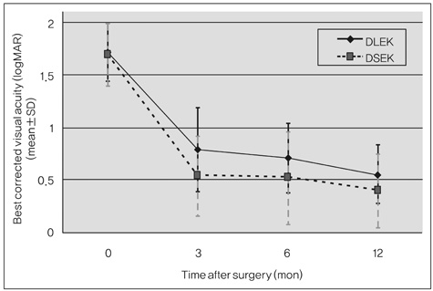
Fig. 1 The best corrected visual acuities (logarithm of the minimum angle of resolution [logMAR] units) in the deep lamellar endothelial keratoplasty (DLEK) and Descemet stripping endothelial keratoplasty (DSEK) groups versus postoperative time. No significant differences were identified between the 2 groups (p>0.05).
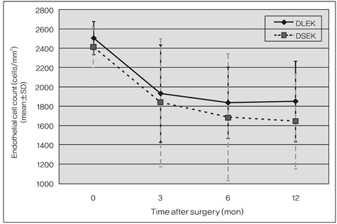
Fig. 2 Longitudinal endothelial cell counts observed over the first postoperative year after deep lamellar endothelial keratoplasty (DLEK) or Descemet stripping endothelial keratoplasty (DSEK). No significant differences were observed between groups at any time point (p>0.05).

Fig. 3 Longitudinal changes in mean group contrast sensitivity function values. No significant differences were observed between groups (p>0.05). Patients in both groups had notably lower contrast sensitivity function values when compared with normal healthy controls. DLEK=deep lamellar endothelial keratoplasty; DSEK=Descemet stripping endothelial keratoplasty.
Reference
-
1. Terry MA, Ousley PJ. Replacing the endothelium without corneal surface incisions or sutures: the first United States clinical series using the deep lamellar endothelial keratoplasty procedure. Ophthalmology. 2003. 110:755–764.2. Terry MA, Ousley PJ. Deep lamellar endothelial keratoplasty visual acuity, astigmatism, and endothelial survival in a large prospective series. Ophthalmology. 2005. 112:1541–1548.3. Melles GR, Wijdh RH, Nieuwendaal CP. A technique to excise the descemet membrane from a recipient cornea (descemetorhexis). Cornea. 2004. 23:286–288.4. Price FW Jr, Price MO. Descemet's stripping with endothelial keratoplasty in 50 eyes: a refractive neutral corneal transplant. J Refract Surg. 2005. 21:339–345.5. Price FW Jr, Price MO. Descemet's stripping with endothelial keratoplasty in 200 eyes: early challenges and techniques to enhance donor adherence. J Cataract Refract Surg. 2006. 32:411–418.6. Melles GR. Posterior lamellar keratoplasty: DLEK to DSEK to DMEK. Cornea. 2006. 25:879–881.7. Terry MA, Hoar KL, Wall J, Ousley P. Histology of dislocations in endothelial keratoplasty (DSEK and DLEK): a laboratory-based, surgical solution to dislocation in 100 consecutive DSEK cases. Cornea. 2006. 25:926–932.8. Allan BD, Terry MA, Price FW Jr, et al. Corneal transplant rejection rate and severity after endothelial keratoplasty. Cornea. 2007. 26:1039–1042.9. Fogla R, Padmanabhan P. Initial results of small incision deep lamellar endothelial keratoplasty (DLEK). Am J Ophthalmol. 2006. 141:346–351.10. Terry MA, Ousley PJ. Rapid visual rehabilitation after endothelial transplants with deep lamellar endothelial keratoplasty (DLEK). Cornea. 2004. 23:143–153.11. Koenig SB, Covert DJ. Early results of small-incision Descemet's stripping and automated endothelial keratoplasty. Ophthalmology. 2007. 114:221–226.12. Koenig SB, Covert DJ, Dupps WJ Jr, Meisler DM. Visual acuity, refractive error, and endothelial cell density six months after Descemet stripping and automated endothelial keratoplasty (DSAEK). Cornea. 2007. 26:670–674.13. Gorovoy MS. Descemet-stripping automated endothelial keratoplasty. Cornea. 2006. 25:886–889.14. Jager MJ, Hermans LJ, Kok JH. Visual results after corneal transplantation. Doc Ophthalmol. 1989. 72:265–271.15. Schraepen P, Koppen C, Tassignon MJ. Visual acuity after penetrating keratoplasty for pseudophakic and aphakic bullous keratopathy. J Cataract Refract Surg. 2003. 29:482–486.16. Holz HA, Meyer JJ, Espandar L, et al. Corneal profile analysis after Descemet stripping endothelial keratoplasty and its relationship to postoperative hyperopic shift. J Cataract Refract Surg. 2008. 34:211–214.17. Yoo SH, Kymionis GD, Deobhakta AA, et al. One-year results and anterior segment optical coherence tomography findings of descemet stripping automated endothelial keratoplasty combined with phacoemulsification. Arch Ophthalmol. 2008. 126:1052–1055.18. Terry MA. Endothelial keratoplasty: clinical outcomes in the two years following deep lamellar endothelial keratoplasty (an American Ophthalmological Society thesis). Trans Am Ophthalmol Soc. 2007. 105:530–563.19. Ousley PJ, Terry MA. Stability of vision, topography, and endothelial cell density from 1 year to 2 years after deep lamellar endothelial keratoplasty surgery. Ophthalmology. 2005. 112:50–57.20. Van Dooren B, Mulder PG, Nieuwendaal CP, et al. Endothelial cell density after posterior lamellar keratoplasty (Melles techniques): 3 years follow-up. Am J Ophthalmol. 2004. 138:211–217.21. Terry MA, Chen ES, Shamie N, et al. Endothelial cell loss after Descemet's stripping endothelial keratoplasty in a large prospective series. Ophthalmology. 2008. 115:488–496. e3.22. Silva CA, Oliveira ES, Sena Junior MP, Souza LB. Contrast sensitivity in deep anterior lamellar keratoplasty versus penetrating keratoplasty. Arq Bras Oftalmol. 2008. 71:71–74.23. Terry MA, Ousley PJ. Deep lamellar endothelial keratoplasty: early complications and their management. Cornea. 2006. 25:37–43.24. Bahar I, Kaiserman I, McAllum P, et al. Comparison of posterior lamellar keratoplasty techniques to penetrating keratoplasty. Ophthalmology. 2008. 115:1525–1533.
- Full Text Links
-
- Actions
-
Cited
- CITED
-
- Close
- Share
-
- Similar articles
-
- Results of Deep Lamellar Endothelial Keratoplasty (DLEK)
- Clinical Evaluation of Full-thickness Deep Lamellar Keratoplasty
- Case Report: Femtosecond Laser-Assisted Small Incision Deep Lamellar Endothelial Keratoplasty
- Descemet Membrane Endothelial Keratoplasty to Treat Graft Failure after Descemet Stripping Endothelial Keratoplasty
- A Case of Anterior Synechiolysis with Lamellar Corneal Dissection in Penetrating Keratoplasty
