Morphologic Feasibility of Pedicle Screw Insertion in Korean
- Affiliations
-
- 1Department of Orthopaedic Surgery, Yeungnam University Hospital, Daegu, Korea. mwahn@med.yu.ac.kr
- KMID: 854596
- DOI: http://doi.org/10.4055/jkoa.2007.42.2.255
Abstract
- PURPOSE: This study examined the morphological characteristics of the thoracic and lumbar vertebrae of normal Koreans and the factors causing breakage of the pedicular wall by measuring the thoracolumbar vertebrae relative to the pedicle screw insertion. MATERIALS AND METHODS: The effect of the pedicle screw shape on the pedicle wall integrity of 56 normal Koreans was examined by performing a computer simulation of the inserting pedicle screws into the pedicle wall by superimposing the graphical images of the screws onto the CT scan images. RESULTS: Because the inner pedicle diameters of the most thoracic vertebrae from T4 to T10 were <5 mm, most pedicles of the thoracic vertebrae were expected to be broken after inserting the 5 mm-diameter cylindrical screws. The pedicles of the thoracic and lumbar vertebrae were classified into 6 groups by performing the cluster analysis using morphometric parameters. Group 1 was labeled "relatively narrow". Group 2 "moderate". Group 3 "wide and angular". Group 4 "severly narrow and short", Group 5 "long", and group 6 "relatively wide and angular". The simulation showed the pedicles of groups 1 and 4 to be too narrow for the 5 mm-diameter cylindrical screws to preserve the pedicular wall integrity. CONCLUSION: The pedicles of the vertebra of Koreans are similar in size to those of Caucasians. Personal morphological characteristics of the pedicles as well as their sizes and levels of the vertebrae are believed to be the significant factors that can cause the breakage of the pedicular wall.
Figure
-
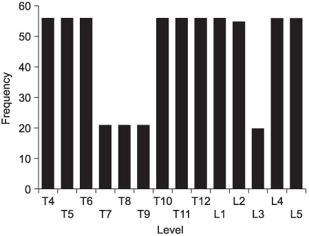
Fig. 1 Distribution of the slices of the axial CAT images.
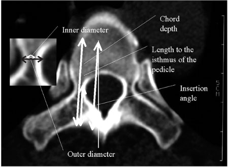
Fig. 2 Morphometric parameters of the vertebrae.

Fig. 3 Status of the pedicular wall during the insertion of screw into the pedicle.
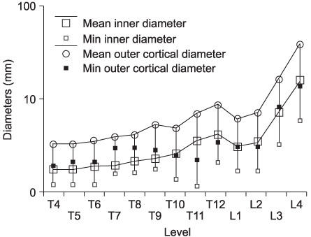
Fig. 4 Inner and outer diameter of the pedicular isthmus from T4 to L5.
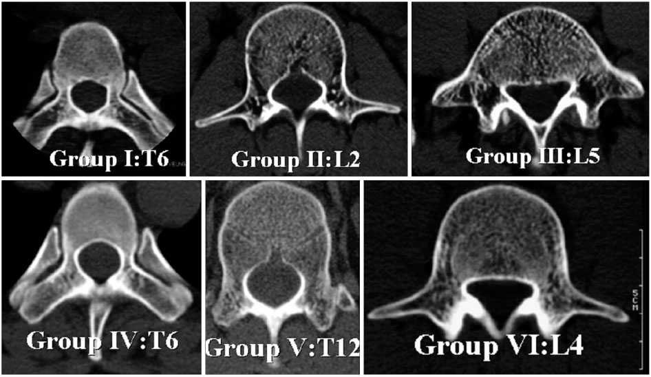
Fig. 5 Morphologic characteristics of each cluster. Group I was characterized by the severe narrowing of the inner and outer pedicular diameters and the short pedicular length. Group II was characterized by the narrow pedicular canal. However, the pedicular length and insertion angle had increased. The pedicular canal and insertion angle had increased the most in Group III. Group IV was characterized by it's the largest decrease in the pedicular canal and length. The pedicular length had increased the most in Group V. Its pedicular canal was relatively wide. In Group VI, the pedicular canal was wide and its length and insertion angle had increased compared with those of the other groups.
Reference
-
1. Alanay A, Cil A, Acaroglu E, et al. Late spinal cord compression caused by pulled-out thoracic pedicle screws: a case report. Spine. 2003. 28:E506–E510.
Article2. Berry JL, Moran JM, Berg WS, Steffee AD. A morphometric study of human lumbar and selected thoracic vertebrae. Spine. 1987. 12:362–367.
Article3. Cheung KM, Ruan D, Chan FL, Fang D. Computed tomographic osteometry of Asian lumbar pedicles. Spine. 1994. 19:1495–1498.
Article4. Cinotti G, Gumina S, Ripani M, Postacchini F. Pedicle instrumentation in the thoracic spine. A morphometric and cadaveric study for placement of screws. Spine. 1999. 24:114–119.5. Datir SP, Mitra SR. Morphometric study of the thoracic vertebral pedicle in an Indian population. Spine. 2004. 29:1174–1181.
Article6. Heini P, Scholl E, Wyler D, Eggli S. Fatal cardiac tamponade associated with posterior spinal instrumentation. A case report. Spine. 1998. 23:2226–2230.7. Hou S, Hu R, Shi Y. Pedicle morphology of the lower thoracic and lumbar spine in a Chinese population. Spine. 1993. 18:1850–1855.
Article8. Kim NH, Lee HM, Chung IH, Kim HJ, Kim SJ. Morphometric study of the pedicles of thoracic and lumbar vertebrae in Koreans. Spine. 1994. 19:1390–1394.
Article9. Kim YJ, Lenke LG, Cheh G, Riew KD. CT scan accuracy of "free hand" pedicle screw placement technique in spinal defomity; an analysis of 789 pedicl screws. 2004. In : Podium presentation, internaional meeting on advanced spine technology annual meeting; July 1-3; Bermuda.10. Krag MH, Beynnon BD, Pope MH, Frymoyer JW, Haugh LD, Weaver DL. An internal fixator for posterior application to short segments of the thoracic, lumbar and lumbosacral spine, disign and testing. Clin Orthop Relat Res. 1986. 203:75–98.11. Krag MH, Weaver DL, Beynnon BD, Haugh LD. Morphometry of the thoracic and lumbar spine related to transpedicular screw placement for surgical spine fixation. Spine. 1988. 13:27–32.12. Levine DS, Dugas JR, Tarantino SJ, Boachie-Adjei O. Chance fracture after pedicle screw fixation. A case report. Spine. 1998. 23:382–385.13. Misenhimer GR, Peek RD, Wiltse LL, Rothman SL, Widell EH Jr. Anatomic analysis of pedicle cortical and cancellous diameter as related to screw size. Spine. 1989. 14:367–372.
Article14. Papin P, Arlet V, Marchesi D, Rosenblatt B, Aebi M. Unusual presentation of spinal cord compression related to misplaced pedicle screws in thoracic scoliosis. Eur Spine J. 1999. 8:156–159.
Article15. Rinella AS, Cahill P, Ghanayem A, et al. Thoracic pedicle expansion after pedicle screw placement in a pediatric cadaveric spine. 2004. In : Podium presentation, Scoliosis research society 2004 annual meeting; September; Buenos aires, Argentina.16. Saillant G. Anatomical study of the vertebral pedicles. Surgical application. Rev Chir Orthop Reparatrice Appar Mot. 1976. 62:151–160.17. Liljenqvist U, Hackenberg L, Link T, Halm H. Pullout strength of pedicle screws versus pedicle and laminar hooks in the thoracic spine. Acta Orthop Belg. 2001. 67:157–163.18. Scoles PV, Linton AE, Latimer B, Lery ME, Digiovanni BF. Vertebral body and posterior element morphology: the normal spine in middle life. Spine. 1988. 13:1082–1086.19. Sjostrom L, Jacobsson O, Karlstrom G, Pech P, Rauschning W. CT analysis of pedicles and screw tracts after implant removal in thoracolumbar fractures. J Spinal Disord. 1993. 6:225–231.20. Suk SI, Lee JH, Yoon KS, Kim WJ. The diameter and changes of the vertebral pedicles after screw insertion. J Kor Soc Spine Surg. 1995. 2:168–176.21. Tan SH, Teo EC, Chua HC. Quantitative three-dimensional anatomy of cervical, thoracic and lumbar vertebrae of Chinese Singaporeans. Eur Spine J. 2004. 13:137–146.22. Ugur HC, Attar A, Uz A, Tekdemir I, Egemen N, Genc Y. Thoracic pedicle: Surgical anatomic evaluation and relations. J Spinal Disord. 2001. 14:39–45.23. Vaccaro AR, Rizzolo SJ, Allardyce TJ, et al. Placement of pedicle screws in the thoracic spine. Part I: morphometric analysis of the thoracic vertebrae. J Bone Joint Surg. m. 1995. 77:1193–1199.24. Vaccaro AR, Rizzolo SJ, Balderston RA, et al. Placement of pedicle screws in the thoracic spine. Part II: An anatomical and radiographic assessment. J Bone Joint Surg. r. 1995. 77:1200–1206.25. Zindrick MR, Knight GW, Sartori MJ, Carnevale TJ, Patwardhan AG, Lorenz MA. Pedicle morphology of the immature thoracolumbar spine. Spine. 2000. 25:2726–2735.
Article26. Zindrick MR, Wiltse LL, Dorrnik A, et al. Analysis of the morphometric characteristics of the thoracic and lumbar pedicles. Spine. 1987. 12:160–166.
Article
- Full Text Links
-
- Actions
-
Cited
- CITED
-
- Close
- Share
-
- Similar articles
-
- Rate of Pedicle Disruption after Screw Fixation
- A Biomechanical Study of Two Kinds of Tapered Pedicle Screws in Osteoporotic Lumber Spine
- Cervical Pedicle Screw Fixation: Anatomic Feasibility of Pedicle Morphology and Radiologic Evaluation of the Anatomical Measurements
- Safety of Pedicle Screws in Adolescent Idiopathic Scoliosis Surgery
- Radiologic Evaluation of Proper Pedicle Screw Placement after Pedicle Screw Fixation in Degenerative Lumbar Disc Disease
