A Study of the Vascular Network of the Iris Using Flat Preparation
- Affiliations
-
- 1Department of Ophthalmology, Hanyang University Hospital, Seoul, Korea. fovea@hanyang.ac.kr
- KMID: 754767
- DOI: http://doi.org/10.3341/kjo.2009.23.4.296
Abstract
- PURPOSE
This study was performed to examine the vascular network of the human iris using flat preparation.
METHODS
The ciliary body-iris structures were separated from human eyeballs, and a portion of the irises were treated with trypsin to remove the pigment granules. These iris tissues were unfolded and placed onto glass slides using flat preparation, and the vascular network of each iris was examined by fluorescein microscopy. The ciliary body-iris structures separated from the remaining eyes were stained with hematoxylin-eosin without trypsin treatment and were examined by light microscopy.
RESULTS
The long posterior ciliary artery formed several branches before entering the iris root, and such branches formed the major arterial circle of the iris with diverse diameters in the vicinity of the iris root and the ciliary process. In the pupillary margin, the iris vasculature network formed a cone shape and then formed an arcade by connecting to adjacent vasculatures. In the vicinity of the collarette, the iris vasculature network formed the minor arterial circle of the iris with diverse diameters perpendicular to the arcade of the iris network located in the pupillary margin. In the pupillary margin, the capillaries were somewhat thick and connected to the irregular traveling iris vein.
CONCLUSIONS
The above findings explain the human iris vascular network and provide a theoretical basis for the sectoral filling of the iris vasculature seen in fluorescein iris angiography.
MeSH Terms
Figure
-

Fig. 1 Light micrograph of the human iris vasculature: the iris was separated from the ciliary process using flat preparation (×40).

Fig. 2 Light micrographs of the human iris vasculature. (A) The branch (arrowheads) of the long posterior ciliary artery (arrow) enters the iris through the ciliary body (×100). (B) The major arterial circle of the iris (arrows) was observed around the iris root, and it ran toward the pupillary margin (flat preparation) (×100).
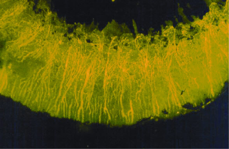
Fig. 3 Fluorescent micrograph of the human iris vasculature: the vascular network of the iris consists of arteries, veins, and capillaries of the iris root and pupillary margin (flat preparation) (×100).

Fig. 4 Fluorescent micrograph of the human iris vasculature: large sized vessels (arrow), probably veins, and relatively small sized vessels, the major circle of the iris (arrowheads), were observed around the iris root (flat preparation) (×200).
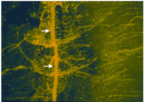
Fig. 5 Fluorescent micrograph of the human iris vasculature: the major arterial circle of the iris (arrows) around the ciliary process is composed of large vessels, which are perpendicular to the iris vessels (flat preparation) (×200).

Fig. 6 Fluorescent micrographs of the human iris vasculature: the vascular network of the pupillary margin forms the arcade, where capillaries continue to veins. The minor arterial circle of the iris (arrows) runs perpendicularly to the arcade (arrowheads) of the vascular network of the pupillary margin (flat preparation) (×400).
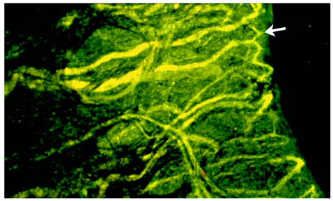
Fig. 7 Fluorescent micrographs of the human iris vasculature: the arcade of the vascular network of the pupillary margin forms an anastomosis (arrow) with adjacent vessels (flat preparation) (×400).
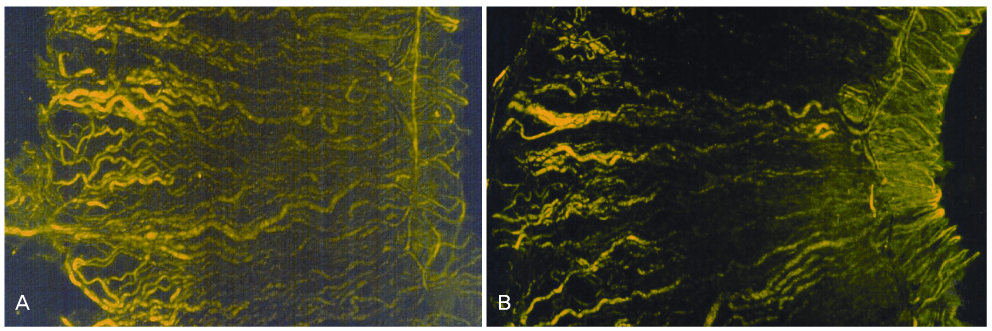
Fig. 8 Fluorescent micrographs of the human iris vasculature: the vascular network and minor arterial circle of the iris were observed between the iris root and the pupillary margin. The iris vessel showed an irregular course and got larger as it approached the iris root (flat preparation) (×100).
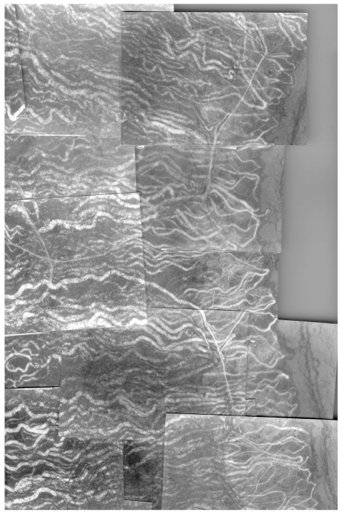
Fig. 9 Fluorescent micrograph of the human iris vasculature: the montage of vascular networks of the iris and the minor arterial circle of the iris. The blood vessels of the minor arterial circle of the iris did not form a complete circle.
Reference
-
1. Hogan MJ, Alvarado JA, Weddell JE. Histology of the Human Eye. 1971. Philadelphia: WB Saunders;202–208. 325–327.2. Johnson D, Liesegang TJ, Brubaker RF. Aqueous humor dynamics in Fuchs' uveitis syndrome. Am J Ophthalmol. 1983. 95:783–787.3. Laurell CG, Zetterstrom C. Inflammation and blood-aqueous barrier disruption. J Cataract Refract Surg. 2000. 26:306–307.4. Carnevalini A, Carassa R, Serini P, et al. Fluorophotometric and fluoroiridographic aspects of the blood-iris barrier in normal subjects. Ocular Fluorometry: Proceedings of the International Society of Ocular Fluorophotometry-ISOF. 1987. Berlin: Kugler & Ghedini;113–118.5. Chignell AH, Easty DL. Iris fluorescein photography followong retinal detachment and in certain ocular ischemic disorders. Trans Ophthalmol Soc UK. 1971. 91:243–259.6. Hayreh SS, Scott WE. Fluorescein Iris Angiography: II. Disturbances in iris circulation following strabismus operation on the various recti. Arch Ophthalmol. 1978. 96:1390–1400.7. Brancato R, Bandello F, Lattanzio R. Iris fluorescein angiography in clinical practice. Surv Ophthalmol. 1997. 42:41–70.8. Hayreh SS, Scott WE. Fluorescein iris angiography: I. Normal pattern. Arch Ophthalmol. 1978. 96:1383–1389.9. Burger PC, Chandler DB, Fryczkowski AW, Klintworth GK. Scanning electron microscopy of microcorrosion casts: applications in ophthalmologic research. Scanning Microsc. 1987. 1:223–231.10. Funk R, Rohen JW. Scanning electron microscopic study on the vasculature of the human anterior eye segment, especially with respect to the ciliary process. Exp Eye Res. 1990. 51:651–661.11. Olver JM, McCartney AC. Anterior segment vascular casting. Eye. 1989. 3:302–307.12. Ringvold A. Light and electron microscopy of the wall of iris vessels in eyes with and without exfoliation syndrome (pseudoexfoliation of the lens capsule). Virchows Arch A Pathol Pathol Anat. 1970. 349:1–9.13. Wang L, Zhang XX, Li ZL. Observation of orbital ocular vascular architecture using plastic casts. Chin Med J (Engl). 1986. 99:645–652.14. Kuwabara T, Cogan DG. Studies in retinal vascular patterns: Part I. Normal architecture. Arch Ophthalmol. 1960. 64:904–911.15. McDonnell PJ, Gritz DC, McDonnell JM, Zarbin MA. Fluorescence of blood-stained cornea. Cornea. 1991. 10:445–449.16. Wilcox LM, Keough EM, Connolly RJ, Hotte CE. The contribution of blood flow by the anterior ciliary arteries to the anterior segment of the primate eye. Exp Eye Res. 1980. 30:167–174.17. Shimizu K, Ujiie K. Structure of Ocular Vessels. 1978. Tokyo: Igaku-Shoin;50–142.18. Amalric P, Rebière P. New indications for fluorescein angiography of the anterior segment. Ann Ocul (Paris). 1971. 204:833–846.
