A Case of Vogt-Koyanagi-Harada Disease in a Patient With Graves Disease
- Affiliations
-
- 1Department of Ophthalmology, College of Medicine, and Medical Research Center, Seoul National University, Seoul, Korea. hgonyu@ snu.ac.kr
- 2Institute of Research of Sensory Organs, Medical Research Center, Seoul National University, Seoul, Korea.
- 3Department of Ophthalmology, Seoul National University Hospital, Seoul, Korea.
- KMID: 754728
- DOI: http://doi.org/10.3341/kjo.2009.23.2.112
Abstract
- A case of Vogt-Koyanagi-Harada disease (VKH) that developed in a 36-year-old woman with Graves' disease was described. The patient was treated with Lugol's solution and presented with bilateral serous retinal detachment. She had also suffered from methimazole-induced hypersensitivity and steroid-induced myopathy. Fluorescein angiography showed multiple leakage points and a lumbar puncture revealed pleocytosis, which was compatible with VKH. High dose steroid pulse therapy was successful. Altered immune regulation associated with drug-induced hypersensitivity may contribute to the development of VKH in patients with Graves' disease.
Keyword
MeSH Terms
-
Adult
Coloring Agents/administration & dosage
Diagnosis, Differential
Dose-Response Relationship, Drug
Drug Therapy, Combination
Female
Fluorescein Angiography
Follow-Up Studies
Fundus Oculi
Glucocorticoids/administration & dosage
Graves Disease/*complications/diagnosis/drug therapy
Humans
Immunosuppressive Agents/administration & dosage
Injections, Intravenous
Iodides/administration & dosage
Ophthalmic Solutions/administration & dosage
Uveomeningoencephalitic Syndrome/*complications/diagnosis/drug therapy
Figure
-
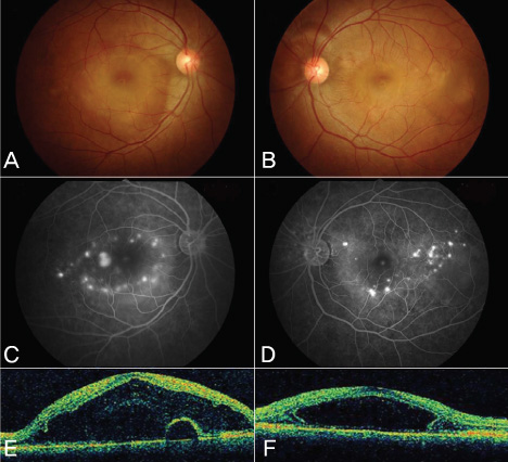
Fig. 1 Fundus photography, fluorescein angiography (FAG) and optical coherence tomography (OCT) performed at the initial visit. At that time, predisolone dosage was 10 mg. (A and B) Fundus photography of both eyes. (C and D) FAG showed multiple leakage points. (E and F) OCT showed subretinal fluid affecting macula.

Fig. 2 Fluorescein angiography (FAG) and indocyanine green angiography (ICG) performed after 1 week of close observation using 5 mg predisolone under the impression of steroid induced central serous retinopathy. Anterior chamber reaction with vitritis developed. (A and B) FAG showed multiple pin point size leakages. (C and D) ICG showed multiple hypofluorescent spots.
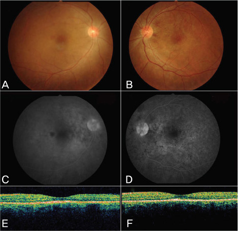
Fig. 3 Fundus photography, fluorescein angiography (FAG) and optical coherence tomography (OCT) performed 4 weeks later. After high-dose steroid (500 mg methylpredisolone) pulse therapy, the patient was given 30 mg predisolone with 75 mg of cyclosporine. (A and B) Fundus photography of both eyes. (C and D) FAG showed the multiple leakages point had disappeared. (E and F) OCT showed that subretinal fluid.
Reference
-
1. Read RW, Holland GN, Rao NA, et al. Revised diagnostic criteria for Vogt-Koyanagi-Harada disease: report of an international committee on nomenclature. Am J Ophthalmol. 2001. 131:647–652.2. Chi HI, Furue M, Ishibashi Y. Vogt-Koyanagi-Harada's syndrome associated with Hashimoto's thyroiditis. J Dermatol. 1994. 21:683–686.3. Gocho K, Kondo I, Yamaki K. Identification of autoreactive T cells in Vogt-Koyanagi-Harada disease. Invest Ophthalmol Vis Sci. 2001. 42:2004–2009.4. Streetman DD, Khanderia U. Diagnosis and treatment of Graves Disease. Ann Phamacother. 2003. 37:1100–1109.
- Full Text Links
-
- Actions
-
Cited
- CITED
-
- Close
- Share
-
- Similar articles
-
- Vogt-Koyanagi-Harada Syndrome Associated with Psoriasis Vulgaris
- A Case of Vogt-Koyanagi-Harada syndrome presenting initially with recurrent vertigo
- A Case of Vogt-Koyanagi-Harada Syndrome
- A Case of Vogt-Koyanagi-Harada's Syndrome
- Photodynamic Therapy with Verteporfin for Subfoveal Choroidal Neovascularization in Vogt-Koyanagi-Harada Syndrome
