Primary Sebaceous Carcinoma of the Corneaoscleral Limbus With Pagetoid Recurrence
- Affiliations
-
- 1Department of Ophthalmology, Seoul National University College of Medicine, Seoul, Korea. kmk9@snu.ac.kr
- 2Seoul National University Hospital Clinical Research Institute, Seoul, Korea.
- 3Department of Ophthalmology, Seoul National University Bundang Hospital, Seongnam, Korea.
- KMID: 754726
- DOI: http://doi.org/10.3341/kjo.2009.23.2.104
Abstract
- We report a sebaceous carcinoma confined to the corneoscleral limbus without involvement of the eyelid. A 60-year-old man, who showed multiple masses on the corneaoscleral limbus and limbal ulceration but with normal eyelids, underwent surgical en-bloc excision of the masses. Histopathologic examination revealed a sebaceous carcinoma. Three weeks after excision, multiple pagetoid recurrences were found along the bulbar conjunctiva 2 mm away from the limbus. After the application of topical mitomycin C, the pagetoid spread regressed completely. After a 2 year follow-up, no other local or systemic recurrences were observed. This report shows that the ulcerative mass which is confined to only the corneoscleral limbus may be a sebaceous carcinoma even without eyelid involvement. Topical mitomycin C may be effective for treating pagetoid spread of sebaceous carcinoma of limbal origin.
MeSH Terms
Figure
-
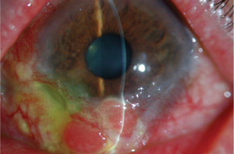
Fig. 1 Slit-lamp examination showed diffuse conjunctival injection and multiple nodular masses confined to inferior limbus and inferior bulbar conjunctiva along the limbus. The masses showed salmon-colored pebbly appearance accompanying peripheral corneal ulceration.
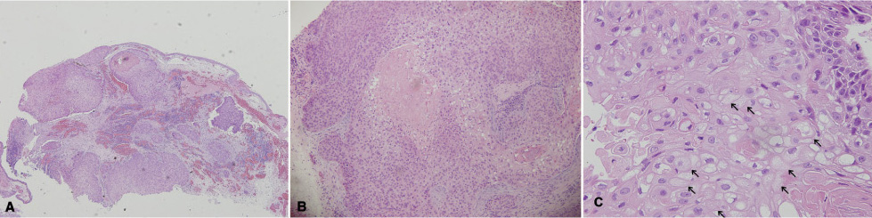
Fig. 2 Light microscopic view of the tumor. (A) At low magnification, the tumor is composed of relatively well-delineated lobules of sebaceous cells (×40). (B) At medium magnification, the tumor cell nest shows comedo-type necrosis (×100). (C) At higher magnification, the large cells with sebaceous differentiation, some of which are vacuolated, have small nuclei and clear cytoplasm (×200).
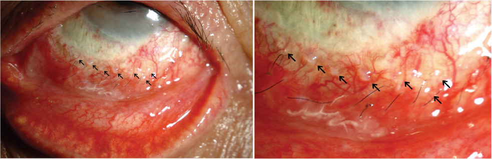
Fig. 3 Slit lamp examination revealed multiple pagetoid spread along with bulbar conjunctiva 2 mm away from the limbus (arrows).
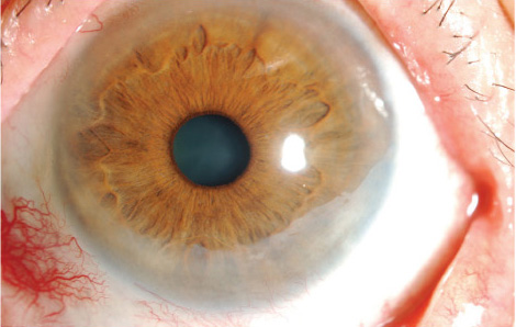
Fig. 4 This photo shows clear conjunctiva and cornea after sebaceous carcinoma regressed completely.
Reference
-
1. Shields JA, Demirci H, Marr BP, et al. Sebaceous carcinoma of the ocular region: a review. Surv Ophthalmol. 2005. 50:103–122.2. Burns SJ, Foss AJ, Butler TK. Outcome of periocular sebaceous gland carcinoma. Ophthal Plast Reconstr Surg. 2005. 21:353–355.3. Boniuk M, Zimmerman LE. Sebaceous carcinoma of the eyelid, eyebrow, caruncle and orbit. Int Ophthalmol Clin. 1972. 12:225–257.4. Tumuluri K, Kourt G, Martin P. Mitomycin C in sebaceous gland carcinoma with pagetoid spread. Br J Ophthalmol. 2004. 88:718–719.5. Jakobiec FA. Sebaceous tumors of the ocular adnexa. Principles and Practice of Ophthalmology. 1994. 3:1745–1770.6. Cook BE, Bartley GB. Treatment options and future prospects for the management of eyelid malignancies An evidence-based update. Ophthalmology. 2001. 108:2088–2098.7. Honavar SG, Shields CL, Maus M, et al. Primary intraepithelial sebaceous gland carcinoma of the palpebral conjunctiva. Arch Ophthalmol. 2001. 119:764–767.8. Margo CE, Grossniklaus HE. Intraepithelial sebaceous neoplasia without underlying invasive carcinoma. Surv Ophthalmol. 1995. 39:293–301.9. Margo CE, Lessner A, Stern GA. Intraepithelial sebaceous carcinoma of the conjunctiva and skin of the eyelid. Ophthalmology. 1992. 99:227–231.10. Theodore FH. Conjunctival carcinoma masquerading as chronic conjunctivitis. Eye Ear Nose Throat Mon. 1967. 46:1419–1420.11. Penn I. Tumors after renal and cardiac transplantation. Hematol Oncol Clin North Am. 1993. 7:431–445.12. Singh SK, Gupta AK, Jha V, et al. Treatment of oropharyngeal cancer in renal transplant recipients without cessation of immunosuppressive therapy. Transplant Proc. 2006. 38:2088–2089.13. Sainz de la Maza M, Foster CS. Peripheral ulcerative keratitis and malignancies. Cornea. 1994. 13:364–367.14. Kuo T. Clear cell carcinoma of the skin. A variant of the squamous cell carcinoma that simulates sebaceous carcinoma. Am J Surg Pathol. 1980. 4:573–583.15. Suster S. Clear cell tumors of the skin. Semin Diagn Pathol. 1996. 13:40–59.16. Margo CE, Groden LR. Primary clear cell carcinoma of the conjunctiva. Arch Ophthalmol. 2008. 126:436–438.17. Kass LG. Role of cryotherapy in treating sebaceous carcinoma of the eyelid. Ophthalmology. 1990. 97:2–4.18. Poothullil AM, Colby KA. Topical medical therapies for ocular surface tumors. Semin Ophthalmol. 2006. 21:161–169.19. Shields CL, Naseripour M, Shields JA, Eagle RC Jr. Topical mitomycin-C for pagetoid invasion of the conjunctiva by eyelid sebaceous gland carcinoma. Ophthalmology. 2002. 109:2129–2133.20. Chalasani R, Giblin M, Conway RM. Role of topical chemotherapy for primary acquired melanosis and malignant melanoma of the conjunctiva and cornea: review of the evidence and recommendations for treatment. Clin Experiment Ophthalmol. 2006. 34:708–714.21. Frucht-Pery J, Sugar J, Baum J, et al. Mitomycin C treatment for conjunctival-corneal intraepithelial neoplasia: a multicenter experience. Ophthalmology. 1997. 104:2085–2093.22. Khong JJ, Muecke J. Complications of mitomycin C therapy in 100 eyes with ocular surface neoplasia. Br J Ophthalmol. 2006. 90:819–822.
