Cancer-associated Nummular Loss of the Retinal Pigment Epithelium
- Affiliations
-
- 1Department of Ophthalmology, Gangnam Sacred Heart Hospital, College of Medicine, Hallym University, Seoul, Korea. hkkimeye@unitel.co.kr
- KMID: 754647
- DOI: http://doi.org/10.3341/kjo.2007.21.4.261
Abstract
- PURPOSE: To report a case of cancer-associated nummular loss of the retinal pigment epithelium. METHODS: A 47-year-old man with a history of hepatocellular carcinoma presented with three weeks of bilateral visual loss. His best-corrected visual acuity was 20/40 in each eye. He had multiple round confluent grayish-brown patches at the level of retinal pigment epithelium, and no pigmented choroidal lesions. Fluorescein angiography showed circular areas of transmission defect and indocyanine green angiography showed early hyperfluorescence, corresponding with the multiple round confluent patches. CONCLUSIONS: We report a case of visual paraneoplastic syndrome which showed nummular loss of the pigment epithelial cells which distinguishes the clinical component of BDUMP syndrome.
Keyword
MeSH Terms
Figure
-

Fig. 1 Initial visit. (A) Fundus photograph. Multiple round confluent grayish-brown patches are noted at the level of retinal pigment epithelium. There were no pigmented choroidal lesions. (B) Fluorescein angiography. Hyperfluorescence due to window defects associated with the widespread retinal pigment epithelium damage is seen corresponding to the lesion. No significant leakage. (C) Optical coherence tomography. OCT image showed zones of retinal pigment epithelium loss alternating with areas of thickened retinal pigment epithelium and deposit of debris that correspond with the multiple round confluent patches.
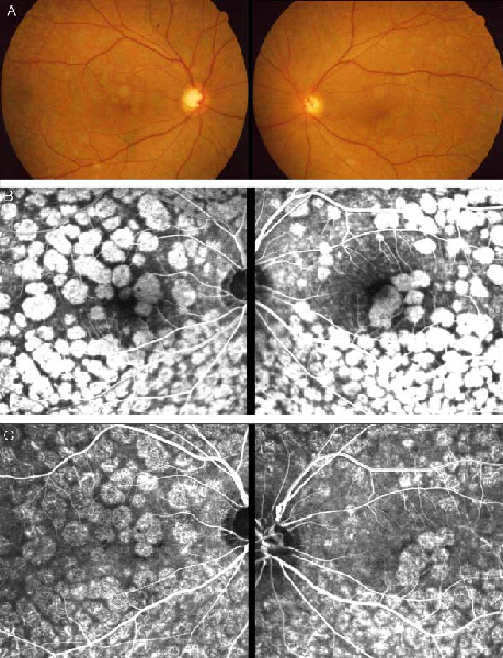
Fig. 2 Two months later. (A) Fundus photograph. (B) Fluorescein angiography. (C) Indocyanin green angiography. Both the number and size of multiple round patches increased.
Reference
-
1. Barr CC, Zimmerman LE, Curtin VT, Front RL. Bilateral diffuse uveal tumors associated with systemic malignant neoplasm. A recently recognized syndrome. Arch Ophthalmol. 1982. 100:249–255.2. Gass JD, Gieser RG, Wilkinson CP, et al. Bilateral diffuse uveal melanocytic proliferation in patients with occult carcinoma. Arch Ophthalmol. 1990. 108:527–533.3. Leys AM, Dierick HG, Sciot RM. Early lesions of bilateral diffuse melanocytic proliferation. Arch Ophthalmol. 1991. 109:1590–1594.4. Borruat FX, Othenin-Girard P, Uffer S, et al. Natural history of diffuse uveal melanocytic proliferation. Case report. Ophthalmology. 1992. 99:1698–1704.5. Yu S, Ikeda T, Ikeda N, et al. Coloration of fundus lesions in bilateral diffuse uveal melanocytic proliferation. Jpn J Ophthalmol. 2003. 47:612–615.6. Chan JW. Paraneoplastic retinopathies and optic neuropathies. Surv Ophthalmol. 2003. 48:12–38.7. Yoon YH, Cho EH, Sohn J, Thirkill CE. An unuasual type of cancer-associated retinopathy in a patient with ovarian cancer. Korean J Ophthalmol. 1999. 13:43–48.8. Saito W, Satoru K, Yoshida K, et al. Bilateral diffuse uveal melanocytic proliferation in a patient with cancer-associated retinopathy. Am J Ophthalmol. 2005. 140:942–945.9. Wu S, Slakter JS, Shields JA, Spaide RF. Cancer-associated nummular loss of the pigment epithelium. Am J Ophthalmol. 2005. 139:933–935.
- Full Text Links
-
- Actions
-
Cited
- CITED
-
- Close
- Share
-
- Similar articles
-
- Growth Patterns of Human Retinal Pigment Epithelium in Vitro
- The Effect of 5-Fluorouraci1 on the Activity of the Retinal Pigment Epithelium in Vitro
- Morphological Changes of Retinal Pigment Epithelium After Experimental Retinal Detachment
- Culture of Bovine Retinal Pigment Epithelium: Topographical Differences of Morphology and Growth Rate in Vitro
- Histologic Study in Transplantation of Cultured Rabbit Retinal Pigment Epithelium
