A Case of Scar Sarcoidosis of The Eyelid
- Affiliations
-
- 1Department of Ophthalmology, Seoul Veterans Hospital, Seoul, Korea.
- 2Department of Ophthalmology, Saumsung Medical Center, School of Medicine Sungkyunkwan University, Seoul, Korea. ydkim@ smc.samsung.co.kr
- KMID: 754590
- DOI: http://doi.org/10.3341/kjo.2006.20.4.238
Abstract
- PURPOSE: We report the case of a patient with scar sarcoidosis that developed along a previous eyelid scar. There was no evidence of ocular or systemic sarcoidosis. METHODS: A 29-year-old man presented with a mass on his right eyelid that had been present for two month. On ocular examination an erythematous, firm, and non-tender mass was diffusely palpable along the upper and lower eyelid scar. We performed an incisional biopsy of the lower lid mass. RESULTS: Histopathologic examination of the mass revealed numerous, noncaseating granulomas with multi-nucleated giant cells. The giant cells contained asteroid bodies and calcium oxalate crystals characteristic of sarcoidosis, although the patient had no other evidence of systemic sarcoidosis. The mass in the upper lid disappeared after intralesional triamcinolone injections. CONCLUSIONS: This case represents a rare occurrence of sarcoidosis that arose in an old eyelid scar. Scar sarcoidosis should be considered in the differential diagnosis of an unusual mass in a scar.
MeSH Terms
-
Triamcinolone/administration & dosage
Sarcoidosis/drug therapy/etiology/*pathology
Male
Injections, Intralesional
Humans
Glucocorticoids/administration & dosage
Follow-Up Studies
Eyelids/injuries
Eyelid Diseases/drug therapy/etiology/*pathology
Eye Injuries/complications
Diagnosis, Differential
Cicatrix/complications/*pathology
Biopsy
Adult
Figure
-
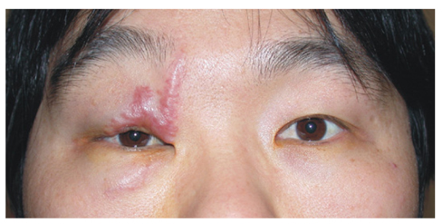
Fig. 1 An erythematous firm mass is visible along the upper and lower eyelid scar.

Fig. 2 A photomicrograph of the biopsy showing the numerous, noncaseating granulomas with multi-nucleated giant cells, surrounded by lymphocytes. (hematoxylin and eosin stain, ×200)
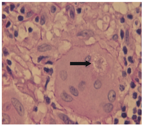
Fig. 3 A photomicrograph of the biopsy showing the giant cells containing asteroid bodies (arrow). (hematoxylin and eosin stain, ×400)

Fig. 4 A photomicrograph of the biopsy showing the calcium oxalate crystals (arrow). (hematoxylin and eosin stain, ×200)
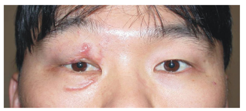
Fig. 5 One month after triamcinolone injection, there was a dramatic resolution of the lesions of the upper lid mass.
Cited by 1 articles
-
A Case of Sarcoidosis Presented as Multiple Conjunctival and Nasal Mucosal Nodule
In-Cheon You, Hyung-Jin Moon, Gwi-Hyeong Mun, Sang-Chul Im, Kyung-Chul Yoon
J Korean Ophthalmol Soc. 2008;49(6):1000-1006. doi: 10.3341/jkos.2008.49.6.1000.
Reference
-
1. Siltzbach LE, James DG, Neville E, et al. Course and prognosis of sarcoidosis around the world. Am J Med. 1985. 57:847–852.2. Brownstein S, Liszauer AD, Carey WD, Nicolle DA. Sarcoidosis of the eyelid skin. Can J Ophthalmol. 1990. 25:256–259.3. Cecchi R, Giomi A. Scar sarcoidosis following herpes zoster. J Eur Acad Dermatol Venereol. 1999. 12:280–282.4. Hancock BW. Cutaneous sarcoidosis in blood donation venepuncture sites. Br Med J. 1972. 23:706–708.5. Murdoch SR, Fenton DA. Sarcoidosis presenting as nodules in both tattoos and scars. Clin Exp Dermatol. 1997. 22:254–255.6. Kormeili T, Neel V, Moy RL. Cutaneous sarcoidosis at sites of previous laser surgery. Cutis. 2004. 73:53–55.7. Hall JG, Cohen KL. Sarcoidosis of the eyelid skin. Am J Ophthalmol. 1995. 119:100–101.8. Callen JP, Mahl CF. Oculocutaneous manifestations observed in multisystem disorders. Dermatol Clin. 1992. 10:709–716.9. Condon KC, O'Sullivan D. Sarcoidosis in long standing facial scars. Br J Plast Surg. 1978. 31:266–267.10. Ingber A, Klinken V. Scar sarcoidosis. Clin Exp Dermatol. 1984. 9:532.
