Korean J Ophthalmol.
2006 Dec;20(4):225-229. 10.3341/kjo.2006.20.4.225.
The Relationship between Optical Coherence Tomography and Scanning Laser Polarimetry Measurements in Glaucoma
- Affiliations
-
- 1Department of Ophthalmology, Hallym University College of Medicine, Anyang, Korea.
- 2Department of Ophthalmology, Kim's Eye Hospital, Myung-Gok Eye Research Institute, Konyang University College of Medicine, Seoul, Korea. yhsohn@konyang.ac.kr
- KMID: 754587
- DOI: http://doi.org/10.3341/kjo.2006.20.4.225
Abstract
- PURPOSE: To investigate the relationship between optical coherence tomography (OCT) and scanning laser polarimetry (SLP) in measuring peripapillary retinal nerve fiber layer (RNFL) thickness in glaucomatous eyes. METHODS: Fifty glaucomatous eyes were evaluated in this study. Evaluations were analyzed two ways. First, parameters of the Stratus OCT (average thickness, superior/ inferior average) and GDx VCC (TSNIT average, nerve fiber indicator (NFI), superior/ inferior average) were correlated using the Pearson's correlation coefficient (r). Secondly, comparison (r) of these parameters was completed using the mean deviation (MD) of visual field defect. RESULTS: The following parameters were found to be significantly correlated (P<0.005). TSNIT average/average thickness (r=0.673), NFI/average thickness (r=-0.742), superior average (r=0.841), and inferior average (r=0.736). In the correlation analysis using the severity of visual field defect, all these parameters had statistically meaningful correlations (P<0.005). CONCLUSIONS: GDx VCC and Stratus OCT are highly correlated in glaucomatous eyes. Therefore, peripapillary RNFL thickness measured by Stratus OCT and GDx VCC may be equally helpful in the diagnosis of glaucoma.
Keyword
MeSH Terms
Figure
-
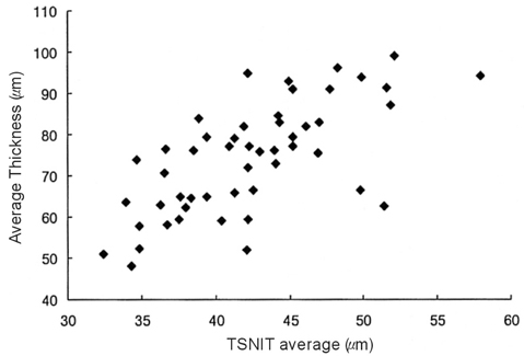
Fig. 1 Scatterplot of TSNIT average versus average thickness.
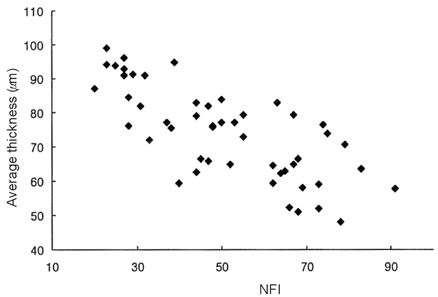
Fig. 2 Scatterplot of NFI (Nerve fiber indicator) versus average thickness.
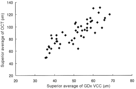
Fig. 3 Scatterplot of superior averages.
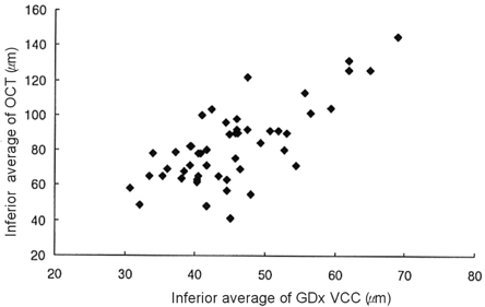
Fig. 4 Scatterplot of inferior averages.
Reference
-
1. Kerrgan-Baumrind LA, Quigley HA, Pease ME, et al. Number of ganglion cells in glaucoma eyes compared with threshold visual field tests in the same persons. Invest Ophthalmol Vis Sci. 2000. 41:741–748.2. Quigley HA, Addicks EM, Green WR. Optic nerve damage in human glaucoma. III. Quantitative correlation of nerve fiber loss and visual field defect in glaucoma, ischemic neuropathy, papilledema, and toxic neuropathy. Arch Ophthalmol. 1982. 100:135–146.3. Jonas JB, Schiro D. Localized wedge shaped defect of the retinal nerve fiber layer in glaucoma. Br J Ophthalmol. 1994. 78:285–290.4. Tuulonen A, Airaksinen PJ. Initial glaucomatous optic disc and retinal nerve fiber layer abnormalities and their progression. Am J Ophthalmol. 1991. 111:485–490.5. Quigley HA, Miller NR, George T. Clinical evaluation of nerve fiber layer atrophy as an indicator of glaucomatous optic nerve damage. Arch Ophthalmol. 1980. 98:1564–1571.6. Huang D, Swanson EA, Lin CP, et al. Optical coherence tomography. Science. 1991. 254:1178–1181.7. Izatt JA, Hee MR, Swanson EA, et al. Micrometer-scale resolution imaging of the anterior eye in vivo with optical coherence tomography. Arch Ophthalmol. 1994. 112:1584–1589.8. Schuman JS, Hee MR, Puliafito CA, et al. Quantification of nerve fiber layer thickness in normal and glaucomatous eyes using optical coherence tomography. Arch Ophthalmol. 1995. 113:586–596.9. Huang XR, Knighton RW. Linear birefringence of the retinal nerve fiber layer measured in vitro with a multispectral imaging micropolarimeter. J Biomed Opt. 2002. 7:199–204.10. Dreher AW, Bailey ED. Assessment of the retinal nerve fiber layer by scanning-laser polarimetry. SPIE. 1993. 1877:266–271.11. Weinreb RN, Dreher AW, Coleman A, et al. Histopathologic validation of Fourier-ellipsometry measurements of retinal nerve fiber layer thickness. Arch Ophthalmol. 1990. 108:557–560.12. Reus NJ, Lemij HG. Scanning laser polarimetry of the retinal nerve fiber layer in perimetrically unaffected eyes of glaucoma patients. Ophthalmology. 2004. 111:2199–2203.13. Medeiros FA, Zangwill LM, Bowed C, et al. Fourier analysis of scanning laser polarimetry measurements with variable corneal compensation in glaucoma. Invest Ophthalmol Vis Sci. 2003. 44:2606–2612.14. Medeiros FA, Zangwill LM, Bowed C, et al. Comparison of scanning laser polarimetry using variable corneal compensation and retinal nerve fiber layer photography for detection of glaucoma. Arch Ophthalmol. 2004. 122:698–704.15. Medeiros FA, Zangwill LM, Bowed C, et al. Evaluation of retinal nerve fiber layer, optic nerve head, and macular thickness measurements for glaucoma detection using optical coherence tomopraphy. Am J Ophthalmol. 2005. 139:44–55.16. Budenz DL, Michael A, Chang RT, et al. Sensitivity and specificity of the Stratus OCT for perimetric glaucoma. Ophthalmology. 2005. 112:3–9.17. Wollstein G, Ishikawa H, Wang J, et al. Comparison of three optic coherence tomography scanning areas for detection of glaucomatous damage. Am J Ophthalmol. 2005. 139:39–43.18. Reus NJ, Lemij HG. Diagnostic accuracy of the GDx VCC for glaucoma. Ophthalmology. 2004. 111:1860–1865.19. Anderson DR. Automated Static Perimetry. 1992. St. Louis: Mosby;53–59.20. Hodapp E, Parrish RK, Anderson DR. Clinical decisions in glaucoma. 1993. St. Louis: Mosby;52–63.21. Dichtl A, Jonas JB, Naumann GO. Retinal nerve fiber layer thickness in human eyes. Graefes Arch Clin Exp Ophthalmol. 1999. 237:474–479.22. Varma R, Skaf M, Barron E. Retinal nerve fiber layer thickness in normal human eyes. Ophthalmology. 1996. 103:2114–2119.23. Greaney MJ, Hoffman DC, Garway-Heath DF, et al. Comparison of optic nerve imaging methods to distinguish normal eyes from those with glaucoma. Invest Ophthalmol Vis Sci. 2002. 43:140–145.24. Bagga H, Greenfield DS, Feuer W, et al. Scanning Laser Polarimetry With Variable Corneal Compensation and Optical Coherence Tomography in Normal and Glaucomatous Eyes. Am J Ophthalmol. 2003. 135:521–529.25. Leung CK, Chan W, Chong KKL, et al. Comparative Study of Retinal Nerve Fiber Layer Measurement by StratusOCT and GDx VCC, I: Correlation Analysis in Glaucoma. Invest Ophthalmol Vis Sci. 2005. 46:3214–3220.26. Colen TP, Tang NEML, Mulder PGH, et al. Sensitivity and Specificity of New GDx Parameters. J Glaucoma. 2004. 13:28–33.27. Kanamori A, Nakamura M, Escano MFT, et al. Evaluation of the Glaucomatous Damage on Retinal nerve fiber layer thickness measured by Optical Coherence Tomography. Am J Ophthalmol. 2003. 135:513–520.28. Blumenthal EZ, Williams JM, Weinreb RN, et al. Reproducibility of nerve fiber layer thickness measurements by use of optical coherence tomography. Ophthalmology. 2000. 107:2278–2282.29. Carpineto P, Ciancaglini M, Zuppardi E, et al. Reliability of nerve fiber layer thickness measurements using optical coherence tomography in normal and glaucomatous eyes. Ophthalmology. 2003. 110:190–195.30. Kook MS, Sung K, Park RH, et al. Reproducibility of scanning laser polarimetry (GDx) of peripapillary retinal nerve fiber layer thickness in normal subjects. Graefes Arch Clin Exp Ophthalmol. 2001. 239:118–121.31. Hoh ST, Ishikawa TC, Greenfield DS, et al. Peripapillary nerve fiber layer thickness measurement reproducibility using scanning laser polarimetry. J Glaucoma. 1998. 7:12–15.32. Sommer A, Katz J, Quigley HA, et al. Clinically detectable nerve fiber layer atrophy precedes the onset of glaucomatous field loss. Arch Ophthalmol. 1991. 109:77–83.33. Reus NJ, Nemij HG. The relationship between standard automated perimetry and GDx VCC measurements. Invest Ophthalmol Vis Sci. 2004. 45:840–845.
- Full Text Links
-
- Actions
-
Cited
- CITED
-
- Close
- Share
-
- Similar articles
-
- Changes in RNFL Thickness According to the Myopia in Patients with Glaucoma and Ocular Hypertension
- The Relationship between the Duration of IOP Elevation during LASIK and Nerve Fiber Layer Thickness Measured by GDx(R)
- Scanning Laser Polarimetry and Optical Coherence Tomography for Detection of Retinal Nerve Fiber Layer Defects
- Diagnostic Outcomes of Patients Suspicious for Glaucoma Referred from The Company Health Screening
- Biometry of Retinal Nerve Fiber Layer Thickness by NFA
