A Korean Family with an Early-Onset Autosomal Dominant Macular Dystrophy Resembling North Carolina Macular Dystrophy
- Affiliations
-
- 1Department of Ophthalmology, Seoul National University College of Medicine, Seoul, Korea. hgonyu@snu.ac.kr
- 2Seoul Artificial Eye Center, Seoul National University Hospital Clinical Research Institute, Seoul, Korea.
- KMID: 754586
- DOI: http://doi.org/10.3341/kjo.2006.20.4.220
Abstract
- PURPOSE: To characterize and report the phenotype of a Korean family with an early-onset autosomal dominant macular dystrophy resembling North Carolina macular dystrophy (NCMD). METHODS: Five members of a Korean family were examined clinically and underwent fundus photography, fluorescein angiography, indocyanine green angiography, optical coherence tomography, full field electroretinogram (ERG), multifocal ERG, electro-oculography (EOG), a color vision test, and a visual field test. RESULTS: Visual acuity ranged from 20/200 to 20/20. Fundus findings demonstrated varying degrees of involvement ranging from drusen only to chorioretinal involvement. Central scotoma corresponded to retinal lesions in two patients. Full field ERG was normal but multifocal ERG showed decreased amplitude and delayed implicit time in the macular area. EOG was normal except in one patient. Color vision tests were also normal. CONCLUSIONS: The phenotype of this Korean family is consistent with NCMD. Linkage analysis is required to confirm the diagnosis.
MeSH Terms
Figure
-
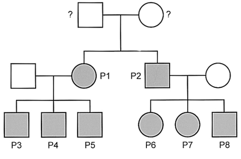
Fig. 1 The pedigree of a Korean family with macular dystrophy demonstrating the characteristics of an autosomal dominant inheritance trait.
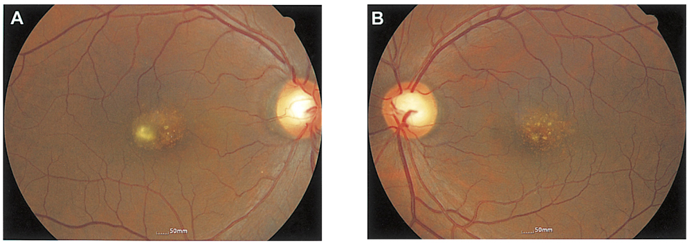
Fig. 2 Fundus photographs of patient 2. There are multiple fine drusen-like lesions in the macular area consistent with stage 1 North Carolina macular dystrophy.
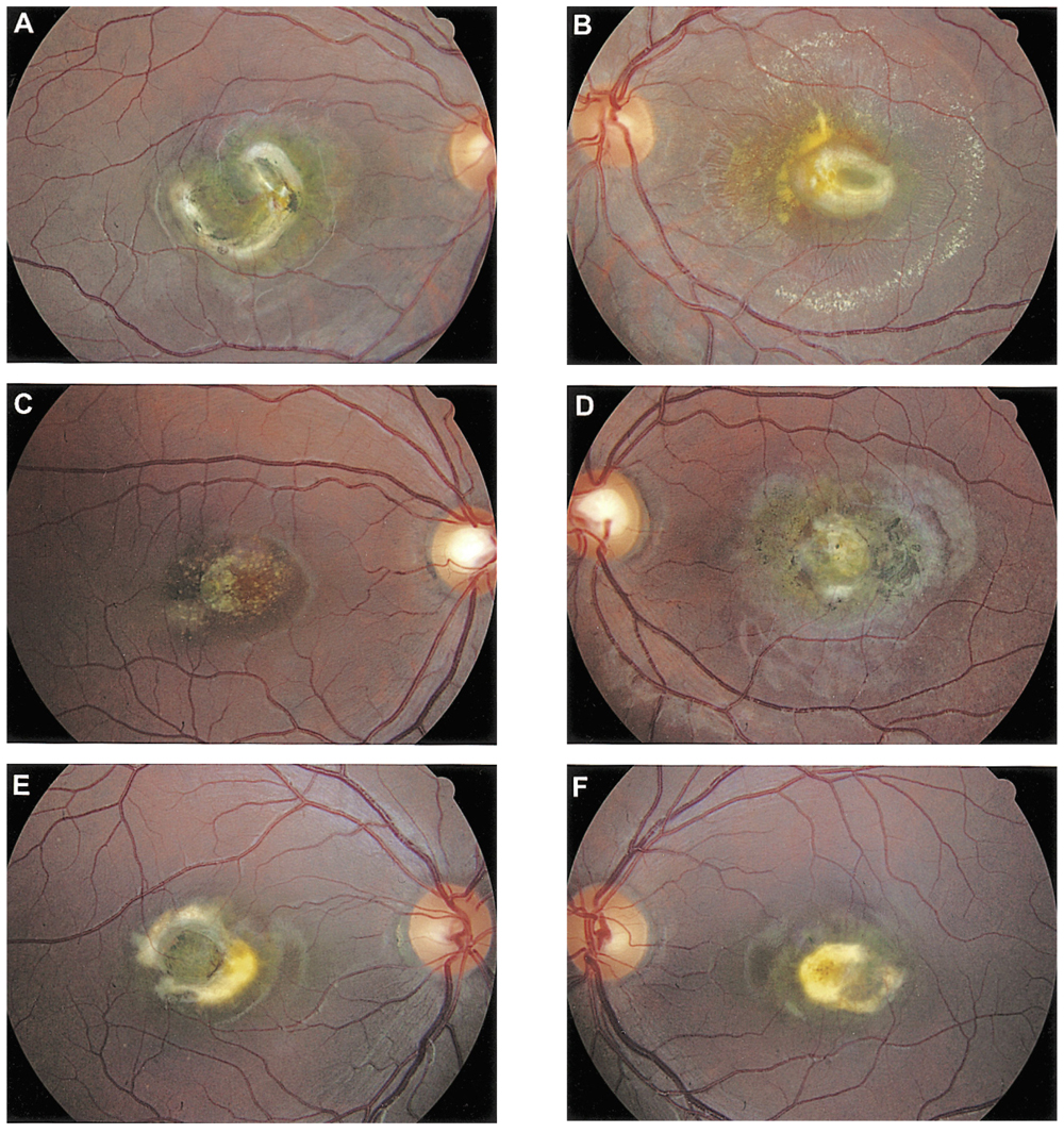
Fig. 3 Fundus photographs of patients 6 (A, B), 7 (C, D), and 8 (E, F). There are confluent drusen-like lesions, depigmented lesions, visible large choroidal vessels, and fibrotic lesions consistent with varying degree of stage 2 North Carolina macular dystrophy.
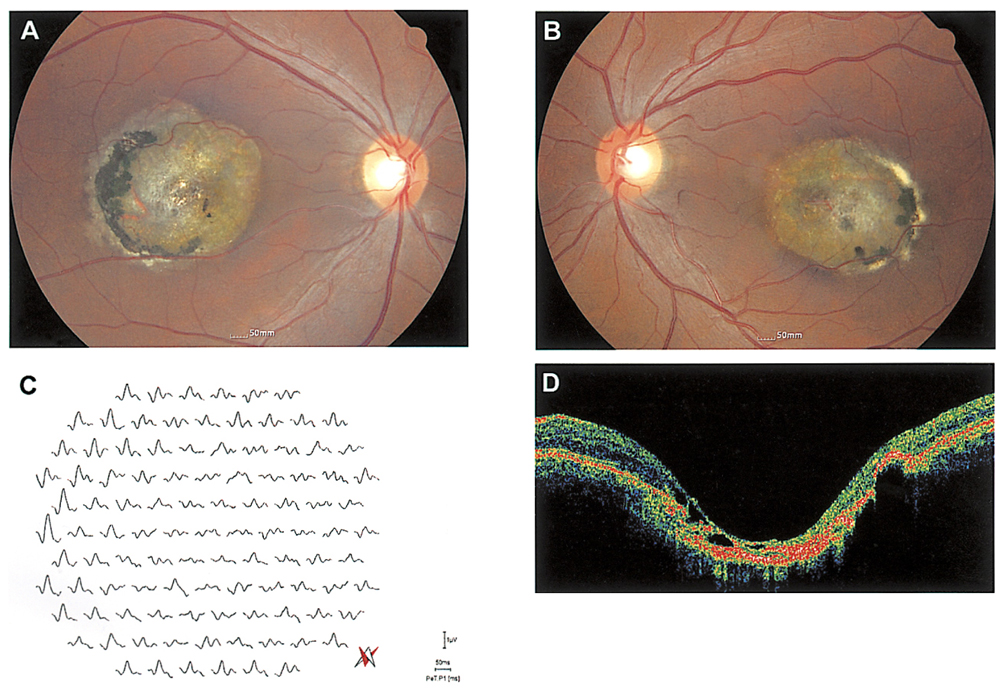
Fig. 4 Fundus photographs of patient 1 (A, B) showing large chorioretinal atrophic lesions with visible choroidal vessels. Multifocal ERG (C) shows abnormal responses in the macular area and normal responses outside the lesion. OCT (D) indicates extensive macular atrophy.
Reference
-
1. Michaelides M, Hunt DM, Moore AT. The genetics of inherited macular dystrophies. J Med Genet. 2003. 40:641–650.2. Lefler WH, Wadsworth JAC, Sidbury J. Hereditary macular degeneration and aminoaciduria. Am J Ophthalmol. 1971. 71:224–230.3. Frank HR, Landers MB 3rd, Williams RJ, Sidbury JB. A new dominant progressive foveal dystrophy. Am J Ophthalmol. 1974. 78:903–916.4. Small KW, Hermsen V, Gurney N, et al. North Carolina macular dystrophy and central areolar pigment epithelial dystrophy. One family, one disease. Arch Ophthalmol. 1992. 110:515–518.5. Small KW, Killian J, McLean WC. North Carolina's dominant progressive foveal dystrophy: how progressive is it? Br J Ophthalmol. 1991. 75:401–406.6. Small KW, Garcia CA, Gallardo G, et al. North Carolina macular dystrophy (MCDR1) in Texas. Retina. 1998. 18:448–452.7. Rabb MF, Mullen L, Yelchits S, et al. A North Carolina macular dystrophy phenotype in a Belizean family maps to the MCDR1 locus. Am J Ophthalmol. 1998. 125:502–508.8. Small KW, Puech B, Mullen L, Yelchits S. North Carolina macular dystrophy phenotype in France maps to the MCDR1 locus. Mol Vis. 1997. 3:1–5.9. Reichel MB, Kelsell RE, Fan J, et al. Phenotype of a British North Carolina macular dystrophy family linked to chromosome 6q. Br J Ophthalmol. 1998. 82:1162–1168.10. Pauleikhoff D, Sauer CG, Muller CR, et al. Clinical and genetic evidence for autosomal dominant North Carolina macular dystrophy in a German family. Am J Ophthalmol. 1997. 124:412–415.11. Scholl HP, Schuster AM, Vonthein R, Zrenner E. Mapping of retinal function in Best macular dystrophy using multifocal electroretinography. Vision Res. 2002. 42:1053–1061.12. Piao CH, Kondo M, Tanikawa A, et al. Multifocal electroretinogram in occult macular dystrophy. Invest Ophthalmol Vis Sci. 2000. 41:513–517.
