Korean J Ophthalmol.
2004 Dec;18(2):106-115. 10.3341/kjo.2004.18.2.106.
Rate of Visual Field Progression in Primary Open-angle Glaucoma and Primary Angle-closure Glaucoma
- Affiliations
-
- 1Department of Ophthalmology, College of Medicine, Chungnam National University, Daejon, Korea.
- 2Department of Ophthalmology, College of Medicine, Kyungpook National University, Daegu, Korea.
- KMID: 754375
- DOI: http://doi.org/10.3341/kjo.2004.18.2.106
Abstract
- To estimate the rate of visual field progression in primary open-angle glaucoma (POAG) and primary angle-closure glaucoma (PACG), we reviewed the medical records of POAG and PACG patients who had a minimum of 5-year longitudinal Goldmann visual field data. I4e and I2e isopters were quantified using grid systems. The rate of change was calculated from the slope of a linear fit to a series of average visual field scores. Twenty-three eyes of POAG patients and 25 of PACG patients were studied. The rate of visual field score change was -2.00 +/- 2.0% per year in the PACG group, and -0.81 +/- 1.0% per year inthe POAG group. In these two patient groups, who were on conventional treatment at two referral hospitals, better visual field on initial presentation yielded faster progression in the POAG group, while the higher average of highest intraocular pressure in each year during follow-up was related to faster progression in the PACG group.
Keyword
MeSH Terms
Figure
-
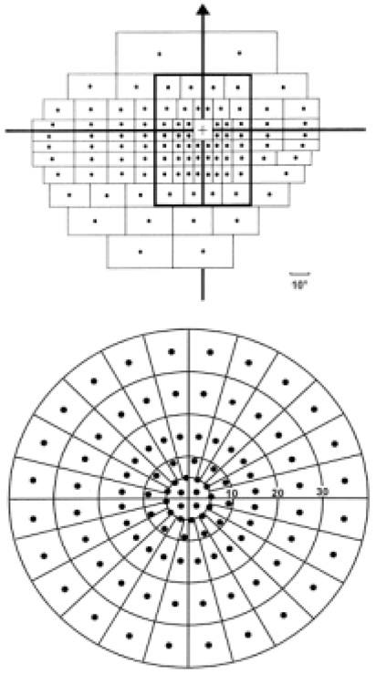
Fig. 1 Manual grid template used to quantify Goldmann visual fields. Top: The grid template made up of 100 dots was used to score I4e isopter of the left eye Goldmann field (adapted from Esterman).3 Bottom: A small grid template also made up of 100 dot was used to score I2e isopter (adapted from Kwon et al).4
Reference
-
1. Armaly MF. Ocular pressure and visual fields. A ten-year follow-up study. Arch Ophthalmol. 1969. 81:25–40.2. Rock WJ, Drance SM, Morgan RW. Visual field screening in glaucoma. An evaluation of the Armaly technique for screening glaucomatous visual fields. Arch Ophthalmol. 1973. 89:287–290.3. Esterman B. Grid for scoring visual fields. II. Perimeter. Arch Ophthalmol. 1968. 79:400–406.4. Kwon YH, Kim CS, Zimmerman MB, Alward WLM, Hayreh SS. Rate of visualfield loss and long-term visual outcome in primary open-angle glaucoma. Am J Ophthalmol. 2001. 132:47–56.5. Botelho PJ, Johnson LN, Arnold AC. The effect of aspirin on the visual outcome of nonarteritic anterior ischemic optic neuropathy. Am J Ophthalmol. 1996. 121:450–451.6. Rasker MT, Enden A, Bakker D, Hoyng PF. Rate of visual field loss in progressive glaucoma. Arch Ophthalmol. 2000. 118:481–488.7. Smith SD, Katz J, Quigley HA. Analysis of progressive change in automated visual fields in glaucoma. Invest Ophthalmol Vis Sci. 1996. 37:1419–1428.8. O'Brien C, Schwartz B, Takamoto T, Wu DC. Intraocular pressure and the rate of visual field loss in chronic open-angle glaucoma. Am J Ophthalmol. 1991. 111:491–500.9. Hart WM, Becker B. The onset and evolution of glaucomatous visual field defects. Ophthalmology. 1982. 89:268–279.10. Mikelberg FS, Schulzer M, Drance SM, Lau W. The rate of progression of scotomas in glaucoma. Am J Ophthalmol. 1986. 101:1–6.11. Chauhan BC, Drance SM. The relationship between intraocular pressure and visual field progression in glaucoma. Graefe's Arch Clin Exp Ophthalmol. 1992. 230:521–526.12. Katz J, Gilbert D, Quigley HA, Sommer A. Estimating progression of visual field loss in glaucoma. Ophthalmology. 1997. 104:1017–1025.13. Quigley HA, Tielsch JM, Katz J, Sommer A. Rate of progression in open-angle glaucoma estimated from cross-sectional prevalence of visual field damage. Am J Ophthalmol. 1996. 122:355–363.14. Quigley HA, Maumenee AE. Long-term follow-up of treated open-angle glaucoma. Am J Ophthalmol. 1979. 87:519–525.15. Weber J, Koll W, Krieglstein GK. Intraocular pressure and the rate of visual field decay in chronic glaucoma. Ger J Ophthalmol. 1993. 2:165–169.16. Hiller R, Kahn HA. Blindness from glaucoma. Am J Ophthalmol. 1975. 80:62–65.17. Anderson DR. The management of elevated intraocular pressure with normal optic disc and visual fields: Therapeutic approach based on high risk factor. Surv Ophthalmol. 1977. 21:479–485.
- Full Text Links
-
- Actions
-
Cited
- CITED
-
- Close
- Share
-
- Similar articles
-
- The Differences of Visual Field Defects in Three Types of Primary Glaucoma
- Visual Field Progression in Patients with Primary Open Angle Glaucoma, Normal Tension Glaucoma, and Primary Angle Closure Glaucoma
- Significance of Peripapillary Atrophy in the Diagnosis of Open-angle Glaucoma
- A Clinical Study on Glaucoma
- Comparison of visual field defects between primary open-angle glaucoma and chronic primary angle-closure glaucoma in the early or moderate stage of the disease
