Korean J Ophthalmol.
2004 Dec;18(2):100-105. 10.3341/kjo.2004.18.2.100.
Relationship between the Extent of Peripheral Anterior Synechiae and the Severity of Visual Field Defects in Primary Angle-closure Glaucoma
- Affiliations
-
- 1Department of Ophthalmology, Korea University College of Medicine, Seoul, Korea.
- KMID: 754374
- DOI: http://doi.org/10.3341/kjo.2004.18.2.100
Abstract
- We investigated the relationship between the circumferential extent of peripheral anterior synechiae (PAS) and the severity of visual field defects in primary angle-closure glaucoma (PACG). Correlations between visual field defects and the extent of PAS were analyzed in 73 eyes; 28 with and 45 without acute attacks. Spearman's correlation coefficient between the severity of visual field defects and the extent of PAS was 0.348 (P = 0.003) in all subjects (n = 73), 0.377 (P = 0.012) in the PACG eyes without acute attacks (n = 45), and 0.338 (P = 0.079) in the eyes with acute attacks (n = 28). Our results showed a statistically significant correlation between the extent of PAS and the severity of visual field damage in PACGoverall, and especially in PACG patients without a history of acute attacks.
MeSH Terms
Figure
-

Fig. 1 Scattergram of visual field defects on PAS for all the eyes (n = 73). Spearman's correlation coefficient is 0.348 (95% CI, 0.128~0.536; P = 0.003). The continuous line is the best-fit regression line.
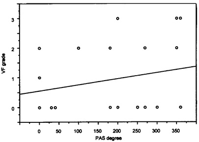
Fig. 2 Scattergram of visual field defects on PAS for all the eyes (n = 73). Spearman's correlation coefficient is 0.348 (95% CI, 0.128~0.536; P = 0.003). The continuous line is the best-fit regression line.

Fig. 3 Scattergram of visual field defects on PAS for the eyes without acute attacks (n = 45). Spearman's correlation coefficient is 0.377 (95% CI, 0.094~0.604; P = 0.012). The continuous line is the best-fit regression line.
Reference
-
1. Wilensky JT, Campbell DG. Albert DM, Jakobiec FA, editors. Primary angle-closure glaucoma. Principles and Practice of Ophthalmology. 2000. 2nd ed. Philadelphia: WB Saunders;2685–2707.2. Gorin G. Shortening of the angle of the anterior chamber in angle-closure glaucoma. Am J Ophthalmol. 1960. 49:141–146.3. Inoue T. Distribution and morphology of peripheral anterior synechia in primary angle-closure glaucoma. J Glaucoma. 1993. 97:78–82.4. Shields MB. Textbook of Glaucoma. 1998. 3rd ed. Baltimore: Williams & Wilkins;198–219.5. Iwata K, Abe H, Sugiyama J. Argon laser iridotomy in primary chronic angle-closure glaucoma. Glaucoma. 1985. 7:103–106.6. Kim YY, Jung HR. Dilated, miotic-resistant pupil and laser iridotomy in primary angle-closure glaucoma. Ophthalmologica. 1997. 211:205–208.7. Salmon JF. Long-term intraocular pressure control after Nd-YAG laser iridotomy in chronic angle-closure glaucoma. J Glaucoma. 1993. 2:291–296.8. Nolan WP, Foster PJ, Devereux JG, Uranchimeg D, Johnson GJ, Baasanhu J. YAG laser iridotomy treatment for primary angle closure in east Asian eyes. Br J Ophthalmol. 2000. 84:1255–1259.9. Katz J. Two eyes or one? The data analyst's dilemma. Ophthalmic Surg. 1988. 19:585–589.10. Leighton DA, Phillips CI, Tsukahara S. Profile of presenting states of eyes in angle-closure glaucoma. Br J Ophthalmol. 1971. 55:577–584.11. Kim YY, Jung HR. Clarifying the nomenclature for primary angle-closure glaucoma. Surv Ophthalmol. 1997. 42:125–136.12. Forbes M. Gonioscopy with corneal indentation. A method for distinguishing between appositional closure and synechial closure. Arch Ophthalmol. 1966. 76:488–492.13. Hodapp E, Parrish RK II, Anderson DR. Clinical Decisions in Glaucoma. 1993. 1st ed. St. Louis: Mosby;52–63.14. Lowe RF. Primary creeping angle-closure glaucoma. Br J Ophthalmol. 1964. 48:544–550.15. Lowe RF. Clinical types of primary angle closure glaucoma. Aust N Z J Ophthalmol. 1988. 16:245–250.16. Pavlin CJ, Ritch R, Foster FS. Ultrasound biomicroscopy in plateau iris syndrome. Am J Ophthalmol. 1992. 113:390–395.17. Pavlin CJ, Harasiewicz K, Foster FS. An ultrasound biomicroscopic dark-room provocative test. Ophthalmic Surg. 1995. 26:253–255.18. Sakuma T, Sawada A, Yamamoto T, Kitazawa Y. Appositional angle closure in eyes with narrow angles: an ultrasound biomicroscopic study. J Glaucoma. 1997. 6:165–169.19. Chandler PA, Grant WM. Lectures on Glaucoma. 1965. 1st ed. Philadelphia: Lea & Febiger;167–185.20. Kronfeld PC. Delayed restoration of the anterior chamber. Am J Ophthalmol. 1954. 38:453–465.21. Harwerth RS, Smith EL 3rd, DeSantis L. Experimental glaucoma: perimetric field defects and intraocular pressure. J Glaucoma. 1997. 6:390–401.22. Newman TB, Browner WS, Cummings SR. Designing a New Study: II. Cross-sectional and Case-controlled Studies. 1988. Baltimore: Williams & Wilkins;367–398.
