Biliary Ascariasis: MR Cholangiography Findings in Two Cases
- Affiliations
-
- 1Department of Radiology, Asan Medical Center, University of Ulsan College of Medicine, Seoul, Korea. tkkim@amc.seoul.kr
- KMID: 754104
- DOI: http://doi.org/10.3348/kjr.2001.2.3.175
Abstract
- We describe the imaging features of two cases of biliary ascariasis. Ultrasonography and CT showed no specific abnormal findings, but MR cholangiography clearly demonstrated an intraductal linear filling defect that led to the correct diagnosis. MR cholangiography is thus a useful technique for the diagnosis of biliary ascariasis.
Keyword
MeSH Terms
Figure
-
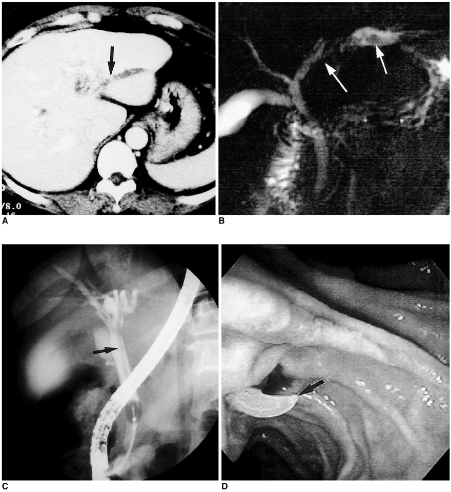
Fig. 1 Biliary ascariasis in a 44-year-old woman. A. Upper abdominal CT scan shows dilation of the intrahepatic bile ducts in the left lobe and a slightly hyperattenuating lesion (arrow) within the dilated bile duct. B. MR cholangiogram shows a linear, tubular hypointense filling defect (white arrows) in the dilated bile duct. C. Endoscopic retrograde cholangiogram shows a linear filling defect (arrow) in the left intrahepatic and common bile duct. D. Photograph obtained during endoscopic extraction of the worm (arrow) shows that this is whitish and extends through the papilla of Vater.

Fig. 2 Biliary ascariasis in a 41-year-old woman. A. Upper abdominal ultrasonogram shows mild dilatation of the common bile duct. B. Contrast-enhanced upper abdominal CT scan shows mild diffuse enlargement of the pancreas. C. MR cholangiogram shows a hypointense tubular filling defect (arrowheads) in the common bile duct. D. Subsequent MR cholangiogram obtained 5 seconds after C demonstrates movement of the worm in the dilated bile duct. E. Endoscopic retrograde cholangiogram shows a linear filling defect (arrow) in the contrast-filled common bile duct.
Reference
-
1. Gutierrez Y, Smith JH. Damjanov I, Linder J, editors. Metazoan diseases. Anderson's Pathology. 1996. St. Louis: Mosby;1014–1015.2. Kubaska SM, Chew FS. Biliary ascariasis. AJR. 1997. 169:492.3. Aslam M, Dore SP, Verbanck J. Ultrasonographic diagnosis of hepatobiliary ascariasis. J Ultrasound Med. 1993. 12:573.4. Ng KK, Wong HF, Kong MS, Chiu LC, Tan CF, Wan YL. Biliary ascariasis: CT, MR cholangiopancreatography, and navigator endoscopic appearance-report of a case of acute biliary obstruction. Abdom Imaging. 1999. 24:470–472.5. Bellini F, Bertolotti J, Conci R, Grumieri G. Hepatobiliary ascaridiasis in childhood: echographic diagnosis. Radiol Med. 1992. 83:619–621.6. Fitoz S, Atasoy C. MR cholangiography in massive hepatobiliary ascariasis. Acta Radiol. 2000. 41:273–274.7. Danaci M, Belet U. MR imaging features of biliary ascariasis. AJR. 1999. 173:503.8. Ferreyra NP, Cerri GG. Ascariasis of the alimentary tract, liver, pancreas and biliary system: its diagnosis by ultrasonography. Hepato-Gastroentero. 1998. 45:932–937.9. Gupta R, Agarwal DK, Choudhuri GD, Saraswat VA, Baijal SS. Biliary ascariasis complicating endoscopic sphincterotomy for choledocholithiasis in India. J Gastroenterol Hepatol. 1998. 13:1072–1073.10. Leung JW, Chung SC. Endoscopic management of biliary ascariasis. Gastrointest Endosc. 1988. 34:318–320.
- Full Text Links
-
- Actions
-
Cited
- CITED
-
- Close
- Share
-
- Similar articles
-
- MR Cholangiography: Usefulness in Obstructive Jaundice
- Reduction in the incidence of biliary and other surgical complications of ascariasis according to the decrease of its national egg prevalence in Korea
- Diagnosis and Treatment of Biliary Ascariasis
- No title available
- MDCT Findings of Biliary Ascariasis: a Case Report
