Interventional Procedures in Superficial Lesions: The Value of 2D with Additional Coronal Reformatted 4D Ultrasonography Guidance
- Affiliations
-
- 1Department of Radiology, Taipei Veterans General Hospital and School of Medicine, National Yang-Ming University, Taipei, Taiwan. cmychang@vghtpe.gov.tw
- 2Department of Orthopedics, Taipei Veterans General Hospital and School of Medicine, National Yang-Ming University, Taipei, Taiwan.
- KMID: 753913
- DOI: http://doi.org/10.3348/kjr.2006.7.1.28
Abstract
OBJECTIVE
We wanted to assess the usefulness of four-dimensional (4D) ultrasonography (US), i.e., real-time three-dimensional US, as an adjunct for performing various US-guided interventional procedures in superficial lesions. MATERIALS AND METHODS: Thirty-three patients were referred for US-guided interventional procedures for superficial lesions, including core biopsy in 19, fine-needle aspiration in eight, therapeutic drug injection in four and needle puncture in two. The procedures were performed under 4D US guidance. We reviewed the pathologic/cytologic results of the core biopsies or needle aspirations, and also the outcomes of drug injection or needle puncture. RESULTS: For all the patients who underwent 4D US-guided core biopsy, the specimens were adequate for making the pathological diagnosis, and specimens were successfully obtained for those patients who underwent 4D US-guided aspiration. The patients treated with 4D US-guided therapeutic drug injection or needle puncture had a good response. No major procedure-related complications occurred. The procedural times were similar to those procedural times with using two-dimensional US. CONCLUSION: Combining the two dimensional and 4D US techniques aids the physician when performing US-guided interventional procedures for the superficial lesions.
MeSH Terms
Figure
-
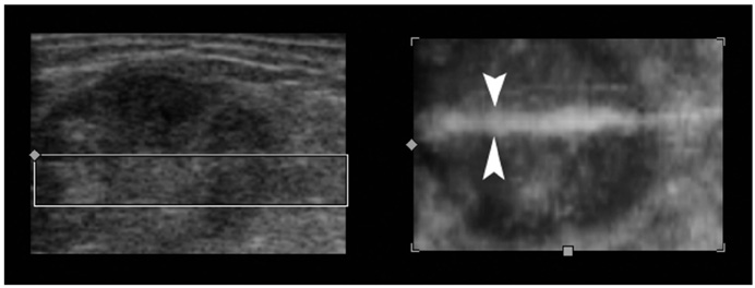
Fig. 1 A 78-year-old woman with a history of surgery for colon cancer developed an enlarged, left inguinal lymph node. During the four-dimensional US-guided biopsy, the needle did not show up on the two-dimensional axial image (left), indicating that it had deviated out of plane. The real-time volume-rendered image in the coronal reformatted plane (right) clearly showed that the needle (arrowheads) was basically in the center of the lesion.
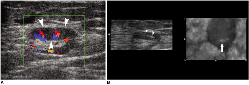
Fig. 2 A 21-year-old man suffering with osteogenic sarcoma in the left distal femur received neoadjuvant chemotherapy and surgical resection of the tumor. An enlarged, left inguinal lymph node was palpated. A. Color Doppler US showed the enlarged node with a prominent cortex of increased vascularity. The proposed optimal site for the needle aspiration was in the cortical portion of the node (arrowheads). B. Four-dimensional US-guided fine-needle aspiration was performed. Axial 2D image (left) showed that the needle (arrowheads) had penetrated into the cortex of the node. Real-time volume-rendered imaging in the coronal reformatted plane (right) showed the needle tip (arrow) in the central cortical portion of the node. By combining information from both images, we confirmed that the needle tip was in the central cortical portion of the node in all three orthogonal planes.
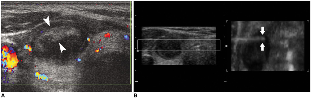
Fig. 3 A 41-year-old woman presented with a nodule on the right side of her neck. A. The image showed the nodule with central cystic change in the right lobe of the thyroid. The solid parts were mainly at the periphery (arrowheads). B. Four-dimensional US-guided fine-needle aspiration of the nodule was performed. The axial two-dimensional image (left) did not show the needle because it had deviated out of the central plane. The real-time volume-rendered image in the coronal reformatted plane (right) showed the needle tip (arrows) in the periphery of the nodule, which was the desired location for tissue sampling.
Reference
-
1. Cho N, Moon WK, Cha JH. Sonographically guided core biopsy of the breast: comparison of 14-gauge automated gun and 11-gauge directional vacuum-assisted biopsy methods. Korean J Radiol. 2005. 6:102–109.2. Lim HK. Radiofrequency thermal ablation of hepatocellular carcinomas. Korean J Radiol. 2000. 1:175–184.3. Lees W. Ultrasound imaging in three and four dimensions. Semin Ultrasound CT MR. 2001. 22:85–105.4. Downey DB, Fenster A, Williams JC. Clinical utility of three-dimensional US. RadioGraphics. 2000. 20:559–571.5. Rose SC, Pretorius DH, Nelson TR, Kinney TB, Huynh TV, Roberts AC, et al. Adjunctive 3D US for achieving portal vein access during transjugular intrahepatic portosystemic shunt procedures. J Vasc Interv Radiol. 2000. 11:611–621.6. Rose SC, Roberts AC, Kinney TB, Pretorius DH, Nelson TR. Three-dimensional ultrasonography for planning percutaneous drainage of complex abdominal fluid collections. J Vasc Interv Radiol. 2003. 14:451–459.7. Smith WL, Surry KJ, Mills GR, Downey DB, Fenster A. Three-dimensional ultrasound-guided core needle breast biopsy. Ultrasound Med Biol. 2001. 27:1025–1034.8. Rose SC, Hassanein TI, Easter DW, Gamagami RA, Bouvet M, Pretorius DH, et al. Value of three-dimensional US for optimizing guidance for ablating focal liver tumors. J Vasc Interv Radiol. 2001. 12:507–515.9. Strasser H, Janetschek G, Horninger W, Bartsch G. Three-dimensional sonographic guidance for interstitial laser therapy in benign prostatic hyperplasia. J Endourol. 1995. 9:497–501.10. Weismann CF, Forstner R, Prokop E, Rettenbacher T. Three-dimensional targeting: a new three-dimensional ultrasound technique to evaluate needle position during breast biopsy. Ultrasound Obstet Gynecol. 2000. 16:359–364.11. Chin JL, Downey DB, Mulligan M, Fenster A. Three-dimensional transrectal ultrasound guided cryoablation for localized prostate cancer in nonsurgical candidates: a feasibility study and report of early results. J Urol. 1998. 159:910–914.12. Won HJ, Han JK, Do KH, Lee KH, Kim KW, Kim SH, et al. Value of four-dimensional ultrasonography in ultrasonographically guided biopsy of hepatic masses. J Ultrasound Med. 2003. 22:215–220.13. Liu JC, Chiou HJ, Chen WM, Chou YH, Chen TH, Chen W, et al. Sonographically guided core needle biopsy of soft tissue neoplasms. J Clin Ultrasound. 2004. 32:294–298.
- Full Text Links
-
- Actions
-
Cited
- CITED
-
- Close
- Share
-
- Similar articles
-
- Ultrasound-guided genitourinary interventions: principles and techniques
- Is the Index Finger and Ring Finger Ratio (2D:4D) Reliable Predictor of Semen Quality?
- A Relationship between 2nd to 4th Digit Length Ratio and Aggression Related-Sports Entries Characteristics in Female Athletics of Korean National Teams
- Value of Coronal Reformatted Images Using Multi-detector Computed Tomography for Nodal Staging in Non Small Cell Lung Cancer Cases
- The Study on The Near Point in Koreans
