Hepatic Parasitic Abscess Caused by Clonorchiasis: Unusual CT Findings of Clonorchiasis
- Affiliations
-
- 1Department of Radiology and Research Institute of Radiology, University of Ulsan College of Medicine, Asan Medical Center, Seoul, Korea. jhbyun@amc.seoul.kr
- 2Department of Pathology, University of Ulsan College of Medicine, Asan Medical Center, Seoul, Korea.
- KMID: 753868
- DOI: http://doi.org/10.3348/kjr.2007.8.1.70
Abstract
- Clonorchiasis is caused by a chronic infestation of liver flukes, Clonorchis sinensis, and these reside mainly in the medium- and small-sized intrahepatic bile ducts. Therefore, diffuse, uniform, minimal or mild dilatation of these bile ducts, particularly in the periphery, without dilatation of the extrahepatic bile duct is the typical finding on several imaging modalities. We report here on the CT findings of an unusual case of hepatic parasitic abscess that was caused by clonorchiasis; this malady mimicked cholangiocarcinoma, and there was no dilatation of the intrahepatic bile ducts.
MeSH Terms
Figure
-
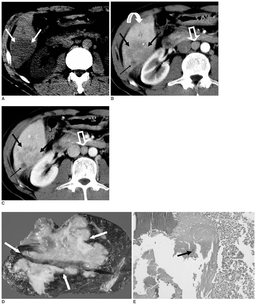
Fig. 1 A 52-year-old man with a hepatic parasitic abscess caused by clonorchiasis. A. The transverse unenhanced CT scan shows a heterogeneous low-attenuation mass (arrows) in the right posteroinferior segment of the liver. B. The contrast-enhanced CT scan during the hepatic arterial phase shows a very ill-margined, lobulated mass (arrows) with heterogeneous, peripheral contrast enhancement and a surrounding transient hepatic attenuation difference (curved arrow) in the right posteroinferior segment of the liver. There is a small portion of lower attenuation in the peripheral area of the mass (thin arrow). A 1.5-cm lymph node, which was proven to be a reactive lymph node, is seen in the aortocaval space (open arrow). C. During the portal venous phase, the CT scan shows an ill-margined, lobulated mass (arrows) with a central filled-in area of contrast enhancement from the peripheral portion of the mass in the right posteroinferior segment of the liver. There is a small portion with poor contrast enhancement in the peripheral area of the mass, and this represents a necrotic area (thin arrow). The intrahepatic bile ducts are not dilated. There is a 1.5-cm lymph node that was proven to be a reactive lymph node in the aortocaval space (open arrow). D. Photograph of the gross specimen shows an ill-defined, lobulated, creamy yellow mass measuring 7×6×5 cm (arrows). The tumor is close to the round ligament and the hepatic capsule. There was no evidence of liver cirrhosis. E. Photomicrograph of the histopathologic specimen demonstrates inflammatory cells such as lymphocytes, neutrophils and macrophages, and a parasitic egg (arrow). The histopathologic diagnosis is parasitic abscess caused by clonorchiasis with a granulomatous reaction. (H&E stain, ×100)
Reference
-
1. Rim HJ. The current pathobiology and chemotherapy of clonorchiasis. Korean J Parasitol. 1986. 24:7–20. [Korean].2. Lim JH. Radiologic findings of clonorchiasis. AJR Am J Roentgenol. 1990. 155:1001–1008.3. Choi BI, Kim HJ, Han MC, Do YS, Han MH, Lee SH. CT findings of clonorchiasis. AJR Am J Roentgenol. 1989. 152:281–284.4. Jeong YY, Kang HK, Kim JW, Yoon W, Chung TW, Ko SW. MR imaging findings of clonorchiasis. Korean J Radiol. 2004. 5:25–30.5. Kim YH. Extrahepatic cholangiocarcinoma associated with clonorchiasis: CT evaluation. Abdom Imaging. 2003. 28:68–71.6. Mirdha BR, Gulati S, Sarkar T, Samantray JC. Acute clonorchiasis in a child. Indian J Gastroenterol. 1988. 17:155.7. Seong NJ, Lee JM, Kim SH, Han JK, Kim YJ, Kim JH, et al. Differentiation between mass-forming type peripheral cholangiocarcinoma and hepatic abscesses: application of artificial neural networks to CT images. J Korean Radiol Soc. 2005. 53:343–352.
- Full Text Links
-
- Actions
-
Cited
- CITED
-
- Close
- Share
-
- Similar articles
-
- Ultrasonographic diagnosis of clonorchiasis
- A Case of Clonorchiasis with Focal Intrahepatic Duct Dilatation Mimicking an Intrahepatic Cholangiocarcinoma
- ERCP Findings In Hepatic Clonorchiasis
- Long-lasting sonographic and histopathological findings in cured clonorchiasis of rabbits
- Imaging diagnosis of clonorchiasis
