Leiomyosarcoma Arising from the Blind End of a Bifid Renal Pelvis
- Affiliations
-
- 1Department of Urology, College of Medicine, Inha University, Incheon, Korea. dhseong@inha.ac.kr
- 2Department of Pathology, College of Medicine, Inha University, Incheon, Korea.
- KMID: 724117
- DOI: http://doi.org/10.3349/ymj.2007.48.3.557
Abstract
- Sarcoma of the kidney is a rare condition. Leiomyosarcoma is the most common of the kidney sarcomas. Renal leiomyosarcoma usually originates from the smooth muscle layers of the kidney, for example, the renal capsule and renal vessels. Renal pelvis neoplasms, however, are primarily transitional cell carcinomas, and renal pelvis leiomyosarcomas are extremely uncommon. Renal pelvis leiomyosarcoma has never been reported in Korea. Moreover, no more than 10 cases have been reported internationally. However, none of these were associated with kidney abnormalities. Here we describe a case of leiomyosarcoma that originated from the blind end of a bifid renal pelvis.
Keyword
MeSH Terms
Figure
-
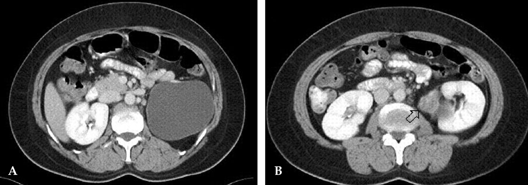
Fig. 1 Computed tomography shows a 10 × 8cm sized cystic lesion in the upper pole of left kidney (A), and a 5 × 3cm sized irregular shaped mass (arrow) with heterogeneous enhancement adjacent to the renal pelvis (B). It is not clarified whether is renal pelvis origin or extrarenal origin.
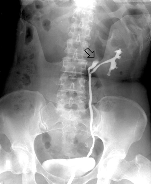
Fig. 2 Retrograde pyelography shows the bifid pelvis of left kidney, with the filling defect (arrow) in the renal pelvis of upper moiety.

Fig. 3 The cut surface (A) shows a well marginated tumor mass at bifid pelvis, the other ureter and a brownish tan cystic change lesion at the upper pole of kidney. Microphotography (B) shows that a tumor mass arises from the smooth muscle layer of the renal pelvis (H & E, × 10). M: tumor mass, P: bifid pelvis, C: cyst, U: another ureter, S: smooth muscle layer of renal pelvis.

Fig. 4 Microphotography shows mitosis (a cell with dark nucleoli in the center) and spindle shaped cells with large nucleus in H-E staining (A)(× 400). Positive staining of the tumor cells for smooth muscle-specific actin reveals that the tumor is originated from smooth muscle tissue in actin staining (B)(× 100) and desmin staining (C)(× 200).
Reference
-
1. Park CH, An JT, Ihm JK, Lee YG, Shim JW, Park HW. A case of renal leiomyosarcoma. Korean J Urol. 2000. 41:333–336.2. Moudouni SM, En-Nia I, Rioux-Leclerq N, Guille F, Lobel B. Leiomyosarcoma of the renal pelvis. Scand J Urol Nephrol. 2001. 35:425–427.
Article3. Todd CS, Michael H, Sutton G. Retroperitoneal leiomyosarcoma: eight cases and a literature review. Gynecol Oncol. 1995. 59:333–337.
Article4. Chen JH, Lee SK. Renal leiomyosarcoma mimicking transitional cell carcinoma. AJR Am J Roentgenol. 1997. 169:312–313.
Article5. Minami H, Ueki O, Tanaka T, Nishida H, Hashimoto T, Kawaguchi K. Case of leiomyosarcoma of the renal pelvis. Int J Urol. 2004. 11:122–124.
Article6. Novick AC, Campbell SC. Walsh PC, Retik AB, Vaughan ED, Wein AJ, editors. Renal tumors. Campbell's urology. 2005. 8th ed. Philadelphia: Saunders;2719–2720.7. Miyajima K, Oda Y, Oshiro Y, Tamiya S, Kinukawa N, Masuda K, et al. Clinicopathological prognostic factors in soft tissue leiomyosarcoma: a multivariate analysis. Histopathology. 2002. 40:353–359.
Article8. Cormier JN, Pollock RE. Soft tissue sarcoma. CA Cancer J Clin. 2004. 54:94–109.9. Spillane AJ. Retroperitoneal sarcoma: time for a change in attitude? ANZ J Surg. 2001. 71:303–308.
