Resurfacing the defect from wide excision of a malignant peripheral nerve sheath tumor based on a thoracodorsal artery perforator free flap: a case report
- Affiliations
-
- 1Department of Plastic and Reconstructive Surgery, Hanyang University College of Medicine, Seoul, Korea
- KMID: 2542563
- DOI: http://doi.org/10.12790/ahm.23.0007
Abstract
- Malignant peripheral nerve sheath tumors (MPNSTs) are rare, aggressive soft tissue sarcomas with a high rate of recurrence and metastasis. Limb salvage surgery with free flap reconstruction is a viable option for selected patients with MPNSTs, but careful consideration should be given to the risk of recurrence. This case report describes a 26-year-old male patient with a recurrent, aggressive, high-grade MPNST who underwent limb salvage surgery with thoracodorsal artery perforator free flap reconstruction. Despite the surgical intervention, local recurrence of the MPNST was detected, and below-knee amputation was ultimately recommended. This case highlights the importance of early, definitive treatment decision-making in cases of aggressive, high-grade MPNSTs. Close postoperative monitoring and early detection of recurrence are crucial for achieving optimal outcomes in patients with MPNSTs undergoing limb salvage surgery with free flap reconstruction.
Figure
-

Fig. 1. A patient presented with malignant peripheral nerve sheath tumor (MPNST) on his right ankle. (A) In 2017, he was diagnosed with MPNST on the medial side of the right ankle and received wide excision and coverage with a thoracodorsal artery perforator free flap, followed by adjuvant radiation therapy. (B) In 2022, he presented with recurrent MPNST on the lateral side of the right ankle. (C) Sagittal view of preoperative T2-weighted magnetic resonance imaging revealed extensive involvement of surrounding structures.
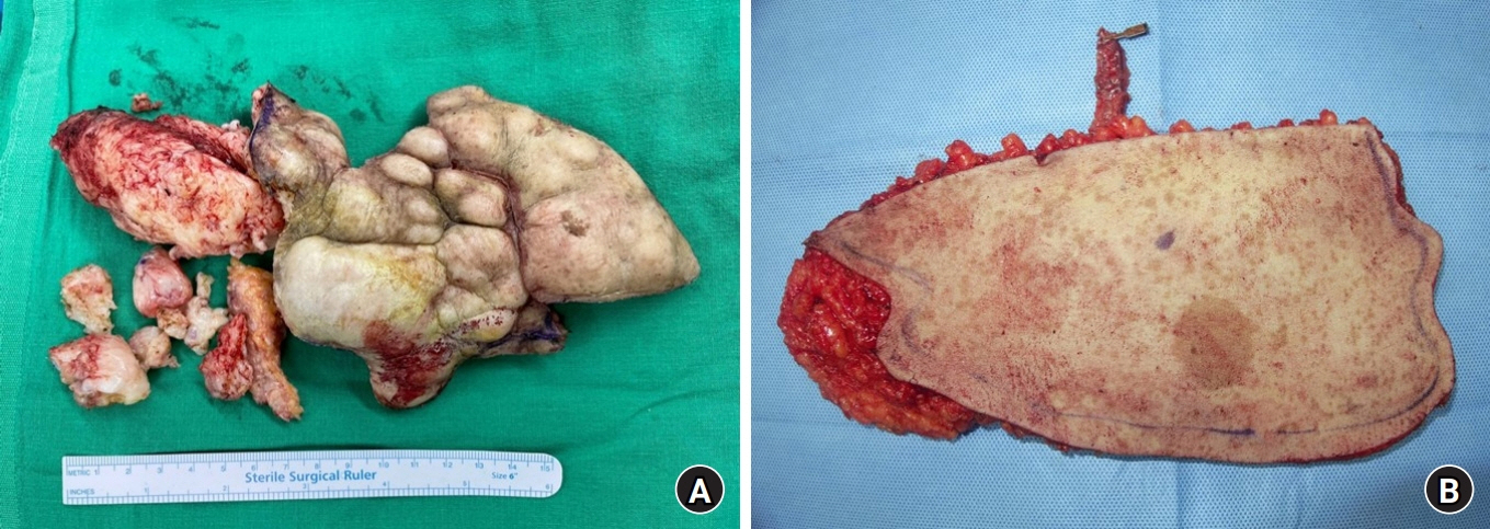
Fig. 2. (A) Recurrent malignant peripheral nerve sheath tumor (MPNST) was removed from the lateral aspect of the right ankle with a positive margin. The main mass measured 14×18 cm. (B) A thoracodorsal artery perforator free flap was harvested from the right side for defect coverage. The flap measured 15×20 cm, with a 12 cm-long pedicle.
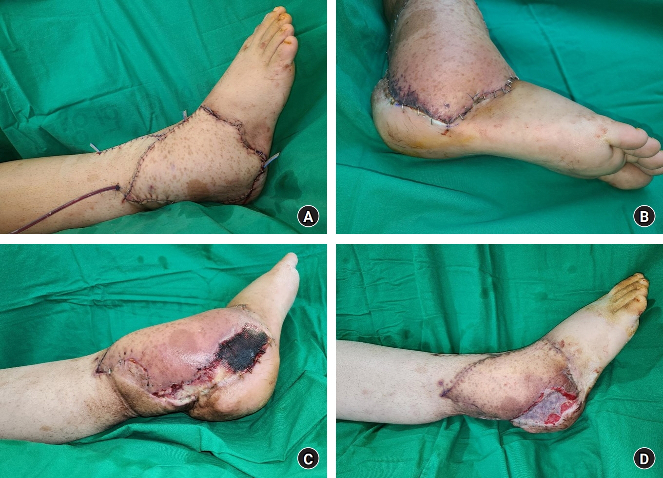
Fig. 3. (A) Free flap transfer was performed successfully without complications. (B) On the 8th postoperative day, the distal portion of the flap showed a bluish color change suggesting arterial insufficiency. (C) By the 25th postoperative day, the final portion with arterial insufficiency measured 3×12 cm. (D) Flap revision was performed to remove the demarcated portion, and the defect was covered with a split-thickness skin graft.
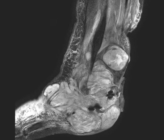
Fig. 4. Sagittal view of postoperative T2-weighted magnetic resonance imaging revealed rapid growth of the known malignant peripheral nerve sheath tumor and the extent of local recurrence.
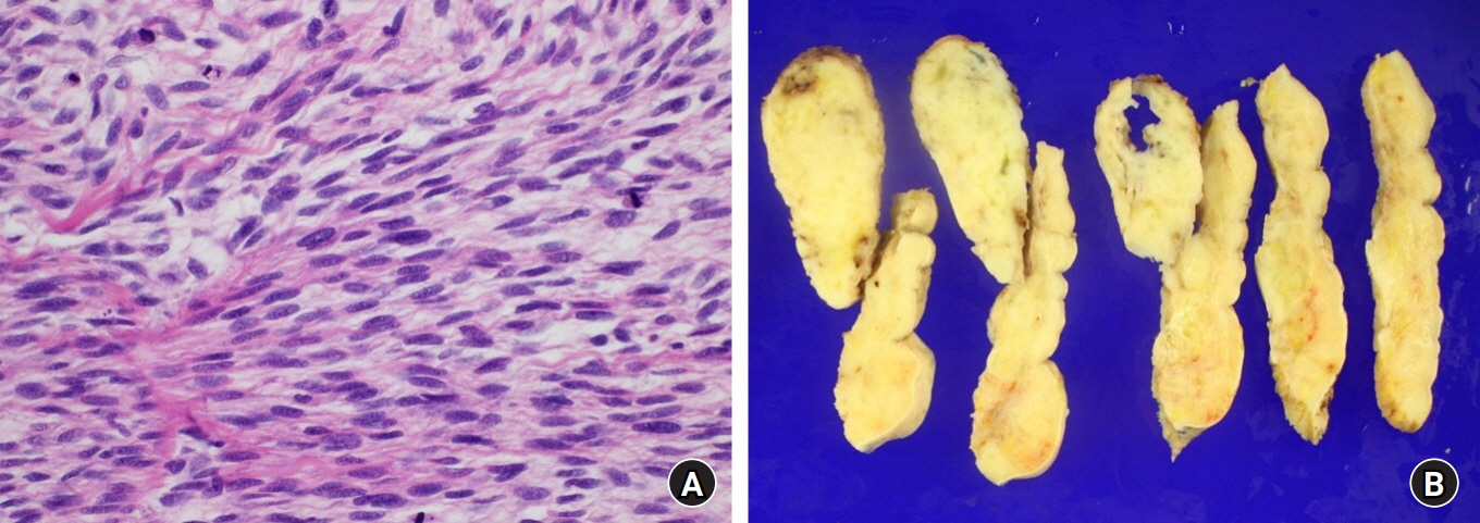
Fig. 5. (A) The histologic image of the resected tumor revealed spindle cells with irregular contours and high mitotic activity, a characteristic of high-grade malignant peripheral nerve sheath tumor ( H&E stain, ×400). (B) Cross-sections of the resected tumor revealed a diffusely located tumor with necrosis, infiltrating the surrounding normal tissue.
Reference
-
References
1. Somatilaka BN, Sadek A, McKay RM, Le LQ. Malignant peripheral nerve sheath tumor: models, biology, and translation. Oncogene. 2022; 41:2405–21.
Article2. Evans DG, Baser ME, McGaughran J, Sharif S, Howard E, Moran A. Malignant peripheral nerve sheath tumours in neurofibromatosis 1. J Med Genet. 2002; 39:311–4.
Article3. Stucky CC, Johnson KN, Gray RJ, et al. Malignant peripheral nerve sheath tumors (MPNST): the Mayo Clinic experience. Ann Surg Oncol. 2012; 19:878–85.
Article4. James AW, Shurell E, Singh A, Dry SM, Eilber FC. Malignant peripheral nerve sheath tumor. Surg Oncol Clin N Am. 2016; 25:789–802.
Article5. Theos A, Korf BR; American College of Physicians; American Physiological Society. Pathophysiology of neurofibromatosis type 1. Ann Intern Med. 2006; 144:842–9.
Article6. Farid M, Demicco EG, Garcia R, et al. Malignant peripheral nerve sheath tumors. Oncologist. 2014; 19:193–201.
Article7. Nakamura JL, Phong C, Pinarbasi E, et al. Dose-dependent effects of focal fractionated irradiation on secondary malignant neoplasms in Nf1 mutant mice. Cancer Res. 2011; 71:106–15.8. Miettinen MM, Antonescu CR, Fletcher CDM, et al. Histopathologic evaluation of atypical neurofibromatous tumors and their transformation into malignant peripheral nerve sheath tumor in patients with neurofibromatosis 1-a consensus overview. Hum Pathol. 2017; 67:1–10.
Article
- Full Text Links
-
- Actions
-
Cited
- CITED
-
- Close
- Share
-
- Similar articles
-
- Reconstruction of the Soft Tissue Defect Using Thoracodorsal Artery Perforator Skin Flap
- Reconstruction of Greater Trochanteric defect using Lumbar Artery Perforator Free Flap: A Case Report
- Reconstruction of a Mangled Hand with a Thoracodorsal Artery Perforator Free Flap: A Report of Two Cases
- Malignant Peripheral Nerve Sheath Tumor of Abdomen
- Thinned Thoracodorsal Perforator-Based Cutaneous Free Flap
