Vascular anatomy and their variations in Situs inversus totalis using postmortem computed tomographic angiography
- Affiliations
-
- 1Division of Forensic Medicine, Graduate School of Medicine, Tottori University, Yonago, Japan
- 2Department of Anatomy, Graduate School of Medicine, Tottori University, Yonago, Japan
- KMID: 2540996
- DOI: http://doi.org/10.5115/acb.22.213
Abstract
- Studies describing the vascular systems and their variations in Situs inversus totalis (SIT) from a whole-body computed tomographic (CT) angiography perspective are lacking. We report a case of SIT in which postmortem CT angiography (PMCTA) was performed as a part of the forensic death investigation and incidentally detected several vascular variations in it. The PMCTA procedure was performed using the multiphase PMCTA protocol. Almost all major vessels were visualized, indeed in a completely reversed pattern. Contrast mixture flow interruptions were noted in the right coronary arterial branches suggesting possible blockage, upon which autopsy revealed >90% vessel occlusions at several locations. As such the cause of death was due to ischemic heart disease. Anomalous origins of the right internal mammary artery; abnormal left thyrocervical trunk and variations in the drainage of testicular veins were noted. Our findings might be helpful to clinicians and add to the body of literature on SIT.
Keyword
Figure
-
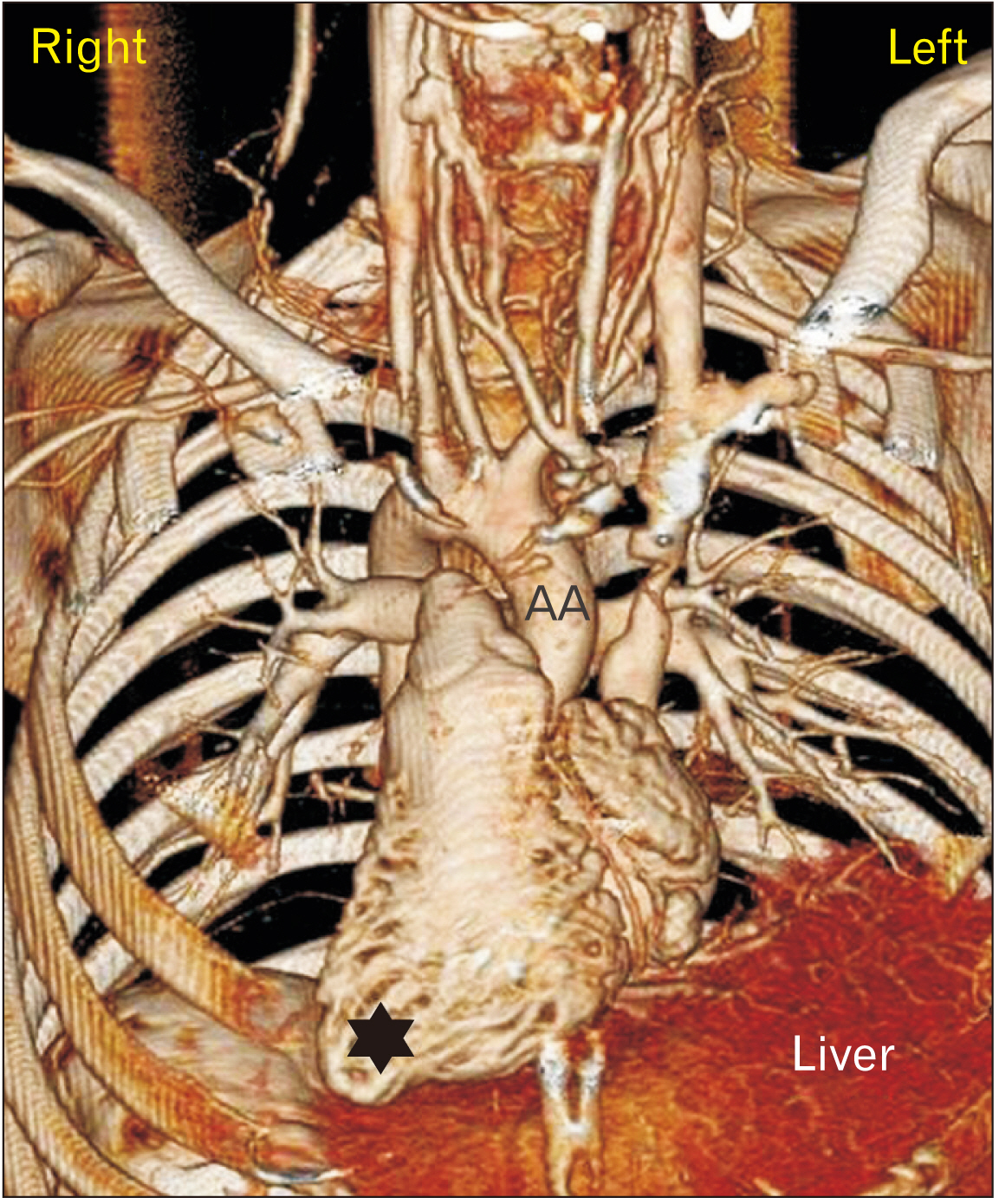
Fig. 1 3D VR reconstruction of the heart and great vessels. A dynamic phase of MPMCTA. Right-sided heart, the apex pointing towards the right (star) and AA emerging from the right ventricle. AA, ascending aorta; VR, virtual reconstruction; MPMCTA, multiphase postmortem computed tomographic angiography; 3D, three-dimensional.

Fig. 2 3D VR reconstruction of coronary arteries. Arterial phase of MPMCTA. Note the contrast flow interruptions shown as filling defects (short arrows). VR, virtual reconstruction; MPMCTA, multiphase postmortem computed tomographic angiography; RCA, right coronary artery; LCA, left coronary artery; CA, circumflex artery; ADA, anterior descending artery; PDA, posterior descending artery; 3D, three-dimensional.

Fig. 3 3D VR reconstruction of the right anterior neck. Arterial phase of MPMCTA. Note the common origin of the right IMA with other arterial branches, namely the TCA and SSA. VR, virtual reconstruction; MPMCTA, multiphase postmortem computed tomographic angiography; IMA, internal mammary artery; TCA, transverse cervical artery; SSA, suprascapular artery; AA, ascending aorta; BcA, brachiocephalic artery; CA, common carotid artery; VA, vertebral artery; ThyCT, thyrocervical trunk; ITA, inferior thyroid artery; ACA, ascending cervical artery; DACA, deep ascending cervical artery; DScA, dorsal scapular artery; SCA, subclavian artery; DA, descending aorta; 3D, three-dimensional.

Fig. 4 3D VR reconstruction of the left anterior neck. Arterial phase of MPMCTA. Note the common origin of the left ThyCT with another atypical arterial trunk, which is comparatively large. VR, virtual reconstruction; MPMCTA, multiphase postmortem computed tomographic angiography; ThyCT, thyrocervical trunk; BcA, Brachiocephalic artery; CA, carotid artery; VA, vertebral artery; IMA, internal mammary artery; ITA, inferior thyroid artery; ACA, ascending cervical artery; DACA, deep ascending cervical artery; SSA, suprascapular artery; DScA, dorsal scapular artery; SpCA, superficial cervical artery; SCA, subclavian artery; 3D, three-dimensional.

Fig. 5 3D VR reconstruction to separate the testicular vessels. A dynamic phase of MPMCTA. Notice dilated & tortuous (varicocele) left testicular vein draining into the left renal vein. Right testicular vein coursing inwards and draining into vesicle venous plexus. Normal testicular arteries. VR, virtual reconstruction; MPMCTA, multiphase postmortem computed tomographic angiography; AO, abdominal aorta; IVC, inferior vena cava; 3D, three-dimensional.
Reference
-
References
1. Lee SE, Kim HY, Jung SE, Lee SC, Park KW, Kim WK. 2006; Situs anomalies and gastrointestinal abnormalities. J Pediatr Surg. 41:1237–42. DOI: 10.1016/j.jpedsurg.2006.03.045. PMID: 16818055.
Article2. Pidvalna U, Mirchuk M, Beshley D, Mateshuk-Vatseba L. 2022; Morphometric characteristics of the aorta and heart in situs inversus totalis. Anat Cell Biol. 55:259–63. DOI: 10.5115/acb.21.252. PMID: 35773223. PMCID: PMC9256485.
Article3. Grabherr S, Doenz F, Steger B, Dirnhofer R, Dominguez A, Sollberger B, Gygax E, Rizzo E, Chevallier C, Meuli R, Mangin P. 2011; Multi-phase post-mortem CT angiography: development of a standardized protocol. Int J Legal Med. 125:791–802. DOI: 10.1007/s00414-010-0526-5. PMID: 21057803.
Article4. Lin AE, Krikov S, Riehle-Colarusso T, Frías JL, Belmont J, Anderka M, Geva T, Getz KD, Botto LD. 2014; Laterality defects in the national birth defects prevention study (1998-2007): birth prevalence and descriptive epidemiology. Am J Med Genet A. 164A:2581–91. DOI: 10.1002/ajmg.a.36695. PMID: 25099286. PMCID: PMC4462240.
Article5. Tubbs RS, Wellons JC 3rd, Salter G, Blount JP, Oakes WJ. 2003; Intracranial anatomic asymmetry in situs inversus totalis. Anat Embryol (Berl). 206:199–202. DOI: 10.1007/s00429-002-0286-1. PMID: 12592571.
Article6. Goldberg A, Southern DA, Galbraith PD, Traboulsi M, Knudtson ML, Ghali WA. 2007; Coronary dominance and prognosis of patients with acute coronary syndrome. Am Heart J. 154:1116–22. DOI: 10.1016/j.ahj.2007.07.041. PMID: 18035084.
Article7. Rathore A, Gowda Somashekar CM, Sadananda KS, Manjunath CN. 2017; Acute myocardial infarction in dextrocardia - a diagnostic and therapeutic challenge. Can dextrocardia be a risk factor? J Cardiol Cases. 17:48–51. DOI: 10.1016/j.jccase.2017.09.003. PMID: 30279853. PMCID: PMC6149624.
Article8. Leigh MW, Pittman JE, Carson JL, Ferkol TW, Dell SD, Davis SD, Knowles MR, Zariwala MA. 2009; Clinical and genetic aspects of primary ciliary dyskinesia/Kartagener syndrome. Genet Med. 11:473–87. DOI: 10.1097/GIM.0b013e3181a53562. PMID: 19606528. PMCID: PMC3739704.
Article9. Paliouras D, Rallis T, Gogakos A, Asteriou C, Chatzinikolaou F, Georgios T, Tsirgogianni K, Tsakiridis K, Mpakas A, Sachpekidis N, Zarogoulidis K, Papaiwannou A, Organtzis J, Karapantzos I, Karapantzou C, Zarogoulidis P, Barbetakis N. 2015; Surgical anatomy of the internal thoracic arteries and their branching pattern: a cadaveric study. Ann Transl Med. 3:212. DOI: 10.3978/j.issn.2305-5839.2015.09.03. PMID: 26488008. PMCID: PMC4583587.10. Murray AC, Rozen WM, Alonso-Burgos A, Ashton MW, Garcia-Tutor E, Whitaker IS. 2012; The anatomy and variations of the internal thoracic (internal mammary) artery and implications in autologous breast reconstruction: clinical anatomical study and literature review. Surg Radiol Anat. 34:159–65. DOI: 10.1007/s00276-011-0886-7. PMID: 21986988.
Article11. López-Muñiz A, García-Fernández C, Hernández LC, Suárez-Garnacho S. 2002; Variants of the thyrocervical trunk and its branches in human bodies. Eur J Anat. 6:109–13.12. González-Castillo A, Rojas S, Ortega M, Rodríguez-Baeza A. 2018; Variations in vascular anatomy and unilateral adrenal agenesis in a female cadaver with situs inversus totalis. Surg Radiol Anat. 40:1169–72. DOI: 10.1007/s00276-018-2060-y. PMID: 29931532.
Article13. Sonje P, Kanasker N, Vatsalaswamy P. 2020; Variations in the origin and draining pattern of gonadal vessels with their surgical significance and developmental correlations. Indian J Anat. 9:39–43. DOI: 10.21088/ija.2320.0022.9120.6.
Article14. Mano Y, Adachi N, Murakami G, Yokoyama T, Dodo Y. 2006; Human situs inversus of the thoracoabdominal structures. Anat Sci Int. 81:7–20. DOI: 10.1111/j.1447-073X.2006.00122.x. PMID: 16526591.
Article15. Kulesza RJ Jr, Kalmey JK, Dudas B, Buck WR. 2007; Vascular anomalies in a case of situs inversus. Folia Morphol (Warsz). 66:69–73. PMID: 17533597.
- Full Text Links
-
- Actions
-
Cited
- CITED
-
- Close
- Share
-
- Similar articles
-
- Laparoscopic cholecystectomy in a case of situs inversus totalis: a review of technical challenges and adaptations
- Neonatal Duodenal Obstruction Associated with Situs Inversus Totalis: A Case Report
- Single Port Laparoscopic Cholecystectomy in a Patient with Situs Inversus Totalis: A Case Report
- Laparoscopic Low Anterior Resection in a Rectal Cancer Patient with Situs Inversus Totalis: A Case Report
- A Case of Pregnancy Complicated by Situs Inversus Totalis Fetus in Overt Diabetic Woman
