Two-Stage Operation Over a Period of 7 Years for a Patient with Macrodactyly: A Case Report
- Affiliations
-
- 1Department of Orthopedic Surgery, Inje University Busan Paik Hospital, Busan, Korea
- 2Department of Pediatrics, Inha University Hospital, Incheon, Korea
- KMID: 2540403
- DOI: http://doi.org/10.14193/jkfas.2023.27.1.24
Abstract
- Macrodactyly of the toe is a rare congenital anomaly characterized by the overgrowth of a digit/digits in the foot and is one of the most difficult conditions to treat. Since the condition alters functionality and appearance, the treatment goal is to restore function and cosmetically enhance the appearance. Various surgical techniques are used for toe macrodactyly, including amputation, debulking, and epiphysiodesis. Herein, we present a case of a six-year-old patient with a second toe macrodactyly who was successfully treated with a twostage operation over a seven-year period. We initially performed an ostectomy of the middle phalanx with a fusion of the proximal and distal phalanges and then performed a soft tissue debulking procedure.
Keyword
Figure
-
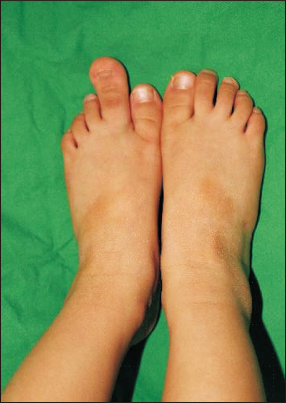
Figure. 1 Gross photograph of a 6-year-old girl with complaints of second left toe enlargement.
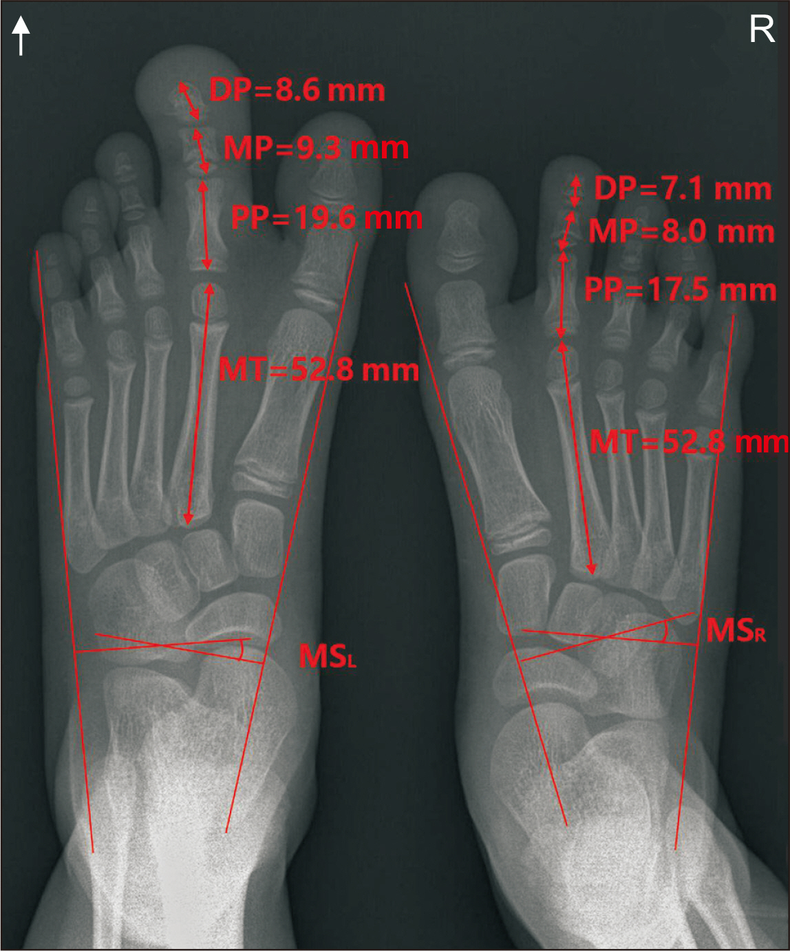
Figure. 2 Measurement of the left foot second phalanges (DP+MP+PP) and the right-side ray (32.6 mm and 37.5 mm, respectively). The left and right second MT lengths measure the same (52.8 mm, each). Metatarsal spread angles of the left and right feet measure 16.9˚ and 17.5˚, respectively. DP: distal phalanx, MP: middle phalanx, PP: proximal phalanx, MT: metatarsal bone, MSL: left side metatarsal spread angle, MSR: right side metatarsal spread angle.
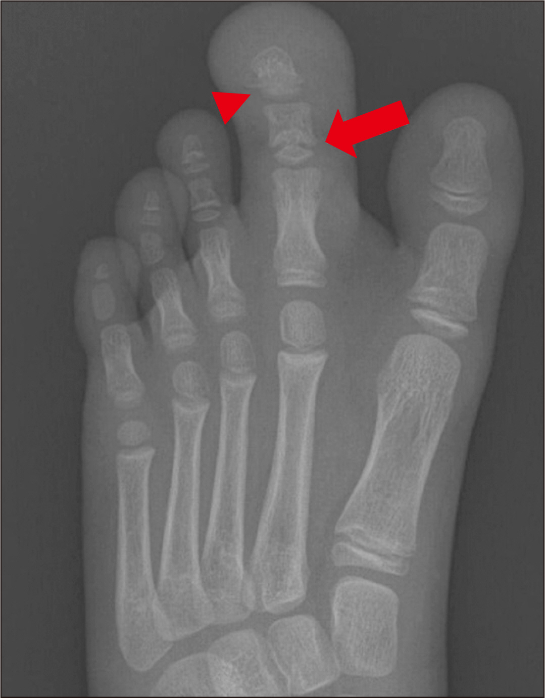
Figure. 3 Tethered proximal physeal line of the left second toe middle phalanx on the initial anterior-posterior view of the foot (standing). The proximal physeal line of the left second to distal phalanx appears closed (marked by an arrow). Other physeal lines of the left foot are normal (marked by a triangle).
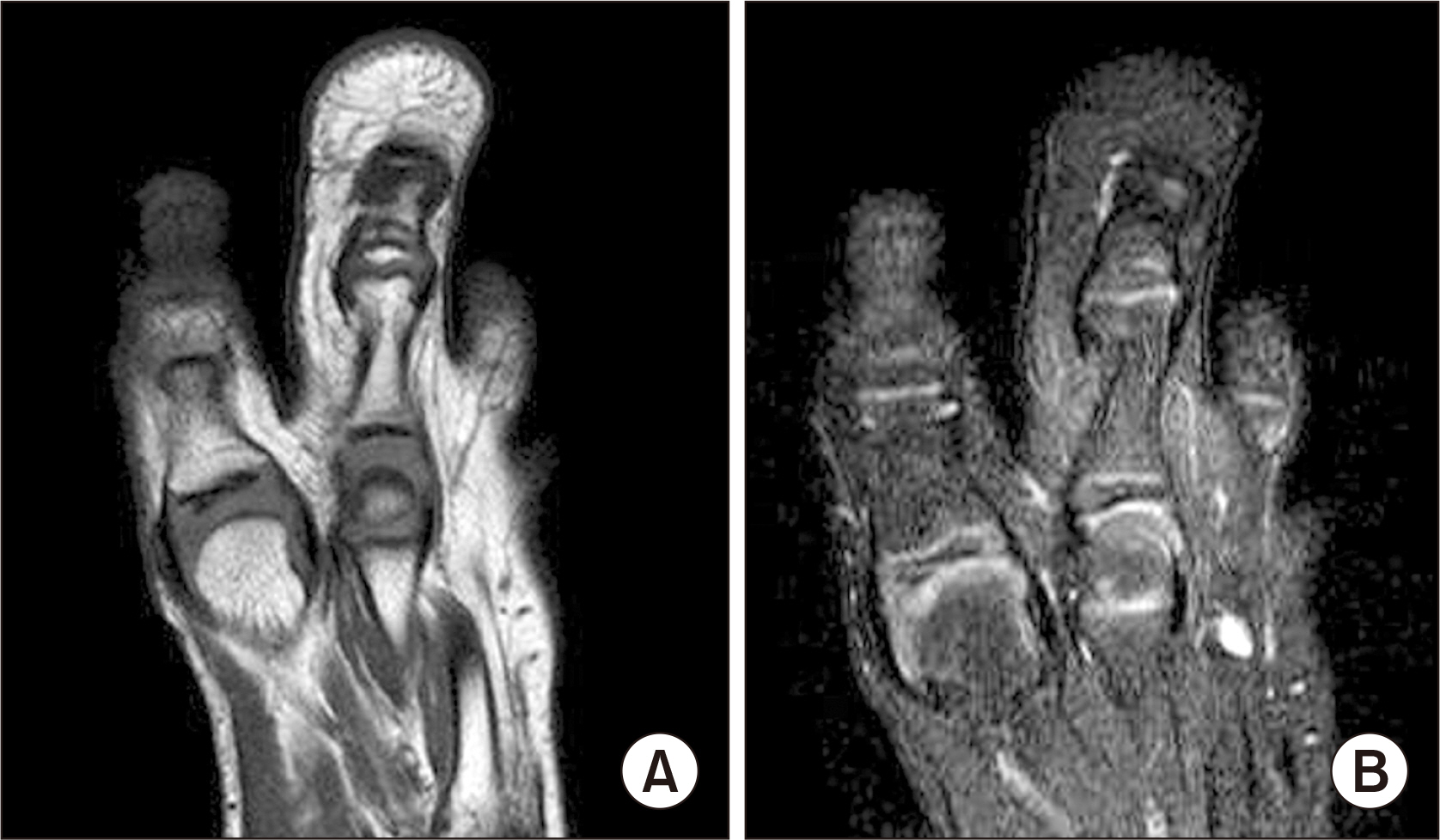
Figure. 4 Preoperative magnetic resonance images of the left foot reveal enlargement of the subcutaneous fat and the presence of bony structures. Definite nerve enlargement is absent. (A) T1-weighted axial view. (B) T2-weighted axial view.

Figure. 5 Radiograph of the patient’s left foot (standing) together with her mother’s right foot. Surgery was performed when the sum of the lengths of the patient's three left second toe phalanges (43.1 mm) was greater than that of her mother's (39.8 mm). DP: distal phalanx, MP: middle phalanx, PP: proximal phalanx, MT: metatarsal bone.
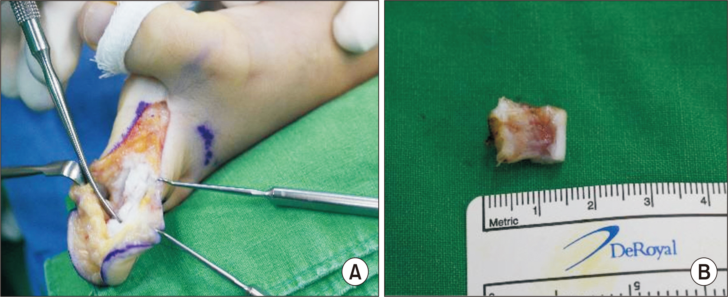
Figure. 6 Intra-operative photographs. (A) Creation of a large incision at the medial side and removal of the thickened adipose tissue. This was followed by ostectomy at the middle phalanx. (B) Removed middle phalanx.

Figure. 7 Radiograph of the foot after the first operation. Ostectomy of middle phalanx followed by a two-K-wire fixation.
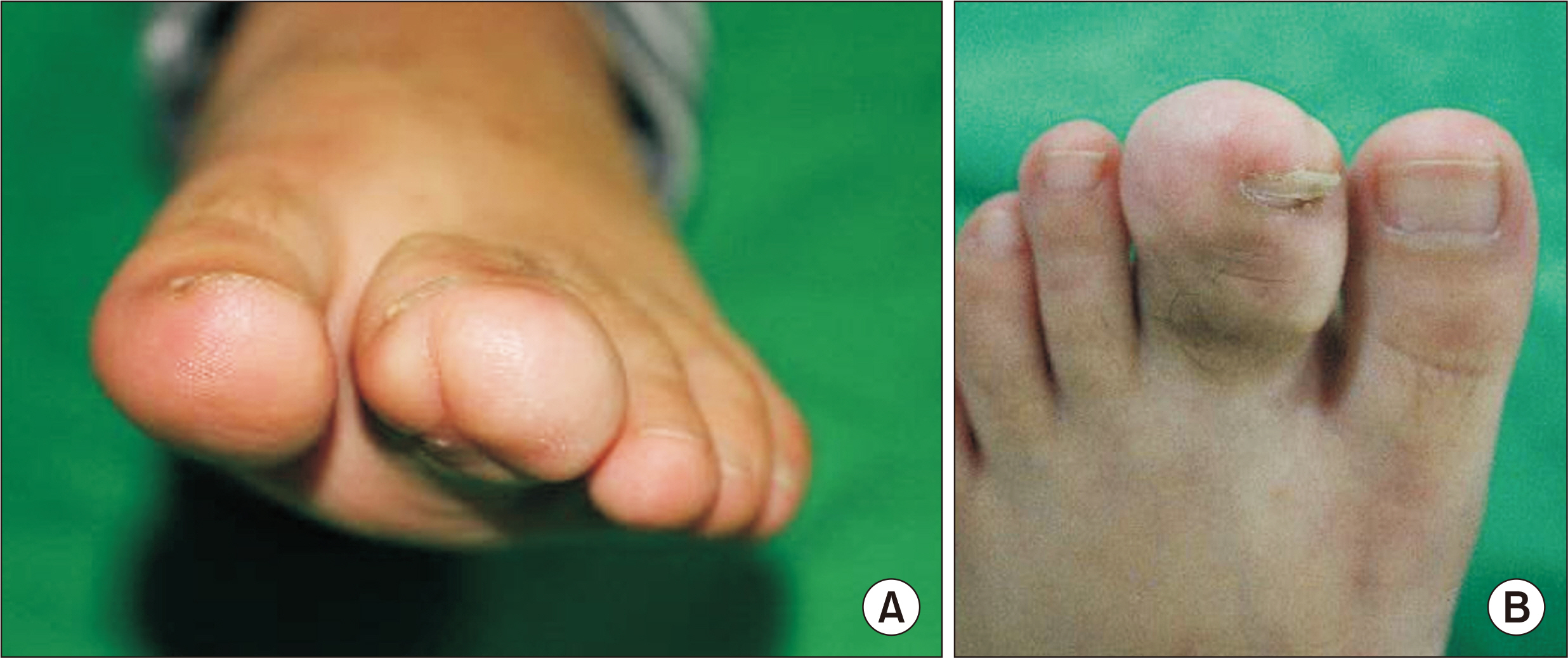
Figure. 8 Outpatient follow-up conducted 3 weeks post surgery. Postoperative width and length appear significantly decreased when compared with those observed preoperatively. (A) Coronal view. (B) Axial view.
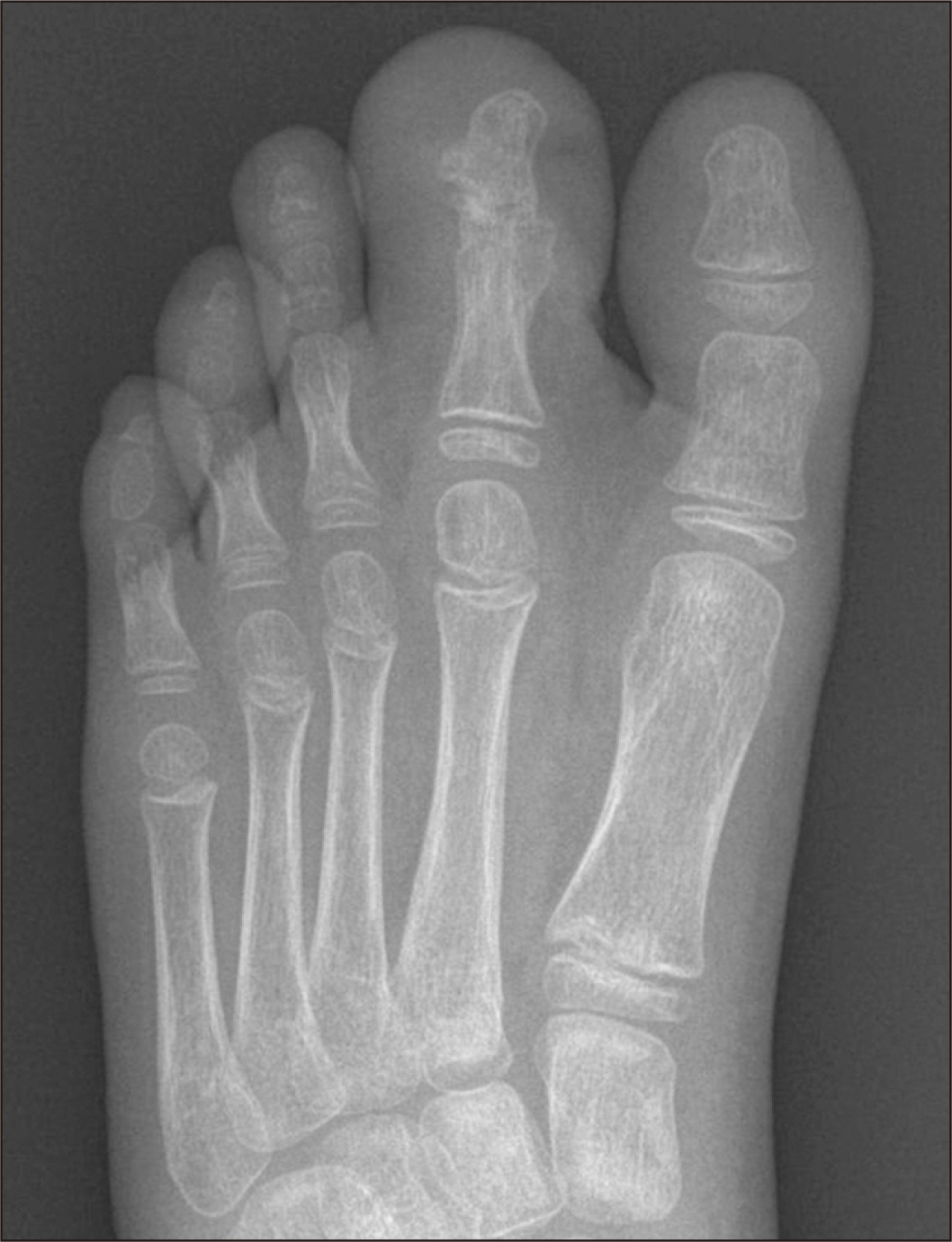
Figure. 9 Plain radiograph image 5 weeks after the operation showing bony union at the fused site, which resulted in k-wire removal.
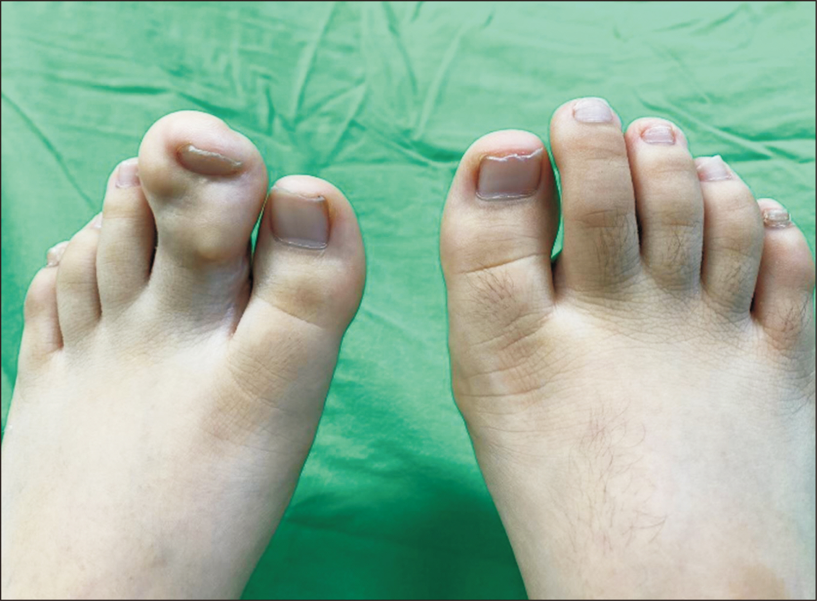
Figure. 10 At the patient’s 7th-year follow-up after the first surgery. The patient experienced difficulty in wearing a shoe. Soft tissue thickening in the outer part of the second toe is observed.
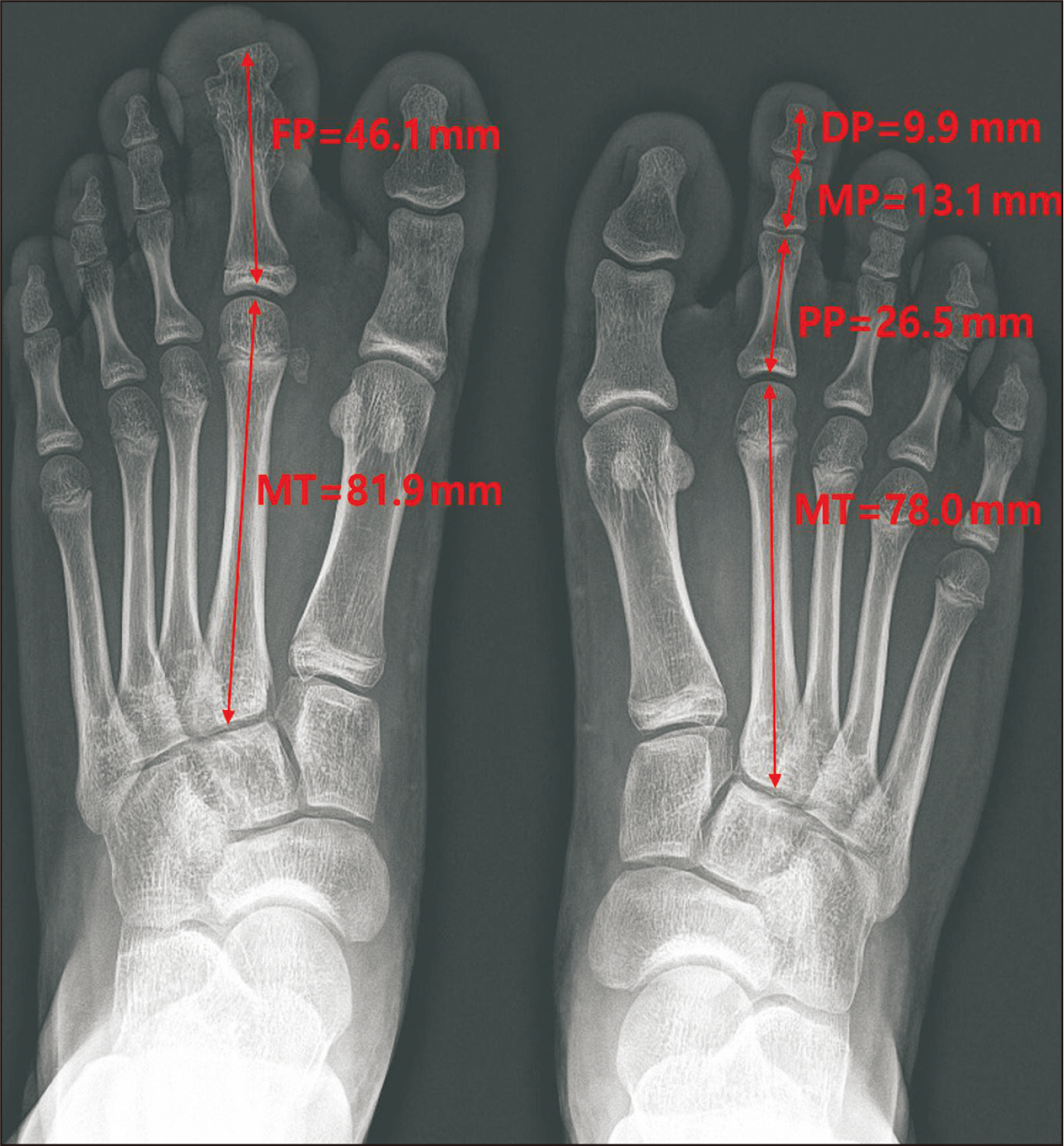
Figure. 11 Foot radiographs obtained at 7th-year follow-up show nearly the same length of both second rays. The left foot’s second fused phalanges measure 46.1 mm, while the right foot second three phalanges (DP+MP+PP) measure 49.5 mm. The left and right second MT measure 81.9 mm and 78.0 mm, respectively. FP: fused phalanges, MT: metatarsal bone, DP: distal phalanx, MP: middle phalanx, PP: proximal phalanx.
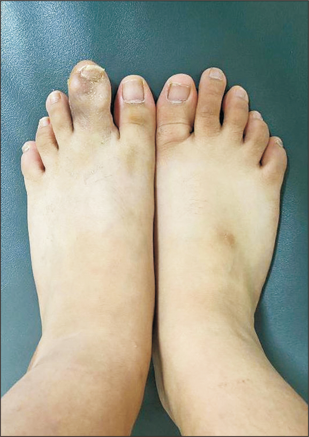
Figure. 12 Final follow-up gross photography at 10 weeks after the second surgery. Cosmetic appearance improved, and both the patient and her parents were satisfied.
Reference
-
1. Barsky AJ. 1967; Macrodactyly. J Bone Joint Surg Am. 49:1255–66. DOI: 10.2106/00004623-196749070-00001. PMID: 4293291.
Article2. Kotwal PP, Farooque M. 1998; Macrodactyly. J Bone Joint Surg Br. 80:651–3. doi: 10.1302/0301-620x.80b4.8489. DOI: 10.1302/0301-620X.80B4.8489. PMID: 9699830.
Article3. Flatt AE. 1994. The care of congenital hand anomalies. 2nd ed. Quality Medical;St. Louis:4. Hardwicke J, Khan MA, Richards H, Warner RM, Lester R. 2013; Macrodactyly - options and outcomes. J Hand Surg Eur Vol. 38:297–303. doi: 10.1177/1753193412451232. DOI: 10.1177/1753193412451232. PMID: 22736742.
Article5. Lee SJ, Lee HJ, Kim PT. 2016; Single stage reduction operation for treatment of toe macrodactyly in skeletally immature patients. J Korean Orthop Assoc. 51:260–5. doi: 10.4055/jkoa.2016.51.3.260. DOI: 10.4055/jkoa.2016.51.3.260.
Article6. Topoleski TA, Ganel A, Grogan DP. 1997; Effect of proximal phalangeal epiphysiodesis in the treatment of macrodactyly. Foot Ankle Int. 18:500–3. doi: 10.1177/107110079701800807. DOI: 10.1177/107110079701800807. PMID: 9278744.
Article7. Tsuge K. 1967; Treatment of macrodactyly. Plast Reconstr Surg. 39:590–9. doi: 10.1097/00006534-196706000-00008. DOI: 10.1097/00006534-196706000-00008. PMID: 6025687.
Article8. Chang CH, Kumar SJ, Riddle EC, Glutting J. 2002; Macrodactyly of the foot. J Bone Joint Surg Am. 84:1189–94. doi: 10.2106/00004623-200207000-00015. DOI: 10.2106/00004623-200207000-00015. PMID: 12107320.
Article9. Kim J, Park JW, Hong SW, Jeong JY, Gong HS, Baek GH. Ray amputation for the treatment of foot macrodactyly in children. Bone Joint J. 2015; 97-B:1364–9. doi: 10.1302/0301-620X.97B10.35660. DOI: 10.1302/0301-620X.97B10.35660. PMID: 26430011.
Article10. Kelly DM. Canale ST, Beaty JH, editors. 2013. Congenital anomalies of the lower extremity. Campbell’s operative orthopaedics. 12th ed. Philadelphia: Elsevier Mosby;p. 980–1078.
Article
