Primary Hepatic Choriocarcinoma with Pregnancy: A Diagnostic and Therapeutic Challenge
- Affiliations
-
- 1Department of Surgical Gastroenterology, Nizam’s Institute of Medical Sciences, Hyderabad, Telangana, India
- 2Department of Surgical Pathology, Nizam’s Institute of Medical Sciences, Hyderabad, Telangana, India
- KMID: 2539578
- DOI: http://doi.org/10.4166/kjg.2022.116
Abstract
- Choriocarcinoma occurs mainly in the gonads, but an extragonadal origin has been reported, albeit infrequently. Primary hepatic choriocarcinoma (PHC) is a rare malignancy, with only 11 cases reported. Most cases reported were in males, with none reported in pregnant females. A 23-year-old primigravida presented with a large liver lesion involving the right lobe of the liver at 28 weeks of pregnancy. Preoperative imaging was suggestive of hepatocellular carcinoma. She underwent a non-anatomical resection of the liver lesion. Surprisingly, her postoperative histopathology revealed a diagnosis of PHC. Her blood workup showed elevated beta human chorionic gonadotrophin. She underwent a termination of her pregnancy at 32 weeks. Before initiating adjuvant chemotherapy four weeks after surgery, a whole-body PET scan revealed multiple bi-lobar liver and pelvic deposits. After a multidisciplinary team discussion, she was started on adjuvant chemotherapy. She is currently under regular follow-up, seven months post-surgery. PHC, one of the vascular lesions of the liver, poses a diagnostic and therapeutic challenge, warranting a multidisciplinary approach.
Keyword
Figure
-

Fig. 1 (A) Coronal MRI images of the abdomen showing a well-encapsulated exophytic liver lesion (white arrow) with a large central scar (asterisk) and fetus (arrowhead). (B) Axial (T2) sections of the exophytic liver lesion with central scar (asterisk).
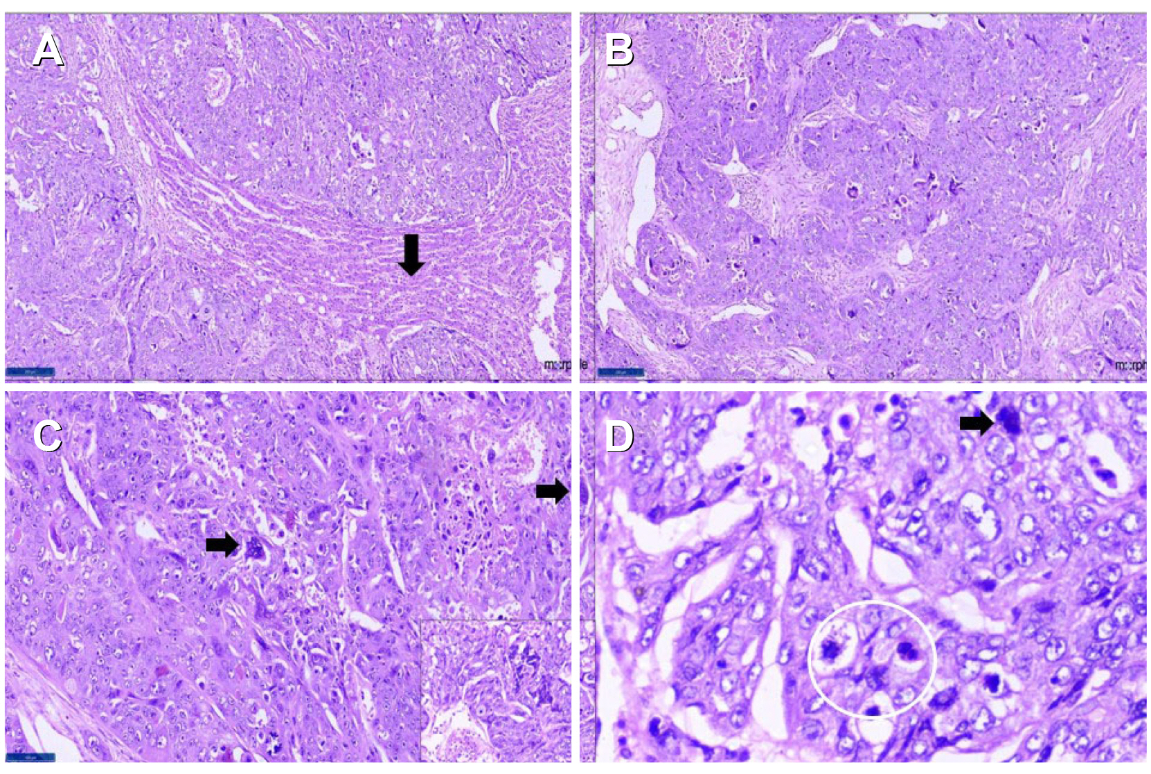
Fig. 2 Photomicrographs showing: (A) tumor cells infiltrating liver parenchyma (normal liver- black arrow). H&E ×40. (B) Infiltrating tumor. H&E ×100. (C) Scattered syncytiotrophoblastic cells (black arrowheads), highlighted within the inset. H&E ×400. (D) Mitotic activity with atypical mitoses (within the white circle), abnormal tumor cell (black arrow). H&E ×400.

Fig. 3 Immunohistochemical panel showed: (Top panel) (A) Positive Sal-like protein-4 (SALL-4); horseradish peroxidase (HRP) Polymer ×100. (B) Cytoplasmic Beta Human Chorionic Gonadotrophin (HCG); HRP Polymer ×100. (C) Positive Pan cytokeratin (pan CK); HRP Polymer ×100. (Bottom panel) The tumor cells were immuno-negative for (D) hepatocyte-specific antigen (HSA) (control hepatocytes positive) (E) Arginase, and (F) D2-40; HRP Polymer ×100.

Fig. 4 PET scan (four weeks after surgery) pre-chemotherapy showing: (A) Multiple 18-fluoro deoxyglucose (FDG) avid lesions noted in both lobes of the liver and perirectal nodules. (B) Post six cycles of chemotherapy showing partial regression in size, number, metabolism of metabolism of hepatic lesions, left iliac, presacral, and gluteal deposits, and complete regression of right lung nodule.
Reference
-
1. Kohler A, Welsch T, Sturm AK, et al. 2018; Primary choriocarcinoma of the liver: a rare, but important differential diagnosis of liver lesions. J Surg Case Rep. 2018:rjy068. DOI: 10.1093/jscr/rjy110.
Article2. Yousefi Z, Saied S, Davachi B, Rezaei A, Mirzamarjani F. Postmenopausal choriocarcinoma: case report and lliterature review. Int J Cancer Manag. 2017; Mar. 20. doi: 10.5812/ijcm.4400. DOI: 10.5812/ijcm.4400.3. Lee E, Cho H. 2019; A case of intraplacental choriocarcinoma with pulmonary metastasis. Case Rep Oncol. 12:802–806. DOI: 10.1159/000503816. PMID: 31762752. PMCID: PMC6873010.
Article4. Ahn Y, Kim JH, Park CS, Kim TE, Hwang S, Lee SG. 2018; Multidisciplinary approach for treatment of primary hepatic choriocarcinoma in adult male patient. Ann Hepatobiliary Pancreat Surg. 22:164–168. DOI: 10.14701/ahbps.2018.22.2.164. PMID: 29896579. PMCID: PMC5981148.
Article5. Sekine R, Hyodo M, Kojima M, et al. 2013; Primary hepatic choriocarcinoma in a 49-year-old man: report of a case. World J Gastroenterol. 19:9485–9489. DOI: 10.3748/wjg.v19.i48.9485. PMID: 24409080. PMCID: PMC3882426.
Article6. Shi H, Cao D, Wei L, Sun L, Guo A. 2010; Primary choriocarcinoma of the liver: a clinicopathological study of five cases in males. Virchows Arch. 456:65–70. DOI: 10.1007/s00428-009-0864-1. PMID: 20013345.
Article7. Jiang F, Xiang Y, Feng FZ, Ren T, Cui ZM, Wan XR. 2014; Clinical analysis of 13 males with primary choriocarcinoma and review of the literature. Onco Targets Ther. 7:1135–1141. DOI: 10.2147/OTT.S62561. PMID: 25018640. PMCID: PMC4074184.
Article8. Martins VF, Moreno F, Vizcaíno JR, Santos J. 2015; Primary gastric choriocarcinoma: A rare case. Int J Surg Case Rep. 14:44–47. DOI: 10.1016/j.ijscr.2015.07.009. PMID: 26218175. PMCID: PMC4573415.
Article9. Xiong Y, Yang MX. 2019; Primary gastric choriocarcinoma with multiple metastases - A case report and literature review of carcinogenesis. Hum Pathol Case Rep. 18:200330. DOI: 10.1016/j.ehpc.2019.200330.
Article10. Qiu J, Jia S, Li G. 2018; Incidence and prognosis factors of extragonadal choriocarcinoma in males: a population-based study. Cancer Manag Res. 10:4565–4573. DOI: 10.2147/CMAR.S175948. PMID: 30410393. PMCID: PMC6197831.
Article
