Korean J Orthod.
2023 Jan;53(1):35-44. 10.4041/kjod22.117.
Does surgically assisted maxillary protraction with skeletal anchorage and Class III elastics affect the pharyngeal airway? A retrospective, long-term study
- Affiliations
-
- 1Department of Orthodontics, School of Dentistry, Marmara University, Istanbul, Turkey
- 2Department of Orthodontics, Bezmialem Vakıf University Faculty of Dentistry, Istanbul, Turkey
- KMID: 2538711
- DOI: http://doi.org/10.4041/kjod22.117
Abstract
Objective
Surgically assisted maxillary protraction is an alternative protocol in severe Class III cases or after the adolescent growth spurt involving increased maxillary advancement. Correction of the maxillary deficiency has been suggested to improve pharyngeal airway dimensions. Therefore, this retrospective study aimed to analyze the airway changes cephalometrically following surgically assisted maxillary protraction with skeletal anchorage and Class III elastics.
Methods
The study population consisted of 15 Class III patients treated with surgically assisted maxillary protraction combined with skeletal anchorage and Class III elastics (mean age: 12.9 ± 1.2 years). Growth changes were initially assessed for a mean of 5.5 ± 1.6 months prior to treatment. Airway and skeletal changes in the control (T0), pre-protraction (T1), post-protraction (T2), and follow-up (T3) periods were monitored and compared using lateral cephalometric radiographs. Statistical significance was set at p < 0.05.
Results
The skeletal or airway parameters showed no statistically significant changes during the control period. Sella to nasion angle, N perpendicular to A, Point A to Point B angle, and Frankfort plane to mandibular plane angle increased significantly during the maxillary protraction period (p < 0.05), but no significant changes were observed in airway parameters (p > 0.05). No statistically significant changes were observed in the airway parameters in the follow-up period either. However, Sella to Gonion distance increased significantly (p < 0.05) during the follow-up period.
Conclusions
No significant changes in pharyngeal airway parameters were found during the control, maxillary protraction, and follow-up periods. Moreover, the significant increases in the skeletal parameters during maxillary protraction were maintained in the long-term.
Figure
-
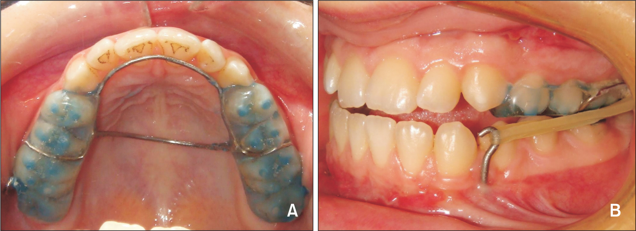
Figure 1 A, The occlusal view of the appliance. B, Intraoral elastic usage.
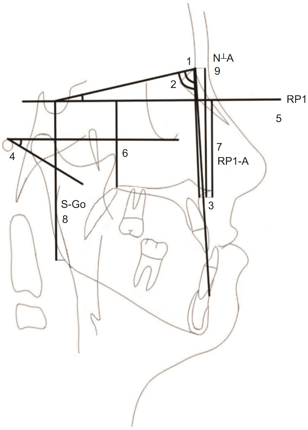
Figure 2 Skeletal measurements. 1, SNA (°); 2, SNB (°); 3, ANB (°); 4, FMA (°); 5, RP1; 6, RP1-PNS (mm); 7, RP1-A (mm); 8, S-Go (mm); 9, N⊥A (mm). See Table 2 for definition of the planes and the measurements.
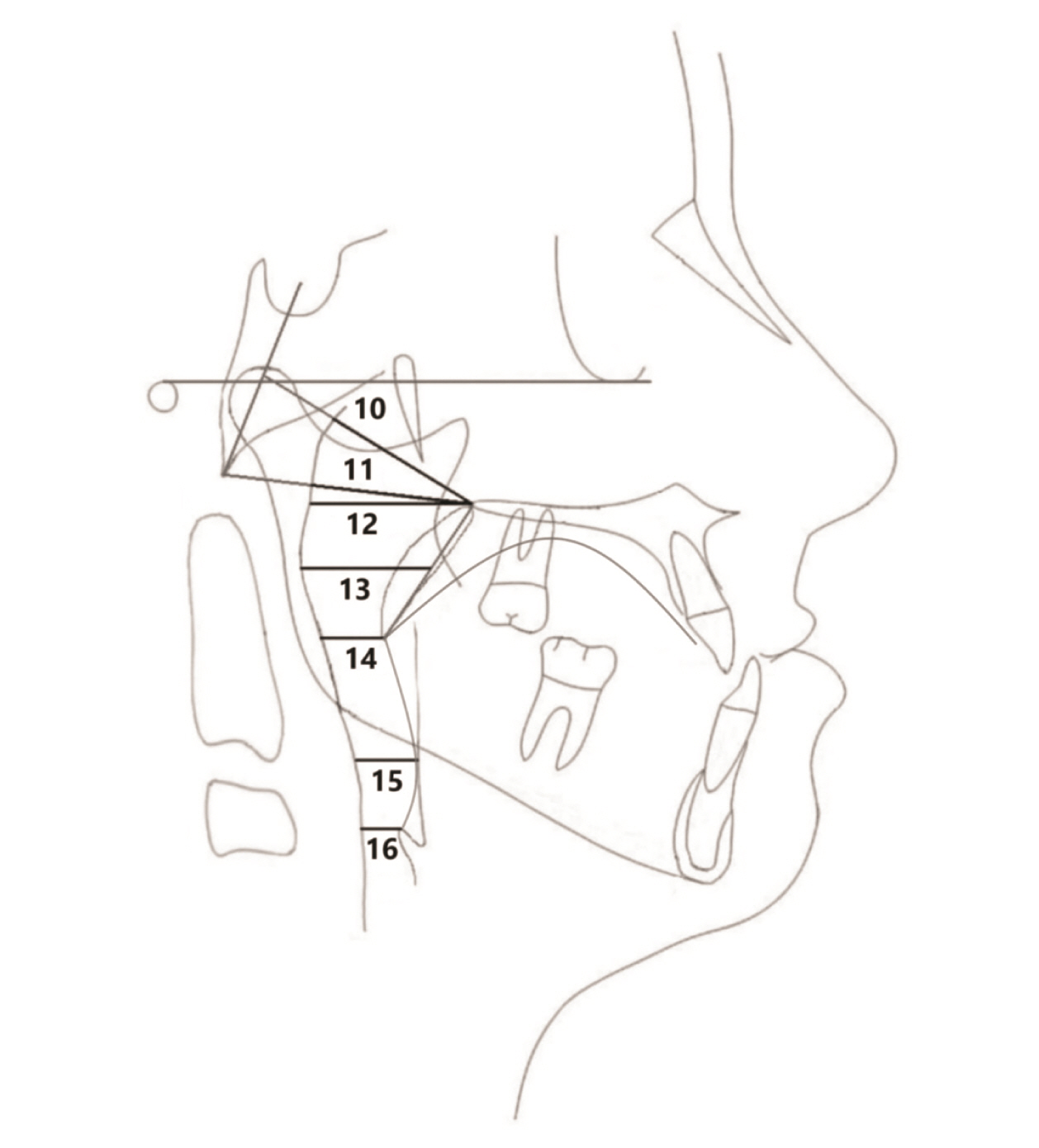
Figure 3 Pharyngeal airway measurements (mm). 10, Ad1-PNS; 11, Ad2-PNS; 12, PAS; 13, SPAS; 14, MAS; 15, IAS; 16, EAS. See Table 2 for definition of the planes and the measurements.

Figure 4 Boxplots of the airway parameters during maxillary protraction period (T2–T1). T1, pre-protraction, before cementation of the acrylic splint; T2, post-protraction, after debonding of the acrylic splint. See Table 2 for definition of the planes and the measurements.
Reference
-
1. Guyer EC, Ellis EE 3rd, McNamara JA Jr, Behrents RG. 1986; Components of class III malocclusion in juveniles and adolescents. Angle Orthod. 56:7–30. DOI: 10.1043/0003-3219(1986)056<0007:COCIMI>2.0.CO;2. PMID: 3485393.2. Ngan P, Moon W. 2015; Evolution of Class III treatment in orthodontics. Am J Orthod Dentofacial Orthop. 148:22–36. DOI: 10.1016/j.ajodo.2015.04.012. PMID: 26124025.
Article3. Baccetti T, Franchi L, McNamara JA Jr. 2004; Cephalometric variables predicting the long-term success or failure of combined rapid maxillary expansion and facial mask therapy. Am J Orthod Dentofacial Orthop. 126:16–22. DOI: 10.1016/j.ajodo.2003.06.010. PMID: 15224054.
Article4. Toffol LD, Pavoni C, Baccetti T, Franchi L, Cozza P. 2008; Orthopedic treatment outcomes in Class III malocclusion. A systematic review. Angle Orthod. 78:561–73. DOI: 10.2319/030207-108.1. PMID: 18416617.5. Nguyen T, Cevidanes L, Paniagua B, Zhu H, Koerich L, De Clerck H. 2014; Use of shape correspondence analysis to quantify skeletal changes associated with bone-anchored Class III correction. Angle Orthod. 84:329–36. DOI: 10.2319/041513-288.1. PMID: 23886012. PMCID: PMC4394197.
Article6. Nevzatoğlu S, Küçükkeleş N. 2014; Long-term results of surgically-assisted maxillary protraction. Aust Orthod J. 30:19–31. PMID: 24968642.7. Yilmaz HN, Garip H, Satilmis T, Kucukkeles N. 2015; Corticotomy-assisted maxillary protraction with skeletal anchorage and Class III elastics. Angle Orthod. 85:48–57. DOI: 10.2319/121513-921.1. PMID: 24913740. PMCID: PMC8634805.
Article8. Küçükkeleş N, Nevzatoğlu S, Koldaş T. 2011; Rapid maxillary expansion compared to surgery for assistance in maxillary face mask protraction. Angle Orthod. 81:42–9. DOI: 10.2319/042210-220.1. PMID: 20936953. PMCID: PMC8926356.
Article9. Conley RS. 2011; Evidence for dental and dental specialty treatment of obstructive sleep apnoea. Part 1: the adult OSA patient and part 2: the paediatric and adolescent patient. J Oral Rehabil. 38:136–56. DOI: 10.1111/j.1365-2842.2010.02136.x. PMID: 20722775.
Article10. Hiyama S, Suda N, Ishii-Suzuki M, Tsuiki S, Ogawa M, Suzuki S, et al. 2002; Effects of maxillary protraction on craniofacial structures and upper-airway dimension. Angle Orthod. 72:43–7. DOI: 10.1043/0003-3219(2002)072<0043:EOMPOC>2.0.CO;2. PMID: 11843272.11. Baccetti T, Franchi L, Mucedero M, Cozza P. 2010; Treatment and post-treatment effects of facemask therapy on the sagittal pharyngeal dimensions in Class III subjects. Eur J Orthod. 32:346–50. DOI: 10.1093/ejo/cjp092. PMID: 19752018.
Article12. Mucedero M, Baccetti T, Franchi L, Cozza P. 2009; Effects of maxillary protraction with or without expansion on the sagittal pharyngeal dimensions in Class III subjects. Am J Orthod Dentofacial Orthop. 135:777–81. DOI: 10.1016/j.ajodo.2008.11.021. PMID: 19524838.
Article13. Kilinç AS, Arslan SG, Kama JD, Ozer T, Dari O. 2008; Effects on the sagittal pharyngeal dimensions of protraction and rapid palatal expansion in Class III malocclusion subjects. Eur J Orthod. 30:61–6. DOI: 10.1093/ejo/cjm076. PMID: 17906307.
Article14. Oktay H, Ulukaya E. 2008; Maxillary protraction appliance effect on the size of the upper airway passage. Angle Orthod. 78:209–14. DOI: 10.2319/122806-535.1. PMID: 18251620.
Article15. Sayinsu K, Isik F, Arun T. 2006; Sagittal airway dimensions following maxillary protraction: a pilot study. Eur J Orthod. 28:184–9. DOI: 10.1093/ejo/cji095. PMID: 16464873.
Article16. Kaygisiz E, Tuncer BB, Yüksel S, Tuncer C, Yildiz C. 2009; Effects of maxillary protraction and fixed appliance therapy on the pharyngeal airway. Angle Orthod. 79:660–7. DOI: 10.2319/072408-391.1. PMID: 19537871.
Article17. Lee JW, Park KH, Kim SH, Park YG, Kim SJ. 2011; Correlation between skeletal changes by maxillary protraction and upper airway dimensions. Angle Orthod. 81:426–32. DOI: 10.2319/082610-499.1. PMID: 21299388. PMCID: PMC8923552.
Article18. Cakirer B, Kucukkeles N, Nevzatoglu S, Koldas T. 2012; Sagittal airway changes: rapid palatal expansion versus Le Fort I osteotomy during maxillary protraction. Eur J Orthod. 34:381–9. DOI: 10.1093/ejo/cjr023. PMID: 21464152.
Article19. Baccetti T, Franchi L, McNamara JA Jr. 2005; The cervical vertebral maturation (CVM) method for the assessment of optimal treatment timing in dentofacial orthopedics. Semin Orthod. 11:119–29. DOI: 10.1053/j.sodo.2005.04.005.
Article20. Mochida M, Ono T, Saito K, Tsuiki S, Ohyama K. 2004; Effects of maxillary distraction osteogenesis on the upper-airway size and nasal resistance in subjects with cleft lip and palate. Orthod Craniofac Res. 7:189–97. DOI: 10.1111/j.1601-6343.2004.00300.x. PMID: 15562581.
Article21. Wolford LM, Karras SC, Mehra P. 2001; Considerations for orthognathic surgery during growth, part 2: maxillary deformities. Am J Orthod Dentofacial Orthop. 119:102–5. DOI: 10.1067/mod.2001.111400. PMID: 11174554.
Article22. Kircelli BH, Pektas ZO. 2008; Midfacial protraction with skeletally anchored face mask therapy: a novel approach and preliminary results. Am J Orthod Dentofacial Orthop. 133:440–9. DOI: 10.1016/j.ajodo.2007.06.011. PMID: 18331946.
Article23. Sar C, Arman-Özçırpıcı A, Uçkan S, Yazıcı AC. 2011; Comparative evaluation of maxillary protraction with or without skeletal anchorage. Am J Orthod Dentofacial Orthop. 139:636–49. DOI: 10.1016/j.ajodo.2009.06.039. PMID: 21536207.
Article24. Celikoglu M, Buyukcavus MH. 2017; Changes in pharyngeal airway dimensions and hyoid bone position after maxillary protraction with different alternate rapid maxillary expansion and construction protocols: a prospective clinical study. Angle Orthod. 87:519–25. DOI: 10.2319/082316-632.1. PMID: 28139938. PMCID: PMC8366701.
Article25. Hwang DM, Lee JY, Choi YJ, Hwang CJ. 2019; Evaluations of the tongue and hyoid bone positions and pharyngeal airway dimensions after maxillary protraction treatment. Cranio. 37:214–22. DOI: 10.1080/08869634.2017.1418644. PMID: 29327661.
Article26. Linder-Aronson S, Leighton BC. 1983; A longitudinal study of the development of the posterior nasopharyngeal wall between 3 and 16 years of age. Eur J Orthod. 5:47–58. DOI: 10.1093/ejo/5.1.47. PMID: 6572594.
Article27. Taylor M, Hans MG, Strohl KP, Nelson S, Broadbent BH. 1996; Soft tissue growth of the oropharynx. Angle Orthod. 66:393–400. DOI: 10.1043/0003-3219(1996)066<0393:STGOTO>2.3.CO;2. PMID: 8893109.28. Ceylan I, Oktay H. 1995; A study on the pharyngeal size in different skeletal patterns. Am J Orthod Dentofacial Orthop. 108:69–75. DOI: 10.1016/S0889-5406(95)70068-4. PMID: 7598107.
Article29. Akcam MO, Toygar TU, Wada T. 2002; Longitudinal investigation of soft palate and nasopharyngeal airway relations in different rotation types. Angle Orthod. 72:521–6. DOI: 10.1043/0003-3219(2002)072<0521:LIOSPA>2.0.CO;2. PMID: 12518943.30. Adobes Martin M, Lipani E, Alvarado Lorenzo A, Bernes Martinez L, Aiuto R, Dioguardi M, et al. 2020; The effect of maxillary protraction, with or without rapid palatal expansion, on airway dimensions: a systematic review and meta-analysis. Eur J Paediatr Dent. 21:262–70. DOI: 10.23804/ejpd.2020.21.04.2. PMID: 33337900.31. Nguyen T, De Clerck H, Wilson M, Golden B. 2015; Effect of Class III bone anchor treatment on airway. Angle Orthod. 85:591–6. DOI: 10.2319/041614-282.1. PMID: 25245416. PMCID: PMC8611741.
Article32. Pamporakis P, Nevzatoğlu Ş, Küçükkeleş N. 2014; Three-dimensional alterations in pharyngeal airway and maxillary sinus volumes in Class III maxillary deficiency subjects undergoing orthopedic facemask treatment. Angle Orthod. 84:701–7. DOI: 10.2319/060513-430.1. PMID: 24417494. PMCID: PMC8650436.
Article33. Onem Ozbilen E, Yilmaz HN, Kucukkeles N. 2019; Comparison of the effects of rapid maxillary expansion and alternate rapid maxillary expansion and constriction protocols followed by facemask therapy. Korean J Orthod. 49:49–58. DOI: 10.4041/kjod.2019.49.1.49. PMID: 30603625. PMCID: PMC6306318.
Article
- Full Text Links
-
- Actions
-
Cited
- CITED
-
- Close
- Share
-
- Similar articles
-
- Maxillary protraction using skeletal anchorage and intermaxillary elastics in Skeletal Class III patients
- Maxillary protraction treatment of skeletal Class III children using miniplate anchorage
- Treatment of skeletal Class III malocclustion with maxillary protraction appliance
- Maxillary protraction using customized mini-plates for anchorage in an adolescent girl with skeletal Class III malocclusion
- Comparison of the effects on the pharyngeal airway space of maxillary protraction appliances according to the methods of anchorage
