J Surg Ultrasound.
2022 Nov;9(2):23-29. 10.46268/jsu.2022.9.2.23.
Automated Breast Ultrasound: The Present and Future
- Affiliations
-
- 1Division of BreastㆍThyroid Surgery, Department of Surgery, Jeonbuk National University Medical School, Jeonju, Korea
- KMID: 2538381
- DOI: http://doi.org/10.46268/jsu.2022.9.2.23
Abstract
- Automated breast ultrasound (ABUS) is a novel imaging method, introduced to overcome the main limitations of traditional hand-held ultrasound, such as the lack of standardization, low reproducibility, small field of view, high operator dependency, and high commitment of physician time. ABUS is a standardized radiologic modality with many advantages in both screening and diagnostic settings. It increases the detection rate of breast cancer, improves workflow, and reduces the examination time. On the other hand, ABUS has some limitations, these include the inability to assess the axilla, vascularization, and elasticity of a lesion. With respect to the interpretation, the disadvantages of ABUS are the artifacts due to poor positioning, lack of contact, or those related to motion or some specific tumors. However, these disadvantages can be diminished by additional attention and training. ABUS can be used in clinical settings and is a promising modality in breast imaging. The purpose of this review is to present a summary of the characteristics and clinical applications of ABUS, and provide future perspectives.
Keyword
Figure
-
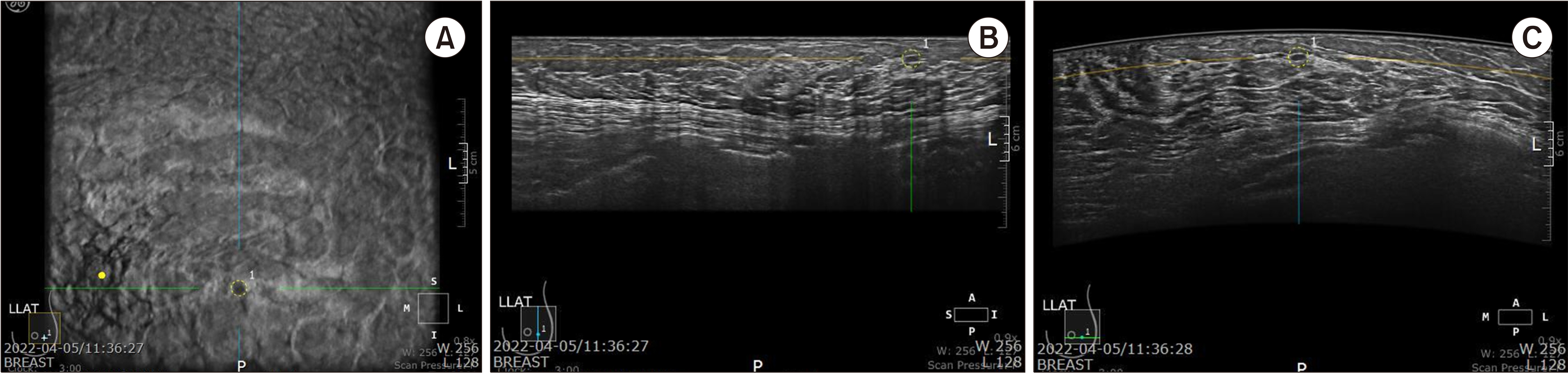
Fig. 1 Automated breast ultrasoundimage of a 40-year-old woman with 0.5 cm sized benign mass. (A) Coronal view (B) Longitudinal view (C) Transverse view The lesion is marked as a dotted circle.
Reference
-
1. International Agency for Research on Cancer. 2022. Cancer today: data visualization tools for exploring the global cancer burden in 2020 [Internet]. International Agency for Research on Cancer;Lyon: Available from: http://gco.iarc.fr/today/home. cited 2022 September 19.2. Chow LW, Yip AY, Ng EL. 2012; Prevention of oncological diseases: primary and secondary prevention. Int J Biol Markers. 27:e337–43. DOI: 10.5301/JBM.2012.10370. PMID: 23250774.
Article3. Drukteinis JS, Mooney BP, Flowers CI, Gatenby RA. 2013; Beyond mammography: new frontiers in breast cancer screening. Am J Med. 126:472–9. DOI: 10.1016/j.amjmed.2012.11.025. PMID: 23561631. PMCID: PMC4010151.
Article4. Kolb TM, Lichy J, Newhouse JH. 2002; Comparison of the performance of screening mammography, physical examination, and breastUS and evaluation of factors that influence them: an analysis of 27,825 patient evaluations. Radiology. 225:165–75. DOI: 10.1148/radiol.2251011667. PMID: 12355001.
Article5. Feig SA. 2004; Adverse effects of screening mammography. Radiol Clin North Am. 42:807–19. DOI: 10.1016/j.rcl.2004.06.013. PMID: 15337417.
Article6. Burkett BJ, Hanemann CW. 2016; A review of supplemental screening ultrasound for breast cancer: certain populations of women with dense breast tissue may benefit. Acad Radiol. 23:1604–9. DOI: 10.1016/j.acra.2016.05.017. PMID: 27374700.7. Berg WA, Blume JD, Cormack JB, Mendelson EB, Lehrer D, Böhm-Vélez M, et al. 2008; Combined screening with ultrasound and mammography vs mammography alone in women at elevated risk of breast cancer. JAMA. 299:2151–63. DOI: 10.1001/jama.299.18.2151. PMID: 18477782. PMCID: PMC2718688.
Article8. Zanotel M, Bednarova I, Londero V, Linda A, Lorenzon M, Girometti R, et al. 2018; Automated breast ultrasound: basic principles and emerging clinical applications. Radiol Med. 123:1–12. DOI: 10.1007/s11547-017-0805-z. PMID: 28849324.
Article9. Giger ML, Inciardi MF, Edwards A, Papaioannou J, Drukker K, Jiang Y, et al. 2016; Automated breast ultrasound in breast cancer screening of women with dense breasts: reader study of mammography-negative and mammography-positive cancers. AJR Am J Roentgenol. 206:1341–50. DOI: 10.2214/AJR.15.15367. PMID: 27043979.
Article10. Brem RF, Tabár L, Duffy SW, Inciardi MF, Guingrich JA, Hashimoto BE, et al. 2015; Assessing improvement in detection of breast cancer with three-dimensional automated breast US in women with dense breast tissue: the SomoInsight Study. Radiology. 274:663–73. DOI: 10.1148/radiol.14132832. PMID: 25329763.
Article11. Chang JM, Moon WK, Cho N, Park JS, Kim SJ. 2011; Radiologists' performance in the detection of benign and malignant masses with 3D automated breast ultrasound (ABUS). Eur J Radiol. 78:99–103. DOI: 10.1016/j.ejrad.2011.01.074. PMID: 21330080.
Article12. Kaplan SS. 2014; Automated whole breast ultrasound. Radiol Clin North Am. 52:539–46. DOI: 10.1016/j.rcl.2014.01.002. PMID: 24792655.
Article13. Nicosia L, Ferrari F, Bozzini AC, Latronico A, Trentin C, Meneghetti L, et al. 2020; Automatic breast ultrasound: state of the art and future perspectives. Ecancermedicalscience. 14:1062. DOI: 10.3332/ecancer.2020.1062. PMID: 32728378. PMCID: PMC7373644.
Article14. Maturo VG, Zusmer NR, Gilson AJ, Smoak WM, Janowitz WR, Bear BE, et al. 1980; Ultrasound of the whole breast utilizing a dedicated automated breast scanner. Radiology. 137:457–63. DOI: 10.1148/radiology.137.2.6254110. PMID: 6254110.
Article15. Kelly KM, Dean J, Lee SJ, Comulada WS. 2010; Breast cancer detection: radiologists' performance using mammography with and without automated whole-breast ultrasound. Eur Radiol. 20:2557–64. DOI: 10.1007/s00330-010-1844-1. PMID: 20632009. PMCID: PMC2948156.
Article16. Giuliano V, Giuliano C. 2013; Improved breast cancer detection in asymptomatic women using 3D-automated breast ultrasound in mammographically dense breasts. Clin Imaging. 37:480–6. DOI: 10.1016/j.clinimag.2012.09.018. PMID: 23116728.
Article17. Wilczek B, Wilczek HE, Rasouliyan L, Leifland K. 2016; Adding 3D automated breast ultrasound to mammography screening in women with heterogeneously and extremely dense breasts: report from a hospital-based, high-volume, single-center breast cancer screening program. Eur J Radiol. 85:1554–63. DOI: 10.1016/j.ejrad.2016.06.004. PMID: 27501888.
Article18. Zhang X, Chen J, Zhou Y, Mao F, Lin Y, Shen S, et al. 2019; Diagnostic value of an automated breast volume scanner compared with a hand-held ultrasound: a meta-analysis. Gland Surg. 8:698–711. DOI: 10.21037/gs.2019.11.18. PMID: 32042678. PMCID: PMC6989916.
Article19. Wang HY, Jiang YX, Zhu QL, Zhang J, Dai Q, Liu H, et al. 2012; Differentiation of benign and malignant breast lesions: a comparison between automatically generated breast volume scans and handheld ultrasound examinations. Eur J Radiol. 81:3190–200. DOI: 10.1016/j.ejrad.2012.01.034. PMID: 22386134.
Article20. Chen L, Chen Y, Diao XH, Fang L, Pang Y, Cheng AQ, et al. 2013; Comparative study of automated breast 3-D ultrasound and handheld B-mode ultrasound for differentiation of benign and malignant breast masses. Ultrasound MedBiol. 39:1735–42. DOI: 10.1016/j.ultrasmedbio.2013.04.003. PMID: 23849390.
Article21. Choi WJ, Cha JH, Kim HH, Shin HJ, Kim H, Chae EY, et al. 2014; Comparison of automated breast volume scanning and hand- held ultrasound in the detection of breast cancer: an analysis of 5,566 patient evaluations. Asian Pac J Cancer Prev. 15:9101–5. DOI: 10.7314/APJCP.2014.15.21.9101. PMID: 25422185.22. Jeh SK, Kim SH, Choi JJ, Jung SS, Choe BJ, Park S, et al. 2016; Comparison of automated breast ultrasonography to handheld ultrasonography in detecting and diagnosing breast lesions. Acta Radiol. 57:162–9. DOI: 10.1177/0284185115574872. PMID: 25766727.
Article23. Hellgren R, Dickman P, Leifland K, Saracco A, Hall P, Celebioglu F. 2017; Comparison of handheld ultrasound and automated breast ultrasound in women recalled after mammography screening. Acta Radiol. 58:515–20. DOI: 10.1177/0284185116665421. PMID: 27565633.
Article24. Niu L, Bao L, Zhu L, Tan Y, Xu X, Shan Y, et al. 2019; Diagnostic performance of automated breast ultrasound in differentiating benign and malignant breast masses in asymptomatic women: a comparison study with handheld ultrasound. J Ultrasound Med. 38:2871–80. DOI: 10.1002/jum.14991. PMID: 30912178.
Article25. Kim H, Cha JH, Oh HY, Kim HH, Shin HJ, Chae EY. 2014; Comparison of conventional and automated breast volume ultrasound in the description and characterization of solid breast masses based on BI-RADS features. Breast Cancer. 21:423–8. DOI: 10.1007/s12282-012-0419-1. PMID: 23086698.
Article26. Zhang X, Lin X, Tan Y, Zhu Y, Wang H, Feng R, et al. 2018; A multicenter hospital-based diagnosis study of automated breast ultrasound system in detecting breast cancer among Chinese women. Chin J Cancer Res. 30:231–9. DOI: 10.21147/j.issn.1000-9604.2018.02.06. PMID: 29861608. PMCID: PMC5953959.
Article27. Xiao YM, Chen ZH, Zhou QC, Wang Z. 2015; The efficacy of automated breast volume scanning over conventional ultrasonography among patients with breast lesions. Int J Gynaecol Obstet. 131:293–6. DOI: 10.1016/j.ijgo.2015.05.036. PMID: 26493011.
Article28. Ibraheem SA, Mahmud R, Mohamad Saini S, Abu Hassan H, Keiteb AS, Dirie AM. 2022; Evaluation of diagnostic performance of automatic breast volume scanner compared to handheld ultrasound on different breast lesions: a systematic review. Diagnostics (Basel). 12:541. DOI: 10.3390/diagnostics12020541. PMID: 35204629. PMCID: PMC8870745.
Article29. Schmachtenberg C, Fischer T, Hamm B, Bick U. 2017; Diagnostic performance of automated breast volume scanning (ABVS) compared to handheld ultrasonography with breast MRI as the gold standard. Acad Radiol. 24:954–61. DOI: 10.1016/j.acra.2017.01.021. PMID: 28336007.
Article30. Girometti R, Zanotel M, Londero V, Bazzocchi M, Zuiani C. 2017; Comparison between automatedbreast volume scanner (ABVS) versus hand-held ultrasound as a second look procedure after magnetic resonance imaging. Eur Radiol. 27:3767–75. DOI: 10.1007/s00330-017-4749-4. PMID: 28120030.
Article31. Chae EY, Shin HJ, Kim HJ, Yoo H, Baek S, Cha JH, et al. 2013; Diagnostic performance of automated breast ultrasound as a replacement for a hand-held second-look ultrasound for breast lesions detected initially on magnetic resonance imaging. Ultrasound MedBiol. 39:2246–54. DOI: 10.1016/j.ultrasmedbio.2013.07.005. PMID: 24035627.
Article32. Kim Y, Kang BJ, Kim SH, Lee EJ. 2016; Prospective study comparing two second-look ultrasound techniques: handheld ultrasound and an automated breast volume scanner. J Ultrasound Med. 2103–12. DOI: 10.7863/ultra.15.11076. PMID: 27503758.33. Van Zelst JC, Platel B, Karssemeijer N, Mann RM. 2015; Multiplanar reconstructions of 3D automated breast ultrasound improve lesion differentiation by radiologists. Acad Radiol. 22:1489–96. DOI: 10.1016/j.acra.2015.08.006. PMID: 26345538.
Article34. Boca Bene I, Ciurea AI, Ciortea CA, Dudea SM. 2021; Pros and cons for Automated Breast Ultrasound (ABUS): a narrative review. J Pers Med. 703. DOI: 10.3390/jpm11080703. PMID: 34442347. PMCID: PMC8400952.35. Wang Q, Li B, Liu Z, Shang H, Jing H, Shao H, et al. 2022; Prediction model of axillary lymph node status using automated breast ultrasound (ABUS) and ki-67 status in early-stage breast cancer. BMC Cancer. 22:929. DOI: 10.1186/s12885-022-10034-3. PMID: 36031602. PMCID: PMC9420256.
Article36. Xiao Y, Zhou Q, Chen Z. 2015; Automated breast volume scanning versus conventional ultrasound in breast cancer screening. Acad Radiol. 22:387–99. DOI: 10.1016/j.acra.2014.08.013. PMID: 25620036.
Article37. Isobe S, Tozaki M, Yamaguchi M, Ogawa Y, Homma K, Satomi R, et al. 2011; Detectability of breast lesions under the nipple using an automated breast volume scanner: comparison with handheld ultrasonography. Jpn J Radiol. 29:361–5. DOI: 10.1007/s11604-010-0555-5. PMID: 21717306.
Article38. Kim SH, Kang BJ, Choi BG, Choi JJ, Lee JH, Song BJ, et al. 2013; Radiologists' performance for detecting lesions and the interobserver variability of automated whole breast ultrasound. Korean J Radiol. 14:154–63. DOI: 10.3348/kjr.2013.14.2.154. PMID: 23482698. PMCID: PMC3590325.
Article39. Meng Z, Chen C, Zhu Y, Zhang S, Wei C, Hu B, et al. 2015; Diagnostic performance of the automated breast volume scanner: a systematic review of inter-rater reliability/agreement and meta-analysis of diagnostic accuracy for differentiating benign and malignant breast lesions. Eur Radiol. 25:3638–47. DOI: 10.1007/s00330-015-3759-3. PMID: 25916389.
Article40. Kang SY, Lee SJ, Youn HJ, Jung SH. 2017; Clinical significance of automated breast ultrasound. J Surg Ultrasound. 4:12–7.41. Zheng FY, Lu Q, Huang BJ, Xia HS, Yan LX, Wang X, et al. 2017; Imaging features of automated breast volume scanner: correlation with molecular subtypes of breast cancer. Eur J Radiol. 86:267–75. DOI: 10.1016/j.ejrad.2016.11.032. PMID: 28027759.
Article42. Jiang J, Chen YQ, Xu YZ, Chen ML, Zhu YK, Guan WB, et al. 2014; Correlation between three-dimensional ultrasound features and pathological prognostic factors in breast cancer. Eur Radiol. 24:1186–96. DOI: 10.1007/s00330-014-3135-8. PMID: 24723231.
Article43. Wang X, Huo L, He Y, Fan Z, Wang T, Xie Y, et al. 2016; Early prediction of pathological outcomes to neoadjuvant chemotherapy in breast cancer patients using automated breast ultrasound. Chin J Cancer Res. 28:478–85. DOI: 10.21147/j.issn.1000-9604.2016.05.02. PMID: 27877006. PMCID: PMC5101221.
Article44. Jiang Y, Inciardi MF, Edwards AV, Papaioannou J. 2018; Interpretation time using a concurrent-read computer-aided detection system for automated breast ultrasound in breast cancer screening of women with dense breast tissue. AJR Am J Roentgenol. 211:452–61. DOI: 10.2214/AJR.18.19516. PMID: 29792747.
Article45. Liao WX, He P, Hao J, Wang XY, Yang RL, An D, et al. 2020; Automatic identification of breast ultrasound image based on supervised block-based region segmentation algorithm and features combination migration deep learning model. IEEE J Biomed Health Inform. 24:984–93. DOI: 10.1109/JBHI.2019.2960821. PMID: 31869809.
Article
- Full Text Links
-
- Actions
-
Cited
- CITED
-
- Close
- Share
-
- Similar articles
-
- Usefulness of Ultrasound-Guided Automated Core Biopsy of Nonpalpable Breast Lesions
- Automated Breast Ultrasound
- Clinical Applications of Automated Breast Ultrasound: Screening for Breast Cancer
- Automated Breast Ultrasound Screening for Dense Breasts
- Assessment of Neoadjuvant Treatment Response Using Automated Breast Ultrasound in Breast Cancer
