Comprehensive Analysis of the Explanted Systemic Sclerosis Heart: Correlation of Pathologic and Imaging Findings
- Affiliations
-
- 1Division of Cardiology, Department of Internal Medicine and Research Institute for Convergence of Biomedical Science and Technology, Pusan National University Yangsan Hospital, Pusan National University School of Medicine, Yangsan, Korea
- 2Department of Forensic Medicine, Pusan National University School of Medicine, Yangsan, Korea
- KMID: 2538106
- DOI: http://doi.org/10.4070/kcj.2022.0266
Figure
-
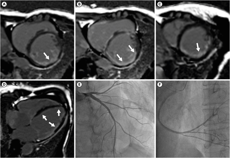
Figure 1 CMRI and coronary angiography of the systemic sclerosis heart. (A-D) Late gadolinium-enhancement CMRI of the explanted heart. An extensive subendocardial enhancement of the left ventricle and multifocal enhancement in the right ventricle was noted (white arrows). (E, F) Coronary angiography of the heart. (E) Left coronary angiography and (F) right coronary angiography revealed no significant epicardial coronary artery stenosis.CMRI = cardiac magnetic resonance imaging.
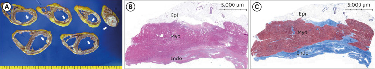
Figure 2 Pathologic findings. (A) Gross appearance of the explanted heart. Cross-sections of the entire heart specimen showed ventricular dilation and prominent endocardial fibrosis in both ventricles (arrow, whitish tissues). (B, C) Low magnification of the inferior wall of the left ventricle showed wall thinning, endocardial thickening with fibrosis, and patchy myocardial fibrosis. (B) Hematoxylin and eosin stain; (C) Masson’s trichrome stain.Endo = endocardium; Epi = epicardium; Myo = myocardium.
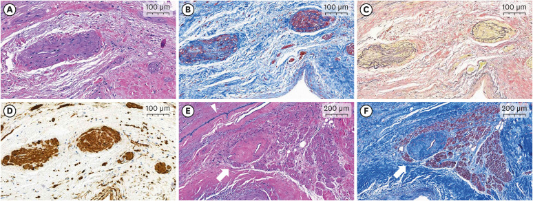
Figure 3 Microvascular alterations in pathologic specimens of the explanted systemic sclerosis heart. (A-D) Microscopic examination reveals fibromuscular dysplastic changes in the small vessels of the subendocardial myocardium. These micro-vasculatures show luminal narrowing due to fibrosis and smooth muscle hyperplasia in the tunica intima and media. Fragmentation or duplication of the internal elastic lamina is also noted. (A) Hematoxylin and eosin stain, ×100; (B) Masson’s trichrome stain, ×100; (C) Verhoeff’s elastic stain, ×100; (D) Immunohistochemical stain for smooth muscle actin, ×100. (E, F) An atrioventricular nodal artery shows luminal narrowing and vascular wall fibrosis. (E) Hematoxylin and eosin stain, ×50; (F) Masson’s trichrome stain, ×50. (E, F) White arrows indicate a nodal artery and (E) blue ink indicates the right side of the interventricular septum (arrowhead).
- Full Text Links
-
- Actions
-
Cited
- CITED
-
- Close
- Share
-
- Similar articles
-
- A Case of Systemic Sclerosis in a Child
- Improved Gastrointestinal Involvement in Systemic Sclerosis after Immunoglobulin Treatment
- A Case of Systemic Sclerosis with Coincidental Lung Cancer
- A case of hypoparathyroidism and hypothyroidism in systemic sclerosis
- A case of acute myocardial infarction associated with coronary vasospasm in a patient with systemic sclerosis
