Automatically Diagnosing Skull Fractures Using an Object Detection Method and Deep Learning Algorithm in Plain Radiography Images
- Affiliations
-
- 1Department of Traumatology, Gil Medical Center, Gachon University College of Medicine, Incheon, Korea
- 2Department of Neurosurgery, Gil Medical Center, Gachon University College of Medicine, Incheon, Korea
- 3Department of Biomedical Engineering, Gil Medical Center, Gachon University College of Medicine, Incheon, Korea
- KMID: 2537848
- DOI: http://doi.org/10.3340/jkns.2022.0062
Abstract
Objective
: Deep learning is a machine learning approach based on artificial neural network training, and object detection algorithm using deep learning is used as the most powerful tool in image analysis. We analyzed and evaluated the diagnostic performance of a deep learning algorithm to identify skull fractures in plain radiographic images and investigated its clinical applicability.
Methods
: A total of 2026 plain radiographic images of the skull (fracture, 991; normal, 1035) were obtained from 741 patients. The RetinaNet architecture was used as a deep learning model. Precision, recall, and average precision were measured to evaluate the deep learning algorithm’s diagnostic performance.
Results
: In ResNet-152, the average precision for intersection over union (IOU) 0.1, 0.3, and 0.5, were 0.7240, 0.6698, and 0.3687, respectively. When the intersection over union (IOU) and confidence threshold were 0.1, the precision was 0.7292, and the recall was 0.7650. When the IOU threshold was 0.1, and the confidence threshold was 0.6, the true and false rates were 82.9% and 17.1%, respectively. There were significant differences in the true/false and false-positive/false-negative ratios between the anteriorposterior, towne, and both lateral views (p=0.032 and p=0.003). Objects detected in false positives had vascular grooves and suture lines. In false negatives, the detection performance of the diastatic fractures, fractures crossing the suture line, and fractures around the vascular grooves and orbit was poor.
Conclusion
: The object detection algorithm applied with deep learning is expected to be a valuable tool in diagnosing skull fractures.
Keyword
Figure
-
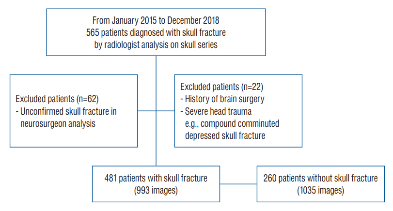
Fig. 1. Patient selection.
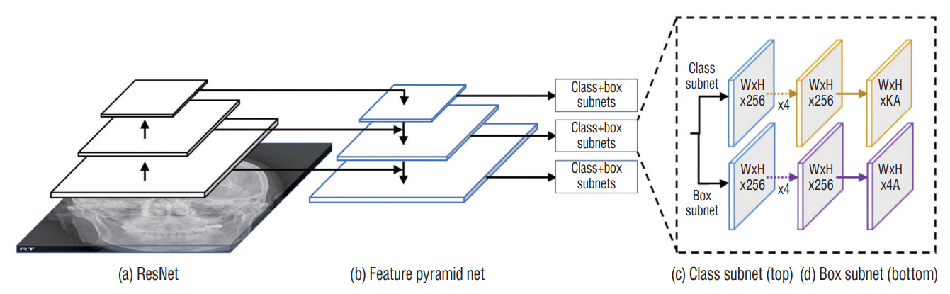
Fig. 2. RetinaNet architecture with (a) ResNet and (b) feature pyramid network as a feature extractor to (c) classify the probability of the skull fracture and (d) regress the bounding box coordinates.
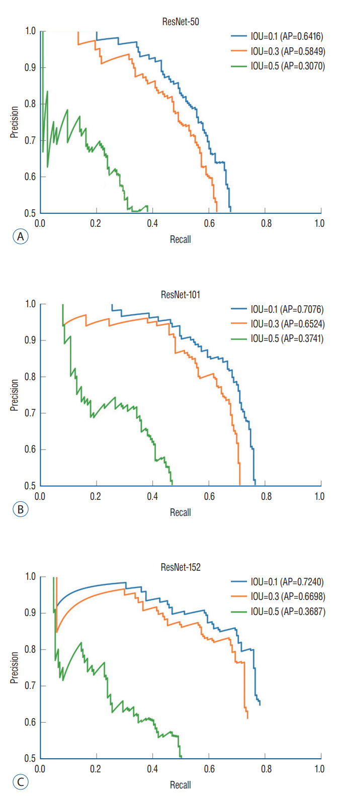
Fig. 3. Precision-recall curve. The diagnostic performance of object detection using RetinaNet was assessed by the precision-recall curve and the average precision (AP). AP is calculated as the area under the precision-recall curve. AP values for ResNet models and intersection over union (IOU) thresholds were analyzed.
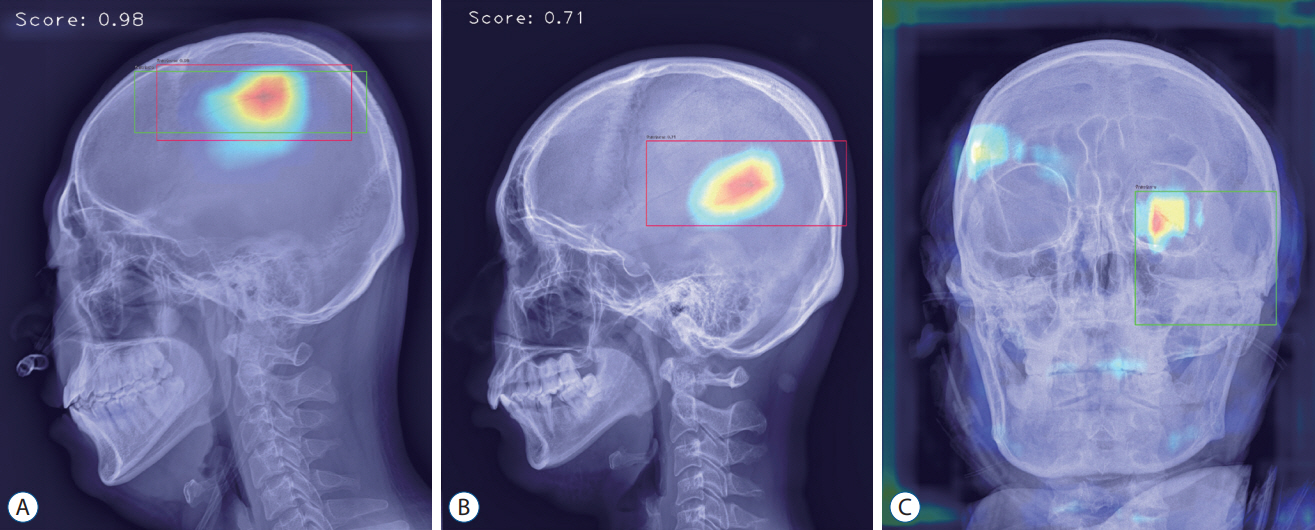
Fig. 4. Analyzed sample images. A : True-positive image detecting parietal bone fracture in lateral view. B : False-positive image detecting vascular groove in lateral view. C : False-negative image not detecting diastatic fracture of left lambdoid suture observed in the left orbit area of anterior-posterior view.
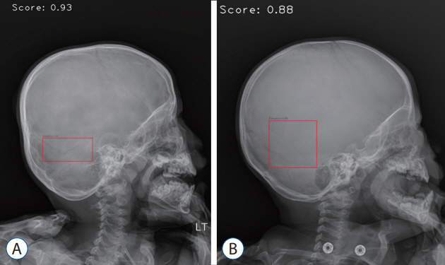
Fig. 5. Pediatric sample images. A : False-positive image detecting lambdoid suture and accessory suture. B : False-positive image detecting vascular groove.
Reference
-
References
1. Amyot F, Arciniegas DB, Brazaitis MP, Curley KC, Diaz-Arrastia R, Gandjbakhche A, et al. A review of the effectiveness of neuroimaging modalities for the detection of traumatic brain injury. J Neurotrauma. 32:1693–1721. 2015.2. Beyaz S, Açıcı K, Sümer E. Femoral neck fracture detection in X-ray images using deep learning and genetic algorithm approaches. Jt Dis Relat Surg. 31:175–183. 2020.3. Bruns JJ Jr, Jagoda AS. Mild traumatic brain injury. Mt Sinai J Med. 76:129–137. 2009.4. Chung SW, Han SS, Lee JW, Oh KS, Kim NR, Yoon JP, et al. Automated detection and classification of the proximal humerus fracture by using deep learning algorithm. Acta orthop. 89:468–473. 2018.5. Everingham M, Van Gool L, Williams CK, Winn J, Zisserman A. The pascal visual object classes (voc) challenge. Int J Comput Vis. 88:303–338. 2010.6. He K, Zhang X, Ren S, Sun J. Deep residual learning for image recognition : Proceedings of the IEEE Conference on Computer Vision and Pattern Recognition. Piscataway: The Institute of Electrical and Electronics Engineers, Inc.;2016. p. 770–778.7. Kim HJ, Roh HG, Lee IW. Craniosynostosis : updates in radiologic diagnosis. J Korean Neurosurg Soc. 59:219–226. 2016.8. Krogue JD, Cheng KV, Hwang KM, Toogood P, Meinberg EG, Geiger EJ, et al. Automatic hip fracture identification and functional subclassification with deep learning. Radiol Artif Intell. 2:e190023. 2020.9. Lakhani P, Sundaram B. Deep learning at chest radiography: automated classification of pulmonary tuberculosis by using convolutional neural networks. Radiology. 284:574–582. 2017.10. Le TH, Gean AD. Neuroimaging of traumatic brain injury. Mt Sinai J Med. 76:145–162. 2009.11. Lin TY, Goyal P, Girshick R, He K, Dollar P. Focal loss for dense object detection : Proceedings of the IEEE International Conference on Computer Vision. Piscataway: The Institute of Electrical and Electronics Engineers, Inc;2017. p. 2980–2988.12. Lin TY, Maire M, Belongie S, Hays J, Perona P, Ramanan D, et al. Microsoft coco: common objects in context : European Conference on Computer Vision. Cham: Springer;2014. p. 740–755.13. Möller TB, Reif E. Pocket Atlas of Radiographic Positioning: Including Positioning for Conventional Angiography, CT, and MRI. Noida: Thieme;2008.14. Nakahara K, Utsuki S, Shimizu S, Iida H, Miyasaka Y, Takagi H, et al. Age dependence of fusion of primary occipital sutures: a radiographic study. Childs Nerv Syst. 22:1457–1459. 2006.15. Ozkaya E, Topal FE, Bulut T, Gursoy M, Ozuysal M, Karakaya Z. Evaluation of an artificial intelligence system for diagnosing scaphoid fracture on direct radiography. Eur J Trauma Emerg Surg. 48:585–592. 2020.16. Rezatofighi H, Tsoi N, Gwak J, Sadeghian A, Reid I, Savarese S. Generalized intersection over union: a metric and a loss for bounding box regression : Proceedings of the IEEE/CVF Conference on Computer Vision and Pattern Recognition. Piscataway: The Institute of Electrical and Electronics Engineers, Inc.;2019. p. 658–666.17. Sanchez T, Stewart D, Walvick M, Swischuk L. Skull fracture vs. accessory sutures: how can we tell the difference? Emerg Radiol. 17:413–418. 2010.18. Sim SY, Yoon SH, Kim SY. Quantitative analysis of developmental process of cranial suture in Korean infants. J Korean Neurosurg Soc. 51:31–36. 2012.19. Taylor CA, Bell JM, Breiding MJ, Xu L. Traumatic brain injury-related emergency department visits, hospitalizations, and deaths - united states, 2007 and 2013. MMWR Surveill Summ. 66:1–16. 2017.20. Weir P, Suttner NJ, Flynn P, McAuley D. Normal skull suture variant mimicking intentional injury. BMJ. 332:1020–1021. 2006.21. Wintermark M, Sanelli PC, Anzai Y, Tsiouris AJ, Whitlow CT; ACR Head Injury Institute, et al. Imaging evidence and recommendations for traumatic brain injury: conventional neuroimaging techniques. J Am Coll Radiol. 12:e1–e14. 2015.22. Zou Z, Shi Z, Guo Y, Ye J. Object detection in 20 years: a survey. Available at : https://doi.org/10.48550/arXiv.1905.05055.
- Full Text Links
-
- Actions
-
Cited
- CITED
-
- Close
- Share
-
- Similar articles
-
- Deep Learning in Dental Radiographic Imaging
- Developing and Evaluating Deep Learning Algorithms for Object Detection: Key Points for Achieving Superior Model Performance
- Diagnostic Performance of a New Convolutional Neural Network Algorithm for Detecting Developmental Dysplasia of the Hip on Anteroposterior Radiographs
- Deep learning based pectoral muscle segmentation on Mammographic Image Analysis Society (MIAS) mammograms
- Automatic Segmentation and Radiologic Measurement of Distal Radius Fractures Using Deep Learning
