Portal vein fenestration: a case report of an unusual portal vein developmental anomaly
- Affiliations
-
- 1Department of Liver Transplant and Hepatobiliary Surgery, Centre for Liver and Biliary Sciences, Max Super Specialty Hospital, Saket, New Delhi, India
- KMID: 2537543
- DOI: http://doi.org/10.4285/kjt.22.0022
Abstract
- Portal vein anatomic variations are common in living donor liver transplantation. Portal vein fenestration, in which a segment of a vessel divides into at least two channels that reunite into a single distal lumen, has not yet been reported in the literature. Failure to identify this anomaly can lead to catastrophic events in donor liver hepatectomy. Herein, we report an unusual portal vein anomaly that was detected intraoperatively in a living liver donor.
Keyword
Figure
-
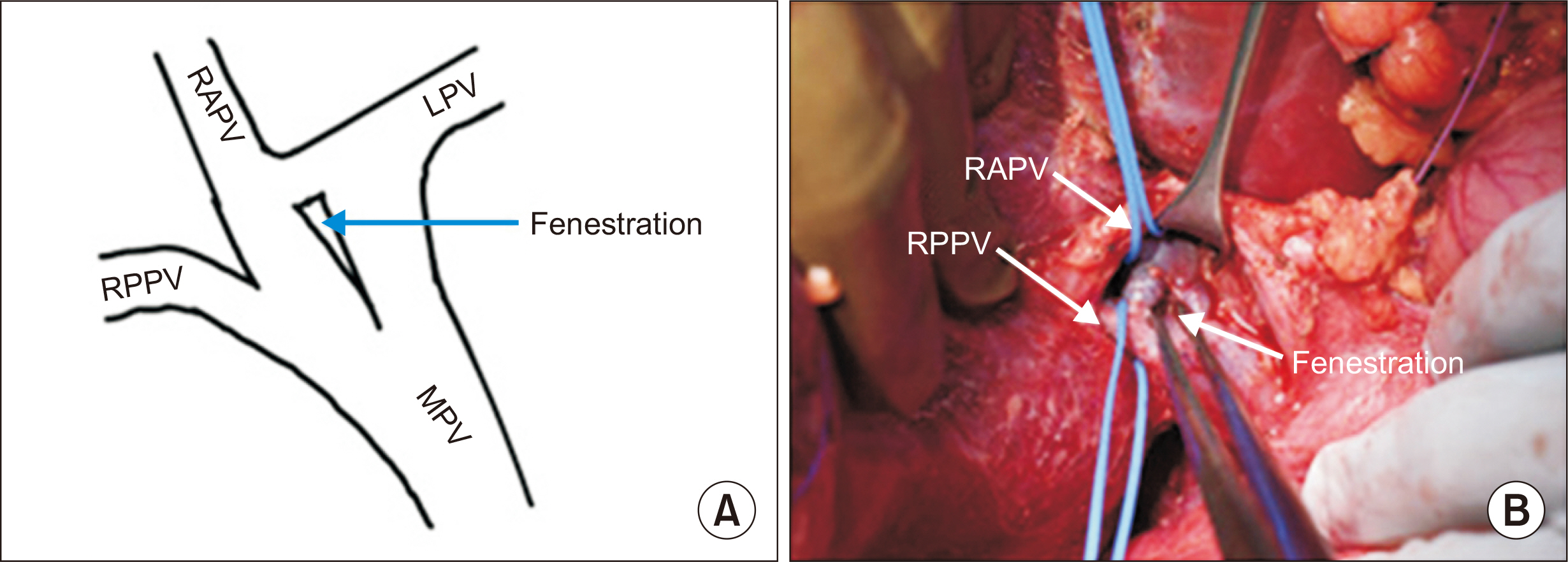
Fig. 1 (A) Schematic illustration of type C portal vein (PV) with PV fenestration in the PV trunk below the confluence of the right anterior PV (RAPV) and left PV (LPV). (B) Intraoperative photograph showing PV fenestration. RPPV, right posterior PV; MPV, main PV.

Fig. 2 (A) Schematic representation of wrongly looped right portal vein. (B) Wrong division of right portal vein compromising the remnant liver portal flow. RPPV, right posterior portal vein; RAPV, right anterior portal vein; LPV, left portal vein; MPV, main portal vein.
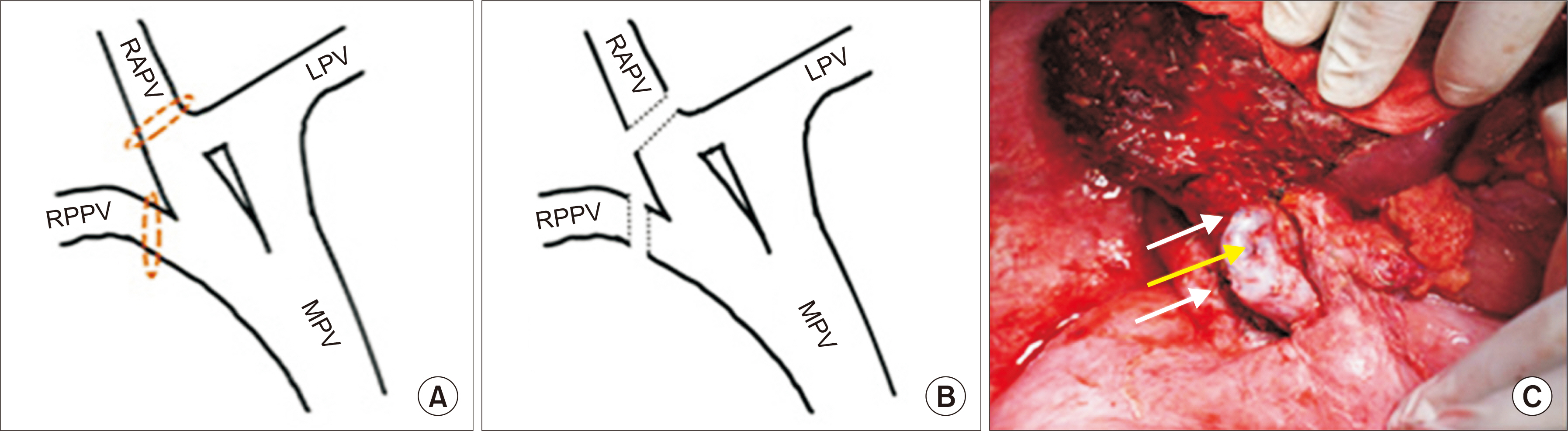
Fig. 3 (A, B) Schematic illustration of the right anterior portal vein (RAPV) and right posterior portal vein (RPPV) looped separately and divided. (C) Intraoperative photograph showing the closed stumps of the RAPV and RPPV (white arrows) and fenestration (yellow arrow). LPV, left portal vein; MPV, main portal vein.
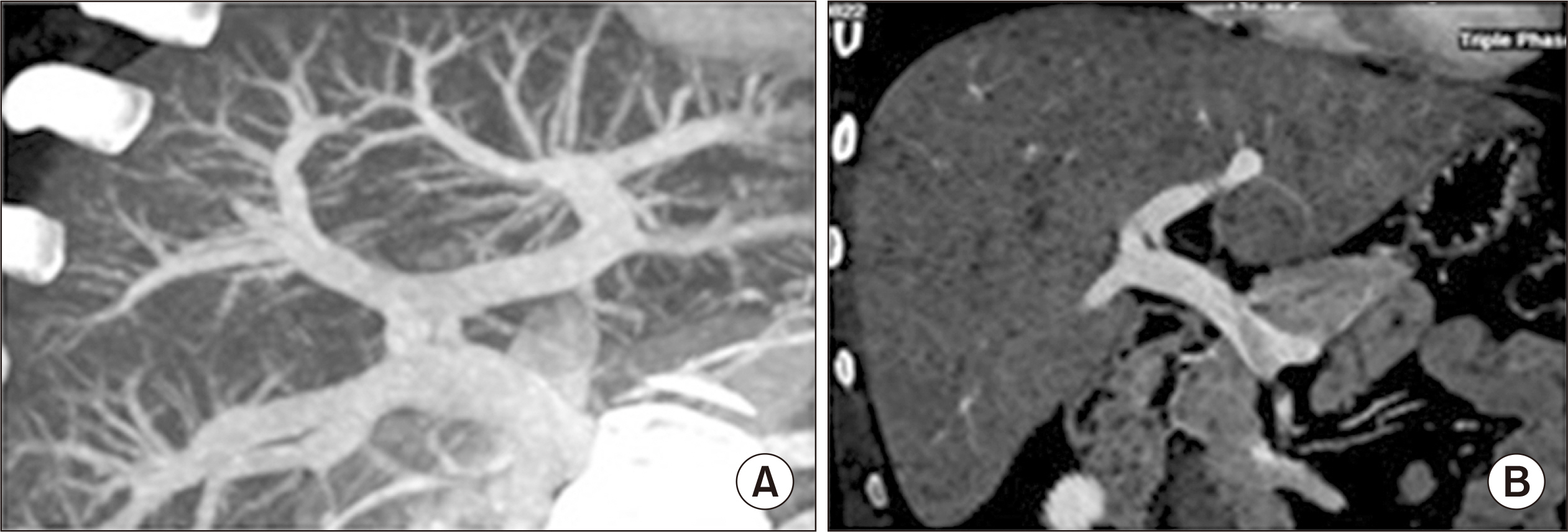
Fig. 4 (A) Preoperative three-dimensional reconstruction of the portal vein in the donor where fenestration was not clearly seen. (B) Coronal oblique reformatted contrast-enhanced computed tomography scan images during the portal phase showing fenestration of the portal vein.
Cited by 1 articles
-
A fenestrated portal vein
Amay Banker, Prashantha Rao, Karthik Ganesan, Ravi Mohanka
Korean J Transplant. 2023;37(2):141-143. doi: 10.4285/kjt.23.0012.
Reference
-
1. Marks C. 1969; Developmental basis of the portal venous system. Am J Surg. 117:671–81. DOI: 10.1016/0002-9610(69)90404-8. PMID: 5791038.2. Nakamura T, Tanaka K, Kiuchi T, Kasahara M, Oike F, Ueda M, et al. 2002; Anatomical variations and surgical strategies in right lobe living donor liver transplantation: lessons from 120 cases. Transplantation. 73:1896–903. DOI: 10.1097/00007890-200206270-00008. PMID: 12131684.3. Yilmaz S, Kayaalp C, Isik B, Ersan V, Otan E, Akbulut S, et al. 2017; Reconstruction of anomalous portal venous branching in right lobe living donor liver transplantation: Malatya approach. Liver Transpl. 23:751–61. DOI: 10.1002/lt.24753. PMID: 28240812.4. Hashimoto Y, Otsuki N, Morimoto K, Saito M, Nibu K. 2012; Four cases of spinal accessory nerve passing through the fenestrated internal jugular vein. Surg Radiol Anat. 34:373–5. DOI: 10.1007/s00276-011-0875-x. PMID: 21938454.5. Dighe M, Vaidya S. 2009; Duplication of the portal vein: a rare congenital anomaly. Br J Radiol. 82:e32–4. DOI: 10.1259/bjr/81921288. PMID: 19168687.6. Cheng YF, Huang TL, Lee TY, Chen TY, Chen CL. 1996; Variation of the intrahepatic portal vein: angiographic demonstration and application in living-related hepatic transplantation. Transplant Proc. 28:1667–8.
- Full Text Links
-
- Actions
-
Cited
- CITED
-
- Close
- Share
-
- Similar articles
-
- Preduodenal Portal Vein Associated with Duodenal Obstruction: A case report
- Duplication of the Portal Vein: A Case Report
- Absent portal vein bifurcation: a rare variant and its clinical significance
- Congenital Absence of the Portal Vein in an Adult Man Presenting with Portosystemic Encephalopathy: A Case Report
- CT Findings of Portal Vein Aneurysm
