Korean J Transplant.
2022 Dec;36(4):289-293. 10.4285/kjt.22.0048.
Successful treatment of renal malakoplakia via the reduction of immunosuppression and antimicrobial therapy after kidney transplantation: a case report
- Affiliations
-
- 1Research Institute for Transplantation, Yonsei University College of Medicine, Seoul, Korea
- 2Department of Surgery, Yonsei University College of Medicine, Seoul, Korea
- 3Department of Pathology, Yonsei University College of Medicine, Seoul, Korea
- KMID: 2537541
- DOI: http://doi.org/10.4285/kjt.22.0048
Abstract
- Malakoplakia is a rare, granulomatous disease that usually affects immunocompromised individuals and is generally associated with poor graft and patient survival. We present a case of renal malakoplakia after kidney transplantation (KT). A 33-year-old female patient with chronic kidney disease underwent living-donor KT at Severance Hospital. The patient was administered 375 mg/m2 rituximab due to high panel reactive antibodies. Immunosuppression was initiated with 1.5 mg/kg anti-thymocyte globulin and intravenous methylprednisolone and maintained with tacrolimus, oral methylprednisolone, and mycophenolate mofetil (MMF). Six months after KT, the patient was hospitalized for a urinary tract infection with an elevated serum creatinine level of 3.14 mg/dL. Renal biopsy revealed malakoplakia involving the renal parenchyma. Upon this diagnosis, the dose of tacrolimus was reduced and MMF was stopped. Fluoroquinolone was used for 16 days, and the trimethoprim/sulfamethoxazole dose was doubled for 6 days. The patient was hospitalized for 3 weeks and closely observed during outpatient visits. Follow-up ultrasonography revealed mass-like lesions of renal malakoplakia, which disappeared 5 months after diagnosis. The serum creatinine level decreased to 1.29 mg/dL 28 months after diagnosis. Our results suggest that renal malakoplakia can be successfully treated by the reduction of immunosuppression and sustained antimicrobial therapy.
Figure
-
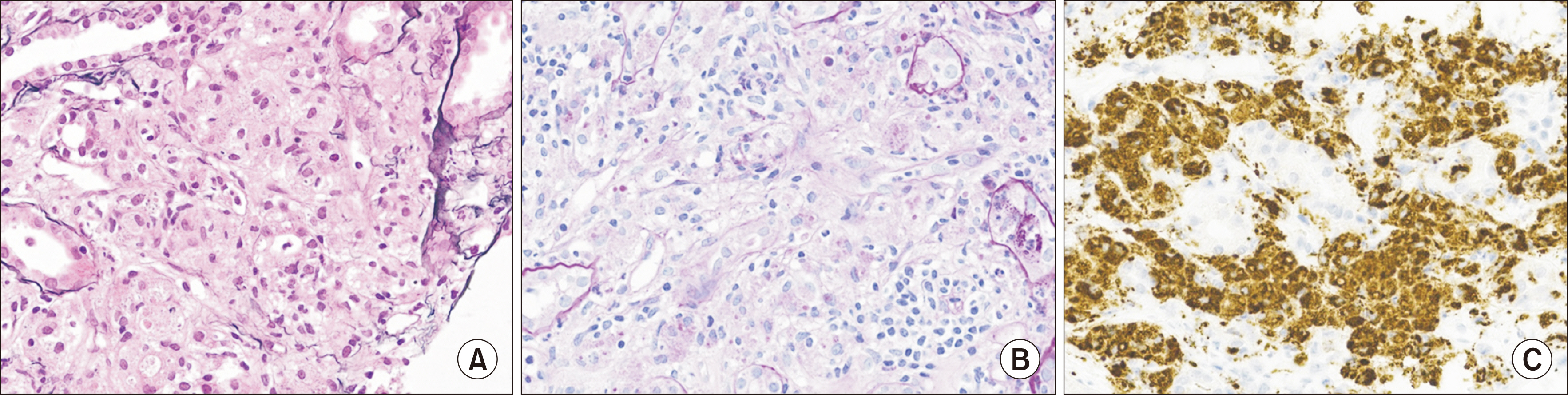
Fig. 1 Microscopic image of malakoplakia. (A) Periodic acid-methenamine silver stain (×400); interstitium showing heavy infiltration of macrophages. (B) Periodic acid-Schiff (PAS) stain (×400); interstitium showing heavy infiltration of macrophages with PAS-positive granules. (C) CD68 stain (×400).

Fig. 2 Serial trough levels of tacrolimus.
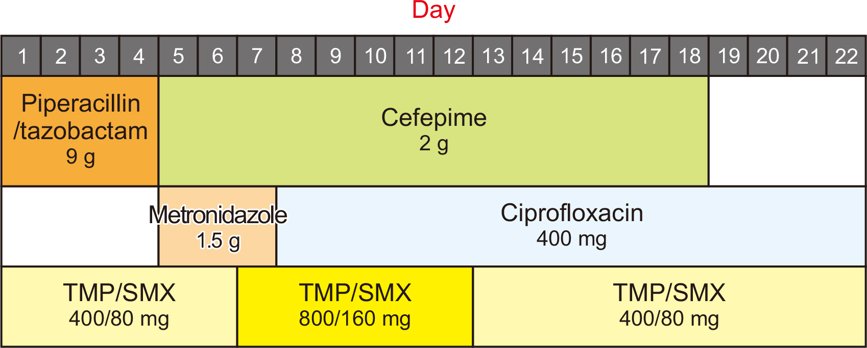
Fig. 3 Change in antibiotics and doses used. TMP/SMX, trimethoprim/sulfamethoxazole.
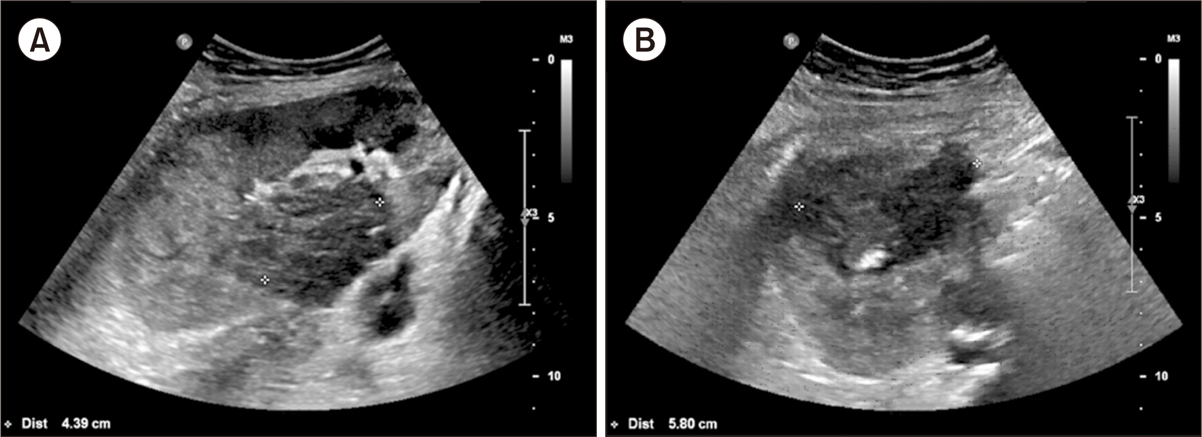
Fig. 4 Ultrasonographic images of malakoplakia in the kidney. (A) Malakoplakia lesion in mid-portion of transplanted kidney, diameter is 4.39 cm. (B) Another malakoplakia lesion in upper pole of transplanted kidney, diameter is 5.80 cm.
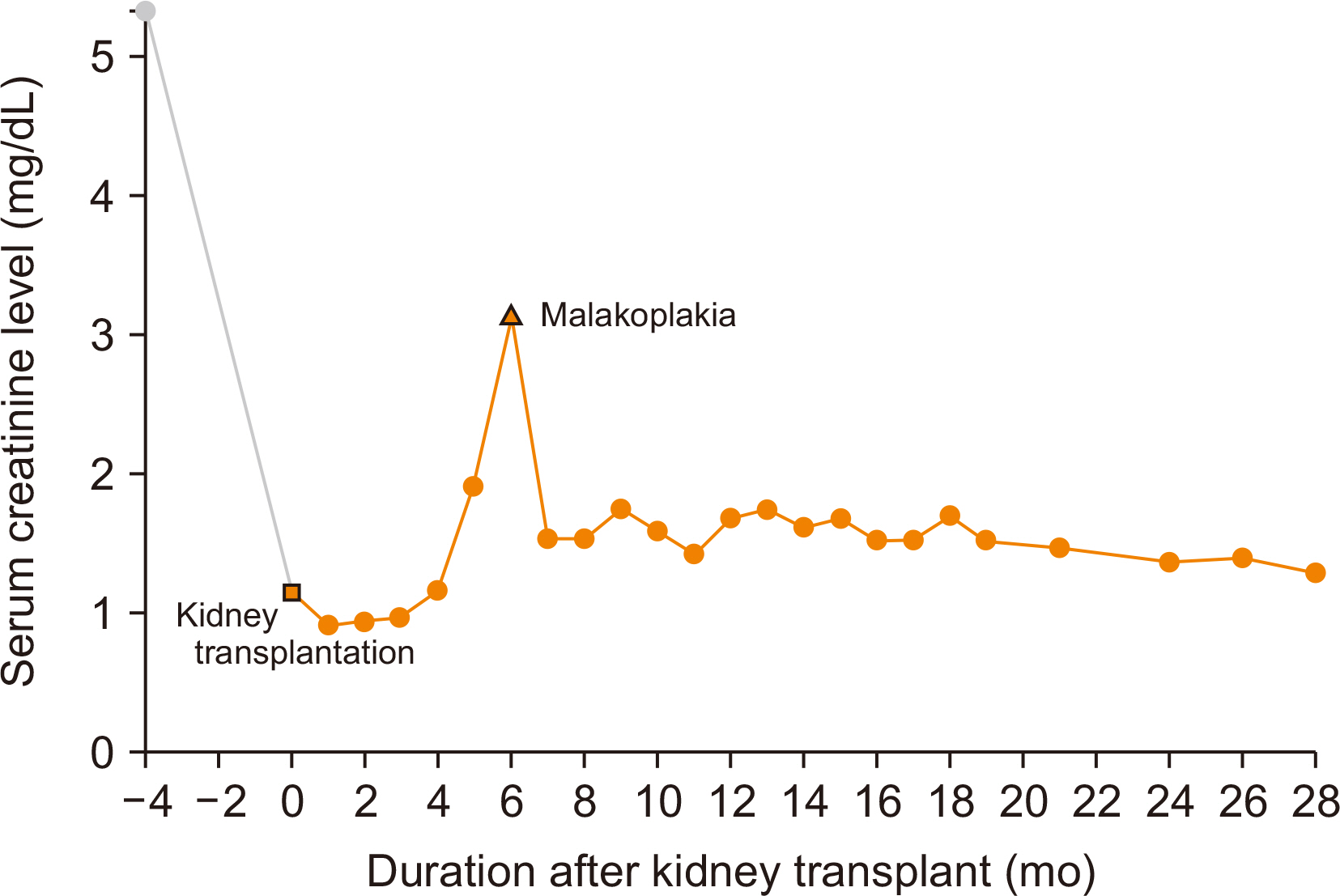
Fig. 5 Serum creatinine levels.
Reference
-
1. McClure J. 1983; Malakoplakia. J Pathol. 140:275–330. DOI: 10.1002/path.1711400402. PMID: 6308195.2. Kobayashi A, Utsunomiya Y, Kono M, Ito Y, Yamamoto I, Osaka N, et al. 2008; Malakoplakia of the kidney. Am J Kidney Dis. 51:326–30. DOI: 10.1053/j.ajkd.2007.08.029. PMID: 18215711.3. Nieto-Ríos JF, Ramírez I, Zuluaga-Quintero M, Serna-Higuita LM, Gaviria-Gil F, Velez-Hoyos A. 2017; Malakoplakia after kidney transplantation: case report and literature review. Transpl Infect Dis. 19:e12731. DOI: 10.1111/tid.12731. PMID: 28561517.4. Kajbafzadeh A, Baharnoori M. 2004; Renal malakoplakia simulating neoplasm in a child: successful medical management. Urol J. 1:218–20. PMID: 17914695.5. Purnell SD, Davis B, Burch-Smith R, Coleman P. 2015; Renal malakoplakia mimicking a malignant renal carcinoma: a patient case with literature review. BMJ Case Rep. 2015:bcr2014208652. DOI: 10.1136/bcr-2014-208652. PMID: 26177998. PMCID: PMC4513562.6. Abdou NI, NaPombejara C, Sagawa A, Ragland C, Stechschulte DJ, Nilsson U, et al. 1977; Malakoplakia: evidence for monocyte lysosomal abnormality correctable by cholinergic agonist in vitro and in vivo. N Engl J Med. 297:1413–9. DOI: 10.1056/NEJM197712292972601. PMID: 200843.7. Albitar S, Genin R, Fen-Chong M, Schohn D, Riviere JP, Serveaux MO, et al. 1997; The febrile patient presenting with acute renal failure and enlarged kidneys: another mode of presentation of malakoplakia. Nephrol Dial Transplant. 12:1724–6. DOI: 10.1093/ndt/12.8.1724. PMID: 9269661.8. Leão CA, Duarte MI, Gamba C, Ramos JF, Rossi F, Galvão MM, et al. 2012; Malakoplakia after renal transplantation in the current era of immunosuppressive therapy: case report and literature review. Transpl Infect Dis. 14:E137–41. DOI: 10.1111/tid.12012. PMID: 23025504.9. Augusto JF, Sayegh J, Croue A, Subra JF, Onno C. 2008; Renal transplant malakoplakia: case report and review of the literature. NDT Plus. 1:340–3. DOI: 10.1093/ndtplus/sfn028. PMID: 25983929. PMCID: PMC4421262.10. Graves AL, Texler M, Manning L, Kulkarni H. 2014; Successful treatment of renal allograft and bladder malakoplakia with minimization of immunosuppression and prolonged antibiotic therapy. Nephrology (Carlton). 19 Suppl 1:18–21. DOI: 10.1111/nep.12194. PMID: 24460630.
- Full Text Links
-
- Actions
-
Cited
- CITED
-
- Close
- Share
-
- Similar articles
-
- Successful treatment of renal malakoplakia with reduction of immunosuppression and antimicrobial therapy after kidney transplantation: case report
- Malakoplakia after kidney transplantation
- Malakoplakia in the Urinary Bladder
- Malakoplakia of the Kidney Extending to the Descending Colon in a Patient with Secondary Adrenal Insufficiency: A Case Report
- ABO-Incompatible Kidney Transplantation
