Terminal bifurcation of the external jugular vein: a rare variation
- Affiliations
-
- 1Department of Basic Medical Sciences, Manipal Academy of Higher Education, Manipal, Karnataka, India
- 2Department of Mathematics, Manipal Institute of Technology, Manipal Academy of Higher Education, Manipal, Karnataka, India
- KMID: 2537470
- DOI: http://doi.org/10.5115/acb.22.084
Abstract
- Variations of external jugular vein are common. Here, we present a rare terminal bifurcation of the left external jugular vein. The left external jugular vein was formed by the union of entire retromandibular vein and posterior auricular vein. One inch above the clavicle, it bifurcated into medial and lateral divisions. The medial division terminated into the internal jugular vein and the lateral division terminated into the subclavian vein. Medial division received a common vein formed by the union of anterior jugular vein and an anonymous vein lying under the sternocleidomastoid muscle. The lateral division received a common vein formed by the union of suprascapular and transverse cervical veins. The knowledge about this variation could be useful to head and neck surgeons, radiologists and plastic surgeons.
Keyword
Figure
-

Fig. 1 Dissection of the left side of the neck showing the variation of the EJV. EJV, external jugular vein; RMV, retromandibular vein; PAV, posterior auricular vein; MD, medial division of the terminal part of the retromandibular vein; LD, lateral division of the terminal part of the retromandibular vein; FV, facial vein; PG, parotid gland; SCM, sternocleidomastoid; AJV, anterior jugular vein; AV, anonymous vein deep to sternocleidomastoid; IJV, internal jugular vein; OH, inferior belly of omohyoid; A, anterior; P, posterior; S, superior; I, inferior.
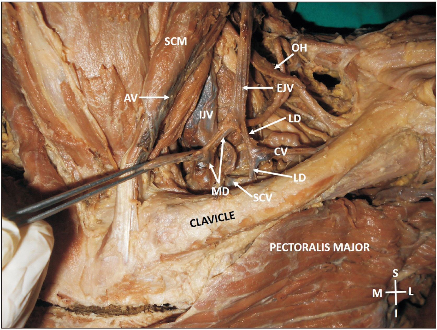
Fig. 2 Dissection of the left side of the neck showing the termination of the EJV. EJV, external jugular vein; MD, medial division of the terminal part of the retromandibular vein; LD, lateral division of the terminal part of the retromandibular vein; CV, common vein formed by the union of suprascapular and transverse cervical veins; SCM, sternocleidomastoid; AV, anonymous vein deep to sternocleidomastoid; IJV, internal jugular vein; OH, inferior belly of omohyoid; SCV, supraclavicular vein; M, medial; S, superior; L, lateral; I, inferior.
Reference
-
References
1. Cvetko E. 2015; A case of left-sided absence and right-sided fenestration of the external jugular vein and a review of the literature. Surg Radiol Anat. 37:883–6. DOI: 10.1007/s00276-014-1398-z. PMID: 25432662.2. Comert E, Comert A. 2009; External jugular vein duplication. J Craniofac Surg. 20:2173–4. DOI: 10.1097/SCS.0b013e3181bf0248. PMID: 19884835.3. Ponnambalam SB, Karuppiah DS. 2020; Unilateral external jugular vein fenestration with variant anatomy of the retromandibular and facial vein. Anat Cell Biol. 53:117–20. DOI: 10.5115/acb.19.172. PMID: 32274258. PMCID: PMC7118259.4. Singh R. 2020; Abnormal formation of external jugular vein and its repercussions. J Craniofac Surg. 31:e354–5. DOI: 10.1097/SCS.0000000000006293. PMID: 32149966.5. Patil J, Kumar N, Swamy RS, D'Souza MR, Guru A, Nayak SB. 2014; Absence of retromandibular vein associated with atypical formation of external jugular vein in the parotid region. Anat Cell Biol. 47:135–7. DOI: 10.5115/acb.2014.47.2.135. PMID: 24987551. PMCID: PMC4076421.6. Chauhan NK, Rani A, Chopra J, Rani A, ivastava AK Sr, Kumar V. 2011; Anomalous formation of external jugular vein and its clinical implication. Natl J Maxillofac Surg. 2:51–3. DOI: 10.4103/0975-5950.85854. PMID: 22442610. PMCID: PMC3304223.7. Vadgaonkar R, Rai R, Ranade AV, Pai MM, Prabhu LV, Ashwin K, Jiji PJ. 2008; An anomalous left external jugular vein draining into right subclavian vein. Bratisl Lek Listy. 109:461–2. DOI: 10.4067/s0717-95022008000400018. PMID: 19166133.8. Shetty P, Nayak SB, Thangarajan R, D'Souza MR. 2016; A rare case of persistent jugulocephalic vein and its clinical implication. Anat Cell Biol. 49:210–2. DOI: 10.5115/acb.2016.49.3.210. PMID: 27722015. PMCID: PMC5052231.9. Ono K, Yoshioka N, Hage D, Ibaragi S, Tubbs RS, Iwanaga J. 2021; Duplication of the external jugular vein: a language barrier of database search in classic anatomical studies. Surg Radiol Anat. 43:1721–8. DOI: 10.1007/s00276-021-02717-6. PMID: 33620594.10. Satheesha NB. 2017; Jugulo-facial venous circle, accessory slip of trapezius and absence of typical facial vein- clinically important anatomical variations. J Anat Soc India. 66(Suppl 2):S11–2. DOI: 10.1016/j.jasi.2017.10.008.11. Nagata T, Masumoto K, Watanabe Y, Katou F. 2012; End-to-side anastomosis to the external jugular vein: preservation of external jugular vein blood flow. Br J Oral Maxillofac Surg. 50:e31–2. DOI: 10.1016/j.bjoms.2011.07.024. PMID: 21871698.12. Ibrahim AE, Adelman DM, Parham C, Hong Z, Villa M, Chahine FM, Ghieh FM. 2019; The external jugular vein used as recipient vessel in head and neck free flap reconstruction: outcomes compared to the internal jugular vein. J Craniofac Surg. 30:178–83. DOI: 10.1097/SCS.0000000000004873. PMID: 30394970.13. Sugiyama S, Iwai T, Tohnai I. 2017; Empty fenestration of the external jugular vein: a rare variant. J Anat Soc India. 66(Suppl 2):S1–3. DOI: 10.1016/j.jasi.2017.10.005.14. Yadav S, Ghosh SK, Anand C. 2000; Variations of superficial veins of head and neck. J Anat Soc India. 49:61–2.
- Full Text Links
-
- Actions
-
Cited
- CITED
-
- Close
- Share
-
- Similar articles
-
- Absence of retromandibular vein associated with atypical formation of external jugular vein in the parotid region
- A Large Subcutaneous Hematoma during Internal Jugular Vein Catheterization: A case report
- A rare case of persistent jugulocephalic vein and its clinical implication
- Unusual Presentation of a Cervical Mass Revealed as External Jugular Venous Aneurysm
- Unilateral external jugular vein fenestration with variant anatomy of the retromandibular and facial vein
