Radiomics Analysis of Magnetic Resonance Proton Density Fat Fraction for the Diagnosis of Hepatic Steatosis in Patients With Suspected NonAlcoholic Fatty Liver Disease
- Affiliations
-
- 1Department of Radiology, Korea University Anam Hospital, Korea University College of Medicine, Seoul, Korea
- 2AI Center, Korea University Anam Hospital, Korea University College of Medicine, Seoul, Korea
- 3Department of Gastroenterology, Korea University Ansan Hospital, Korea University College of Medicine, Ansan, Korea
- 4Department of Pathology, Korea University Anam Hospital, Korea University College of Medicine, Seoul, Korea
- KMID: 2537043
- DOI: http://doi.org/10.3346/jkms.2022.37.e339
Abstract
- Background
This study aimed to assess the diagnostic feasibility of radiomics analysis based on magnetic resonance (MR)-proton density fat fraction (PDFF) for grading hepatic steatosis in patients with suspected non-alcoholic fatty liver disease (NAFLD).
Methods
This retrospective study included 106 patients with suspected NAFLD who underwent a hepatic parenchymal biopsy. MR-PDFF and MR spectroscopy were performed on all patients using a 3.0-T scanner. Following whole-volume segmentation of the MRPDFF images, 833 radiomic features were analyzed using a commercial program. Radiologic features were analyzed, including median and mean values of the multiple regions of interest and variable clinical features. A random forest regressor was used to extract the important radiomic, radiologic, and clinical features. The model was trained using 20 repeated 10-fold cross-validations to classify the NAFLD steatosis grade. The area under the receiver operating characteristic curve (AUROC) was evaluated using a classifier to diagnose steatosis grades.
Results
The levels of pathological hepatic steatosis were classified as low-grade steatosis (grade, 0–1; n = 82) and high-grade steatosis (grade, 2–3; n = 24). Fifteen important features were extracted from the radiomic analysis, with the three most important being wavelet-LLL neighboring gray tone difference matrix coarseness, original first-order mean, and 90th percentile. The MR spectroscopy mean value was extracted as a more important feature than the MR-PDFF mean or median in radiologic measures. Alanine aminotransferase has been identified as the most important clinical feature. The AUROC of the classifier using radiomics was comparable to that of radiologic measures (0.94 ± 0.09 and 0.96 ± 0.08, respectively).
Conclusion
MR-PDFF-derived radiomics may provide a comparable alternative for grading hepatic steatosis in patients with suspected NAFLD.
Keyword
Figure
-
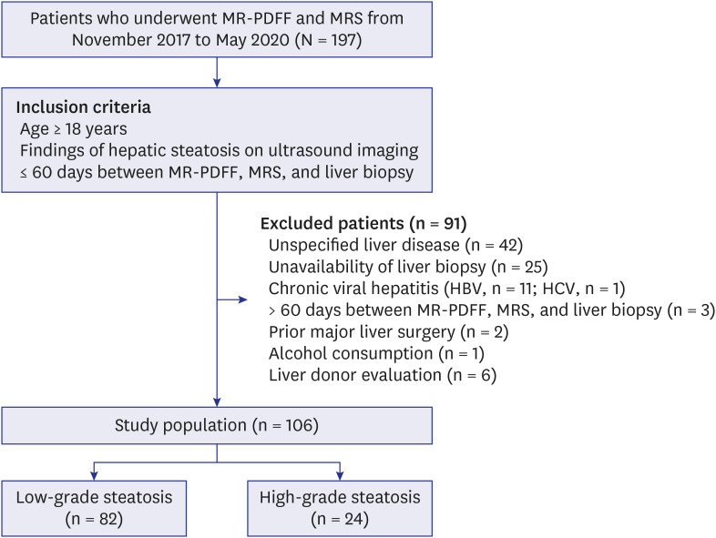
Fig. 1 Flow diagram for the study population.MR-PDFF = magnetic resonance-proton density fat fraction, MRS = magnetic resonance spectroscopy, HBV = hepatitis B virus, HCV = hepatitis C virus.

Fig. 2 A 30-year-old woman with non-alcoholic fatty liver disease. The work screen shows the completion of the whole-volume segmentation of the fat fracture image of the magnetic resonance-proton density fat fraction using a commercial program. Vascular structures such as portal veins (asterisk) are excluded and only the liver parenchyma indicated in green is accurately segmented.

Fig. 3 A 33-year-old man with non-alcoholic fatty liver disease (hepatic steatosis grade 1 and 20% steatosis on histopathology). The liver steatosis values on the MR-PDFF are measured to be approximately 19.0% (A) and 21.3% (B). The value of liver steatosis on MRS is 19.6% (C, D). The MR-PDFF and MRS values measured at similar locations in segment 4a of the liver are almost identical (A, C).MR-PDFF = magnetic resonance-proton density fat fraction, MRS = magnetic resonance spectroscopy.

Fig. 4 Radiomics pipeline.MR-PDFF = magnetic resonance-proton density fat fraction, DM = diabetes mellitus, ALT = alanine aminotransferase, AST = aspartate aminotransferase, TG = triglyceride, LDL = low-density lipoprotein, BMI = body mass index, HDL = high-density lipoprotein, GGT = gamma-glutamyl transferase, ALP = alkaline phosphatase, MRS = magnetic resonance spectroscopy, AUROC = area under the receiver operating characteristic curve.
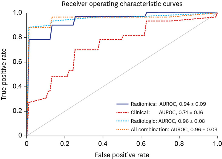
Fig. 5 Receiver operating characteristic curve for prediction of the grade of liver steatosis.AUROC = area under the receiver operating characteristic curve.
Reference
-
1. Younossi ZM, Blissett D, Blissett R, Henry L, Stepanova M, Younossi Y, et al. The economic and clinical burden of nonalcoholic fatty liver disease in the United States and Europe. Hepatology. 2016; 64(5):1577–1586. PMID: 27543837.2. Stål P. Liver fibrosis in non-alcoholic fatty liver disease - diagnostic challenge with prognostic significance. World J Gastroenterol. 2015; 21(39):11077–11087. PMID: 26494963.3. Mitra S, De A, Chowdhury A. Epidemiology of non-alcoholic and alcoholic fatty liver diseases. Transl Gastroenterol Hepatol. 2020; 5(5):16. PMID: 32258520.4. Kang SH, Lee HW, Yoo JJ, Cho Y, Kim SU, Lee TH, et al. KASL clinical practice guidelines: management of nonalcoholic fatty liver disease. Clin Mol Hepatol. 2021; 27(3):363–401. PMID: 34154309.5. Lee YB, Ha Y, Chon YE, Kim MN, Lee JH, Park H, et al. Association between hepatic steatosis and the development of hepatocellular carcinoma in patients with chronic hepatitis B. Clin Mol Hepatol. 2019; 25(1):52–64. PMID: 30360031.6. Jang WY, Chung WJ, Jang BK, Hwang JS, Lee HJ, Hwang MJ, et al. Changes in characteristics of patients with liver cirrhosis visiting a tertiary hospital over 15 years: a retrospective multi-center study in Korea. J Korean Med Sci. 2020; 35(29):e233. PMID: 32715667.7. Bravo AA, Sheth SG, Chopra S. Liver biopsy. N Engl J Med. 2001; 344(7):495–500. PMID: 11172192.8. Kleiner DE, Brunt EM, Van Natta M, Behling C, Contos MJ, Cummings OW, et al. Design and validation of a histological scoring system for nonalcoholic fatty liver disease. Hepatology. 2005; 41(6):1313–1321. PMID: 15915461.9. Bedossa P, Dargère D, Paradis V. Sampling variability of liver fibrosis in chronic hepatitis C. Hepatology. 2003; 38(6):1449–1457. PMID: 14647056.10. Runge JH, Smits LP, Verheij J, Depla A, Kuiken SD, Baak BC, et al. MR Spectroscopy-derived proton density fat fraction is superior to controlled attenuation parameter for detecting and grading hepatic steatosis. Radiology. 2018; 286(2):547–556. PMID: 28915103.11. Imajo K, Kessoku T, Honda Y, Tomeno W, Ogawa Y, Mawatari H, et al. Magnetic resonance imaging more accurately classifies steatosis and fibrosis in patients with nonalcoholic fatty liver disease than Transient elastography. Gastroenterology. 2016; 150(3):626–637.e7. PMID: 26677985.12. Reeder SB, Cruite I, Hamilton G, Sirlin CB. Quantitative assessment of liver fat with magnetic resonance imaging and spectroscopy. J Magn Reson Imaging. 2011; 34(4):729–749. PMID: 22025886.13. Sellers R. MR LiverLab. Updated 2016. Accessed April 5, 2022. http://clinical-mri.com/wp-content/uploads/2016/11/How_I_do_it_LiverLab_Sellers_RSNA_MAGNETOM_Flash.pdf .14. Caussy C, Reeder SB, Sirlin CB, Loomba R. Noninvasive, quantitative assessment of liver fat by MRI-PDFF as an endpoint in NASH trials. Hepatology. 2018; 68(2):763–772. PMID: 29356032.15. Gillies RJ, Kinahan PE, Hricak H. Radiomics: images are more than pictures, they are data. Radiology. 2016; 278(2):563–577. PMID: 26579733.16. Park HJ, Park B, Lee SS. Radiomics and deep learning: hepatic applications. Korean J Radiol. 2020; 21(4):387–401. PMID: 32193887.17. Sim KC, Kim MJ, Cho Y, Kim HJ, Park BJ, Sung DJ, et al. Diagnostic feasibility of magnetic resonance elastography radiomics analysis for the assessment of hepatic fibrosis in patients with nonalcoholic fatty liver disease. J Comput Assist Tomogr. 2022; 46(4):505–513. PMID: 35483092.18. Kim YK, Kwon OS, Her KH. The grade of nonalcoholic fatty liver disease is an independent risk factor for gallstone disease: an observational Study. Medicine (Baltimore). 2019; 98(27):e16018. PMID: 31277096.19. Qayyum A, Nystrom M, Noworolski SM, Chu P, Mohanty A, Merriman R. MRI steatosis grading: development and initial validation of a color mapping system. AJR Am J Roentgenol. 2012; 198(3):582–588. PMID: 22357996.20. Chalasani N, Wilson L, Kleiner DE, Cummings OW, Brunt EM, Unalp A, et al. Relationship of steatosis grade and zonal location to histological features of steatohepatitis in adult patients with non-alcoholic fatty liver disease. J Hepatol. 2008; 48(5):829–834. PMID: 18321606.21. Yokoo T, Serai SD, Pirasteh A, Bashir MR, Hamilton G, Hernando D, et al. Linearity, bias, and precision of hepatic proton density fat fraction measurements by using MR imaging: a meta-analysis. Radiology. 2018; 286(2):486–498. PMID: 28892458.22. Zhao YZ, Gan YG, Zhou JL, Liu JQ, Cao WG, Cheng SM, et al. Accuracy of multi-echo Dixon sequence in quantification of hepatic steatosis in Chinese children and adolescents. World J Gastroenterol. 2019; 25(12):1513–1523. PMID: 30948914.23. Jeon SK, Lee JM, Joo I, Park SJ. Quantitative ultrasound radiofrequency data analysis for the assessment of hepatic steatosis in nonalcoholic fatty liver disease using magnetic resonance imaging proton density fat fraction as the reference standard. Korean J Radiol. 2021; 22(7):1077–1086. PMID: 33739636.24. Hamilton G, Yokoo T, Bydder M, Cruite I, Schroeder ME, Sirlin CB, et al. In vivo characterization of the liver fat 1H MR spectrum. NMR Biomed. 2011; 24(7):784–790. PMID: 21834002.25. Ajmera V, Park CC, Caussy C, Singh S, Hernandez C, Bettencourt R, et al. Magnetic resonance imaging proton density fat fraction associates with progression of fibrosis in patients with nonalcoholic fatty liver disease. Gastroenterology. 2018; 155(2):307–310.e2. PMID: 29660324.26. Middleton MS, Heba ER, Hooker CA, Bashir MR, Fowler KJ, Sandrasegaran K, et al. Agreement between magnetic resonance imaging proton density fat fraction measurements and pathologist-assigned steatosis grades of liver biopsies from adults with nonalcoholic steatohepatitis. Gastroenterology. 2017; 153(3):753–761. PMID: 28624576.27. Middleton MS, Van Natta ML, Heba ER, Alazraki A, Trout AT, Masand P, et al. Diagnostic accuracy of magnetic resonance imaging hepatic proton density fat fraction in pediatric nonalcoholic fatty liver disease. Hepatology. 2018; 67(3):858–872. PMID: 29028128.28. Chen J, Talwalkar JA, Yin M, Glaser KJ, Sanderson SO, Ehman RL. Early detection of nonalcoholic steatohepatitis in patients with nonalcoholic fatty liver disease by using MR elastography. Radiology. 2011; 259(3):749–756. PMID: 21460032.29. Amadasun M, King R. Textural features corresponding to textural properties. IEEE Trans Syst Man Cybern. 1989; 19(5):1264–1274.30. van Griethuysen JJ, Fedorov A, Parmar C, Hosny A, Aucoin N, Narayan V, et al. Computational radiomics system to decode the radiographic phenotype. Cancer Res. 2017; 77(21):e104–e107. PMID: 29092951.31. Çinarer G, Emiroğlu BG, Yurttakal AH. Prediction of glioma grades using deep learning with wavelet radiomic features. Appl Sci. 2020; 10(18):6296.32. Kniep HC, Madesta F, Schneider T, Hanning U, Schönfeld MH, Schön G, et al. Radiomics of brain MRI: utility in prediction of metastatic tumor type. Radiology. 2019; 290(2):479–487. PMID: 30526358.33. Breiman L. Random forests. Mach Learn. 2001; 45(1):5–32.34. Donner A, Zou G. Testing the equality of dependent intraclass correlation coefficients. Statistician. 2002; 51(3):367–379.35. Dzyubak B, Li J, Chen J, Mara KC, Therneau TM, Venkatesh SK, et al. Automated analysis of multiparametric magnetic resonance imaging/magnetic resonance elastography exams for prediction of nonalcoholic steatohepatitis. J Magn Reson Imaging. 2021; 54(1):122–131. PMID: 33586159.36. Park HJ, Lee SS, Park B, Yun J, Sung YS, Shim WH, et al. Radiomics analysis of gadoxetic acid-enhanced MRI for staging liver fibrosis. Radiology. 2019; 290(2):380–387. PMID: 30615554.37. Lan GY, Guo Y, Zhang XY, Cai XL, Shi Y. Value of radiomic analysis of data from magnetic resonance elastography for diagnosing fibrosis stages in patients with hepatitis B/C. Chin J Acad Radiol. 2019; 1(2):74–84.38. Naganawa S, Enooku K, Tateishi R, Akai H, Yasaka K, Shibahara J, et al. Imaging prediction of nonalcoholic steatohepatitis using computed tomography texture analysis. Eur Radiol. 2018; 28(7):3050–3058. PMID: 29404772.39. Achmad E, Yokoo T, Hamilton G, Heba ER, Hooker JC, Changchien C, et al. Feasibility of and agreement between MR imaging and spectroscopic estimation of hepatic proton density fat fraction in children with known or suspected nonalcoholic fatty liver disease. Abdom Imaging. 2015; 40(8):3084–3090. PMID: 26205992.40. Idilman IS, Keskin O, Celik A, Savas B, Elhan AH, Idilman R, et al. A comparison of liver fat content as determined by magnetic resonance imaging-proton density fat fraction and MRS versus liver histology in non-alcoholic fatty liver disease. Acta Radiol. 2016; 57(3):271–278. PMID: 25855666.41. Di Martino M, Pacifico L, Bezzi M, Di Miscio R, Sacconi B, Chiesa C, et al. Comparison of magnetic resonance spectroscopy, proton density fat fraction and histological analysis in the quantification of liver steatosis in children and adolescents. World J Gastroenterol. 2016; 22(39):8812–8819. PMID: 27818597.42. Idilman IS, Aniktar H, Idilman R, Kabacam G, Savas B, Elhan A, et al. Hepatic steatosis: quantification by proton density fat fraction with MR imaging versus liver biopsy. Radiology. 2013; 267(3):767–775. PMID: 23382293.43. Schindhelm RK, Diamant M, Dekker JM, Tushuizen ME, Teerlink T, Heine RJ. Alanine aminotransferase as a marker of non-alcoholic fatty liver disease in relation to type 2 diabetes mellitus and cardiovascular disease. Diabetes Metab Res Rev. 2006; 22(6):437–443. PMID: 16832839.44. Du SX, Lu LL, Geng N, Victor DW, Chen LZ, Wang C, et al. Association of serum ferritin with non-alcoholic fatty liver disease: a meta-analysis. Lipids Health Dis. 2017; 16(1):228. PMID: 29197393.45. Iqbal U, Perumpail BJ, Akhtar D, Kim D, Ahmed A. The epidemiology, risk profiling and diagnostic challenges of nonalcoholic fatty liver disease. Medicines (Basel). 2019; 6(1):41. PMID: 30889791.46. Kawaguchi K, Sakai Y, Terashima T, Shimode T, Seki A, Orita N, et al. Decline in serum albumin concentration is a predictor of serious events in nonalcoholic fatty liver disease. Medicine (Baltimore). 2021; 100(31):e26835. PMID: 34397849.47. Lambin P, Leijenaar RT, Deist TM, Peerlings J, de Jong EE, van Timmeren J, et al. Radiomics: the bridge between medical imaging and personalized medicine. Nat Rev Clin Oncol. 2017; 14(12):749–762. PMID: 28975929.48. Zwanenburg A, Vallières M, Abdalah MA, Aerts HJ, Andrearczyk V, Apte A, et al. The image biomarker standardization initiative: standardized quantitative radiomics for high-throughput image-based phenotyping. Radiology. 2020; 295(2):328–338. PMID: 32154773.
- Full Text Links
-
- Actions
-
Cited
- CITED
-
- Close
- Share
-
- Similar articles
-
- Noninvasive serum biomarkers for liver steatosis in nonalcoholic fatty liver disease: Current and future developments
- Quantitative Ultrasound Radiofrequency Data Analysis for the Assessment of Hepatic Steatosis in Nonalcoholic Fatty Liver Disease Using Magnetic Resonance Imaging Proton Density Fat Fraction as the Reference Standard
- Hepatic Steatosis Index in the Detection of Fatty Liver in Patients with Chronic Hepatitis B Receiving Antiviral Therapy
- Quantitative Evaluation of Hepatic Steatosis Using Advanced Imaging Techniques: Focusing on New Quantitative Ultrasound Techniques
- Evaluation of Portal Venous Velocity with Doppler Ultrasound in Patients with Nonalcoholic Fatty Liver Disease
