Mandibular full-arch rehabilitation with increased vertical dimension of a patient with severe tooth wear
- Affiliations
-
- 1Divison of Prosthodontics, Department of Dentistry, Asan Medical Center, Seoul, Republic of Korea
- 2Divison of Prosthodontics, Department of Dentistry, Asan Medical Center, College of Medicine, University of Ulsan, Seoul, Republic of Korea
- KMID: 2535021
- DOI: http://doi.org/10.14368/jdras.2022.38.3.162
Abstract
- In the case of patients with pathological tooth wear, it is necessary to evaluate the loss of the vertical dimension. Setting the appropriate vertical dimension is important to rehabilitate the patient’s stable intermaxillary relationship. A 77-year-old female patient’s chief complaint was the pain of the mandibular incisors and the lack of molars. At the first visit, pathologic tooth wear of the mandibular incisors were observed. After diagnosis and evaluation, loss of vertical dimension was not observed, but insufficient intermaxillary space for prosthetic restoration was confirmed. Mandibular rehabilitation was performed with vertical dimension increase and this showed satisfactory results both functionally and aesthetically.
Keyword
Figure
-
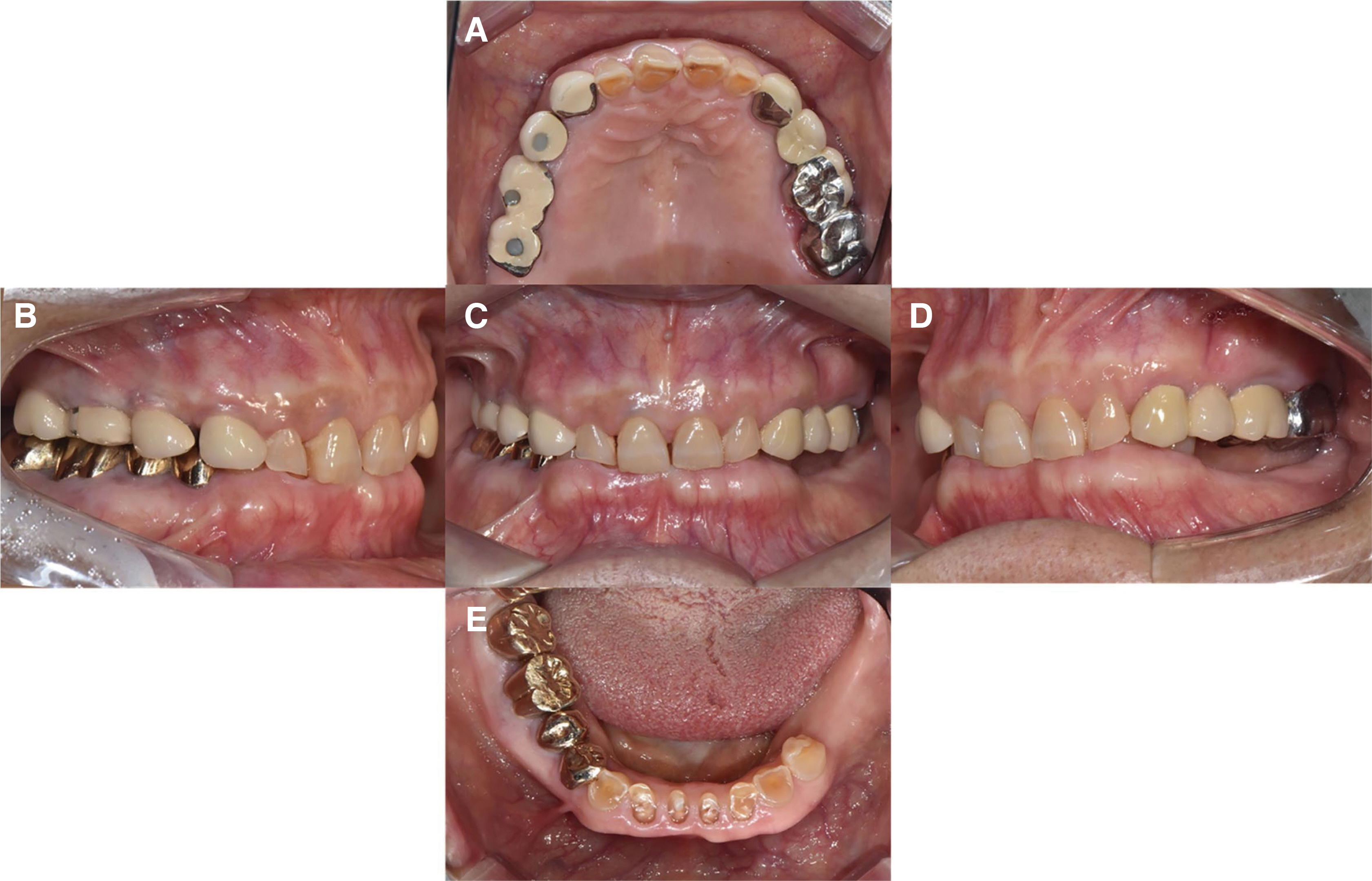
Fig. 1 Intraoral photograph before treatment. (A) Upper view, (B) Right view, (C) Front view, (D) Left view, (E) Lower view.

Fig. 2 Panoramic radiograph before treatment.

Fig. 3 Vertical dimension analysis. (A) Frontal view; upper facial height is measured from the pupil of the eye (IPL: interpupillary line) to the rima oris (ICL: intercomissural line), lower facial height is measured from the base of the nose to the lower border of the chin (B) Lateral view, (C) Distance between the vestibules.
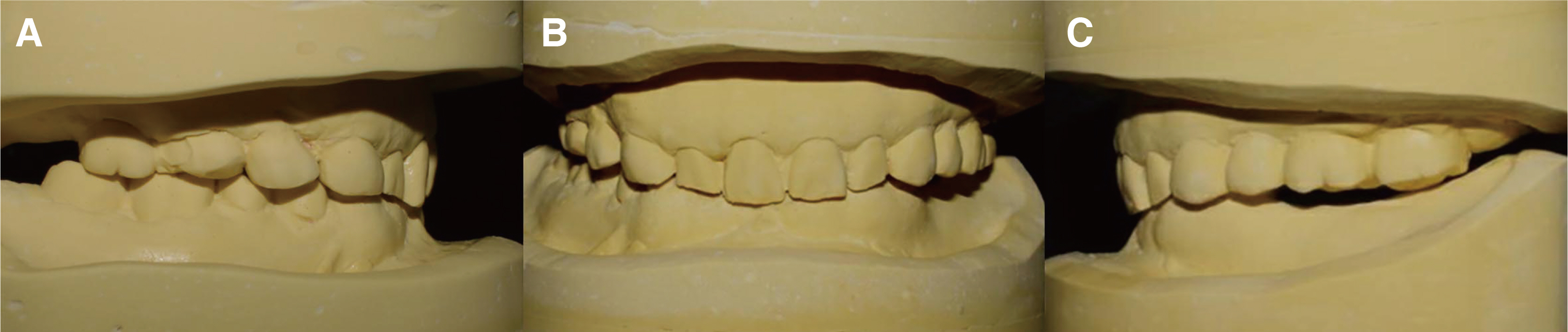
Fig. 4 Diagnostic model. (A) Right side view, (B) Frontal view, (C) Left side view.

Fig. 5 Diagnostic wax up model with increased vertical dimension. (A) Right side view, (B) Frontal view, (C) Left side view, (D) Frontal view of mandible, (E) Lower view of mandible.
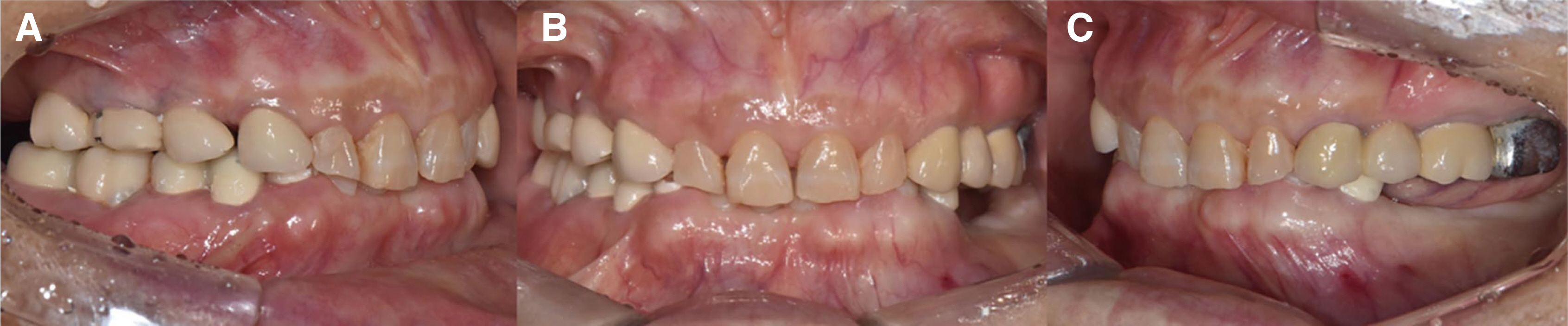
Fig. 6 Provisional restoration. (A) Right side view, (B) Frontal view, (C) Left side view.
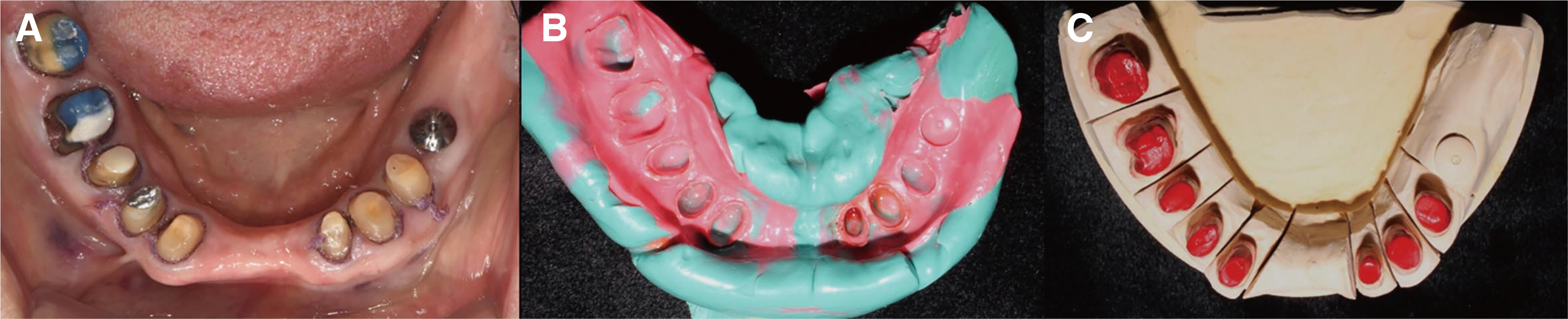
Fig. 7 (A) Tooth preparation, (B) Definitive impression taking, (C) Die preparation.

Fig. 8 (A) Facebow transfer, (B) Maxillary cast mounting using facebow transfer record.

Fig. 9 (A) Full contour wax up, (B) Cut back, (C) Metal coping try in.

Fig. 10 Definitive prosthesis. (A) Upper view, (B) Right view, (C) Front view, (D) Left view, (E) Lower view.
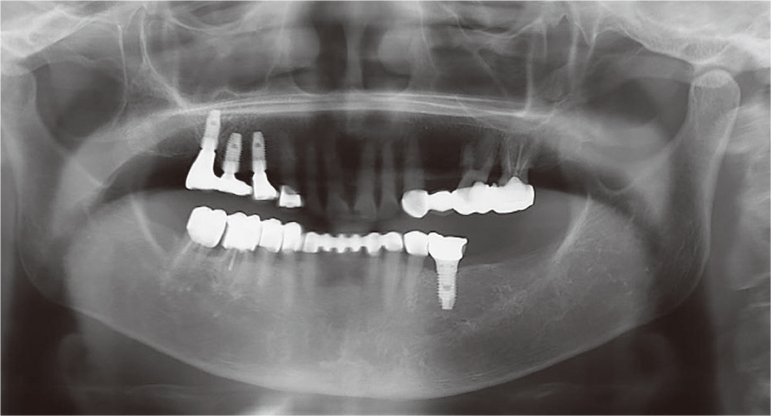
Fig. 11 Panoramic radiograph after treatment.
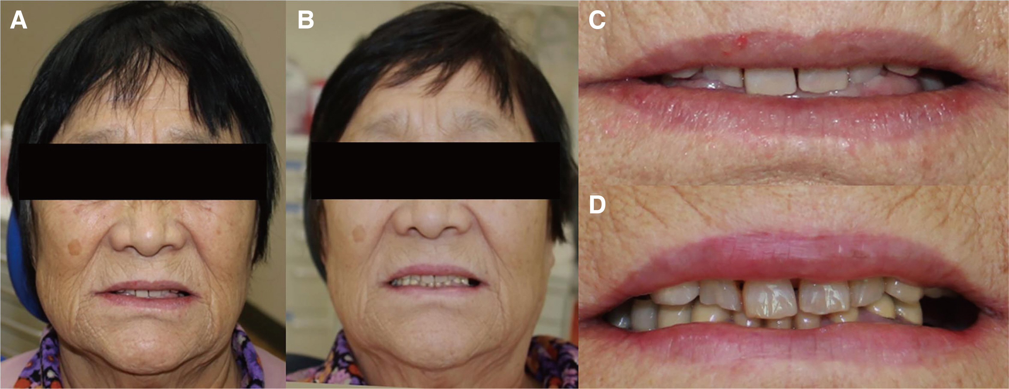
Fig. 12 Extra-oral photograph. (A) Facial photo before treatment, (B) Facial photo after treatment, (C) Smile photo before treatment, (D) Smile photo after treatment.
Reference
-
References
1. Dawson PE. 2007. Functional occlusion: from TMJ to smile design. Mosby;St. Louis: p. 430–52.2. Turner KA, Missirlian DM. 1984; Restoration of the extremely worn dentition. J Prosthet Dent. 52:467–74. DOI: 10.1016/0022-3913(84)90326-3. PMID: 6389829.
Article3. Johnson A, Wildgoose DG, Wood DJ. 2002; The determination of freeway space using two different methods. J Oral Rehabil. 29:1010–3. DOI: 10.1046/j.1365-2842.2002.00950.x. PMID: 12421334.
Article4. Willis FM. 1935; Features of the face involved in full denture prosthesis. Dent Cosmos. 77:851–4.5. Park JH, Jeong CM, Jeon YC, Lim JS. 2005; A study on the occlusal plane and the vertical dimension in Korean adults with natural dentition. J Korean Acad Prosthodont. 43:41–51.6. Silverman MM. 2001; The speaking method in measuring vertical dimension. 1952. J Prosthet Dent. 85:427–31. DOI: 10.1067/mpr.2001.116139. PMID: 11357066.7. Carpentieri J, Greenstein G, Cavallaro J. 2019; Hierarchy of restorative space required for different types of dental implant prostheses. J Am Dent Assoc. 150:695–706. DOI: 10.1016/j.adaj.2019.04.015. PMID: 31352966.
Article8. Donachie MA, Walls AW. 1995; Assessment of tooth wear in an ageing population. J Dent. 23:157–64. DOI: 10.1016/0300-5712(95)93573-K. PMID: 7782527.
Article9. Luke DA, Lucas PW. 1983; The significance of cusps. J Oral Rehabil. 10:197–206. DOI: 10.1111/j.1365-2842.1983.tb00113.x. PMID: 6575161.
Article10. Ingle JI. 1960; Alveolar osteoporosis and pulpal death associated with compulsive bruxism. Oral Surg Oral Med Oral Pathol. 13:1371–81. DOI: 10.1016/0030-4220(60)90302-9. PMID: 13717699.
Article11. Rivera-Morales WC, Mohl ND. 1991; Relationship of occlusal vertical dimension to the health of the masticatory system. J Prosthet Dent. 65:547–53. DOI: 10.1016/0022-3913(91)90298-B. PMID: 2066895.
Article
- Full Text Links
-
- Actions
-
Cited
- CITED
-
- Close
- Share
-
- Similar articles
-
- Full mouth rehabilitation of the patient with severe tooth loss and tooth wear with vertical dimension gaining: A case report
- Full mouth rehabilitation of patient with decreased occlusal vertical dimension due to severely worn dentition and posterior bite collapse
- Full-mouth rehabilitation of the patient with severe tooth wear using all ceramic restorations
- Oral rehabilitation of excessive tooth wear patient usingzirconia fixed prosthesis with increased vertical dimension
- A case of full mouth rehabilitation in patient with loss of vertical dimension and deep bite due to tooth wear
