Spheno-Orbital Plasmacytoma as an Initial Presentation of Multiple Myeloma
- Affiliations
-
- 1Department of Neurosurgery, Pusan National University Hospital, Pusan National University College of Medicine, Busan, Korea
- KMID: 2534726
- DOI: http://doi.org/10.14791/btrt.2022.0026
Abstract
- Intracranial plasmacytoma is a rare neoplasm and a subtype of malignant plasma cell tumor. Most patients with plasma cell tumors are diagnosed with multiple myeloma, but 5%–10% of patients are not. This report includes descriptions of radiologic and clinical findings in a patient with intracranial plasmacytoma. Intracranial extra-axial plasmacytomas can be easily misdiagnosed as meningioma in radiologic and clinical findings. A 69-year-old woman presented with exophthalmos and diplopia, and MRI indicated meningioma. Thus, she underwent gross total resection, and her pathologic diagnosis was plasmacytoma. Exophthalmos and diplopia were fully recovered. She was finally diagnosed with multiple myeloma based on systemic evaluation and treated with targeted chemotherapy. MRI conducted at 3 months after surgery showed no local recurrence or remnant tumor. Although intracranial plasmacytomas are difficult to distinguish from meningiomas in preoperative evaluation, gross total resection is recommended for the same purposes as meningiomas. If the pathologic diagnosis is a plasmacytoma, it is essential to have a systemic evaluation for multiple myeloma.
Keyword
Figure
-
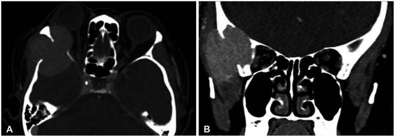
Fig. 1 Preoperative CT scanning. Axial (A) and coronal (B) views of brain CT revealed a mass lesion which has destroyed the lateral wall of the right orbit, zygomatic process of right frontal bone, and squamous part of the right temporal bone and extend to temporal muscle.
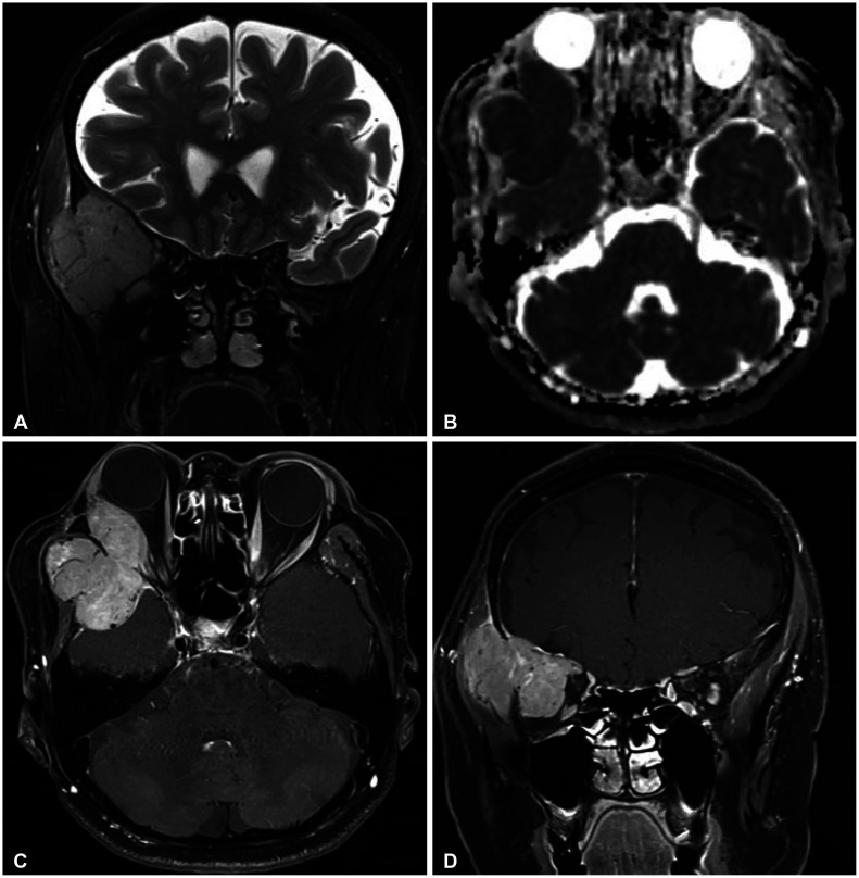
Fig. 2 Preoperative MRI. A: T2-weighted MR coronal image shows a lobulated extra-axial mass extend to exocranial space. B: Apparent diffusion coefficient map MR axial image shows low signal intensity, which indicate more aggressive tumor behavior. C and D: T1-weighted enhanced MR axial image demonstrates a homogenous hyperintense mass and thickened dura mate, which meningioma-like radiological appearance.
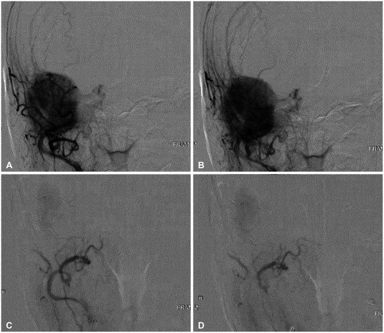
Fig. 3 Digital subtraction angiography. A and B: Pre-embolization angiography shows tumor-like vascular brush. C and D: Post-embolization angiography shows decreased blood flow.
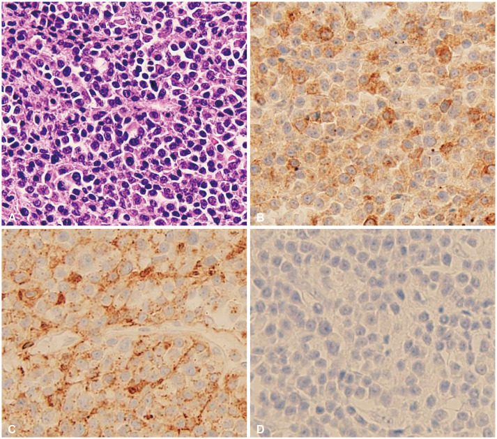
Fig. 4 Pathologic examination of the surgical specimen (×400). A: Hematoxylin-eosin staining shows high cellularity of well-differentiated plasma cells. B and C: Immunohistochemical staining shows diffuse staining CD79a and CD138. D: Epithelial membrane antigen staining shows negative staining.
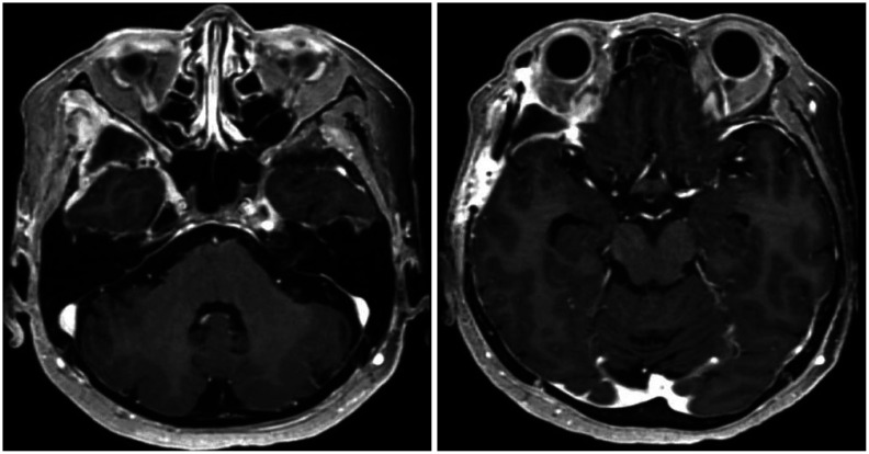
Fig. 5 Postoperative MRI. T1-weighted enhanced MR axial images shows no local recurrence or remnant tumor.
Reference
-
1. Soutar R, Lucraft H, Jackson G, Reece A, Bird J, Low E, et al. Guidelines on the diagnosis and management of solitary plasmacytoma of bone and solitary extramedullary plasmacytoma. Clin Oncol (R Coll Radiol). 2004; 16:405–413. PMID: 15487132.
Article2. Dimopoulos MA, Moulopoulos A, Delasalle K, Alexanian R. Solitary plasmacytoma of bone and asymptomatic multiple myeloma. Hematol Oncol Clin North Am. 1992; 6:359–369. PMID: 1582978.
Article3. Kilciksiz S, Karakoyun-Celik O, Agaoglu FY, Haydaroglu A. A review for solitary plasmacytoma of bone and extramedullary plasmacytoma. ScientificWorldJournal. 2012; 2012:895765. PMID: 22654647.
Article4. Rahmah N, Brotoarianto H, Andor E, Kusnarto G, Muttaqin Z, Hongo K. Dural plasmacytoma mimicking meningioma in a young patient with multiple myeloma. Biomed Imaging Interv J. 2009; 5:e5. PMID: 21611030.
Article5. Seetahal-Maraj P, Knight P, Ramnarine N. Proptosis secondary to a solitary plasmacytoma of the sphenoid bone: a case report on a rare skull base tumour. Egypt J Neurosurg. 2022; 37:4.
Article6. Sahin F, Saydam G, Ertan Y, Calli C, Dönmez A, Tombuloglu M. Dural plasmacytoma mimicking meningioma in a patient with multiple myeloma. J Clin Neurosci. 2006; 13:259–261. PMID: 16459088.
Article7. Hasturk AE, Basmaci M, Erten F, Cesur N, Yilmaz ER, Kertmen H. Solitary dural plasmacytoma mimicking meningioma and invading calvarium. J Craniofac Surg. 2013; 24:e175–e177. PMID: 23524828.
Article8. Singh AD, Chacko AG, Chacko G, Rajshekhar V. Plasma cell tumors of the skull. Surg Neurol. 2005; 64:434–438. discussion 438-9. PMID: 16253694.
Article9. Bindal AK, Bindal RK, van Loveren H, Sawaya R. Management of intracranial plasmacytoma. J Neurosurg. 1995; 83:218–221. PMID: 7616264.
Article10. Dimopoulos MA, Kiamouris C, Moulopoulos LA. Solitary plasmacytoma of bone and extramedullary plasmacytoma. Hematol Oncol Clin North Am. 1999; 13:1249–1257. PMID: 10626148.
Article11. Haegelen C, Riffaud L, Bernard M, Carsin-Nicol B, Morandi X. Dural plasmacytoma revealing multiple myeloma. Case report. J Neurosurg. 2006; 104:608–610. PMID: 16619666.
- Full Text Links
-
- Actions
-
Cited
- CITED
-
- Close
- Share
-
- Similar articles
-
- Rapid Progression of Solitary Plasmacytoma to Multiple Myeloma in Lumbar Vertebra
- A Case of Cutaneous Plasmacytoma Treated with Thalidomide
- Multiple Myeloma Presented With Unilateral Ptosis: A Case Report
- Clinical Application of (18)F-FDG PET in Multiple Myeloma
- A case of plasmacytoma of the testis associated with multiple myeloma
