Cell-cell contacts via N-cadherin induce a regulatory renin secretory phenotype in As4.1 cells
- Affiliations
-
- 1Department of Physiology, Asan Medical Center, University of Ulsan College of Medicine, Seoul 05505, Korea
- 2Department of Internal Medicine, Asan Medical Center, University of Ulsan College of Medicine, Seoul 05505, Korea
- 3Department of Internal Medicine, The Catholic University of Korea, Seoul St. Mary’s Hospital, Seoul 06591, Korea
- 4Department of Physiology, Jeonbuk National University Medical School, Jeonju 54907, Korea
- KMID: 2534524
- DOI: http://doi.org/10.4196/kjpp.2022.26.6.479
Abstract
- The lack of a clonal renin-secreting cell line has greatly hindered the investigation of the regulatory mechanisms of renin secretion at the cellular, biochemical, and molecular levels. In the present study, we investigated whether it was possible to induce phenotypic switching of the renin-expressing clonal cell line As4.1 from constitutive inactive renin secretion to regulated active renin secretion. When grown to postconfluence for at least two days in media containing fetal bovine serum or insulin-like growth factor-1, the formation of cell-cell contacts via N-cadherin triggered downstream cellular signaling cascades and activated smooth muscle-specific genes, culminating in phenotypic switching to a regulated active renin secretion phenotype, including responding to the key stimuli of active renin secretion. With the use of phenotype-switched As4.1 cells, we provide the first evidence that active renin secretion via exocytosis is regulated by phosphorylation/dephosphorylation of the 20 kDa myosin light chain. The molecular mechanism of phenotypic switching in As4.1 cells described here could serve as a working model for full phenotypic modulation of other secretory cell lines with incomplete phenotypes.
Keyword
Figure
-

Fig. 1 Growth curve and cadherin expression before (lane 1) and after confluence (lane 2). (A) Growth curve. Cells were plated (3 × 10³ cells/well) onto 24-well plates (day 0), cultured for 10 days and counted on a hemocytometer every 2–3 days (n = 3). (B) Phase contrast microscopy of ~70% and 100% confluent cells (×200). Scale bar: 60 μm. (C) Expression of cadherins. Protein (40 μg) obtained from cell lysates of cells grown to ~70% confluence (lane 1) or from cells on day 2 postconfluence (lane 2) were resolved by 7% acrylamide SDS-PAGE, followed by Western blotting with antibodies against epithelial cadherin (E-cad), placental cadherin (P-Cad), or neural cadherin (N-Cad). The numbers on the left indicate the molecular masses of standard proteins in kilo Daltons (kDa). The density of the bands was compared by unpaired Student’s tests (n = 3 for each group). β-actin was used as the protein loading control for each gel. NS, no significant difference. (D) N-cad expression at ~70% confluence (upper panel) and 100% confluence (lower panel). Cell nuclei were stained with propidium iodide (DAPI). Scale bar: 10 μm.
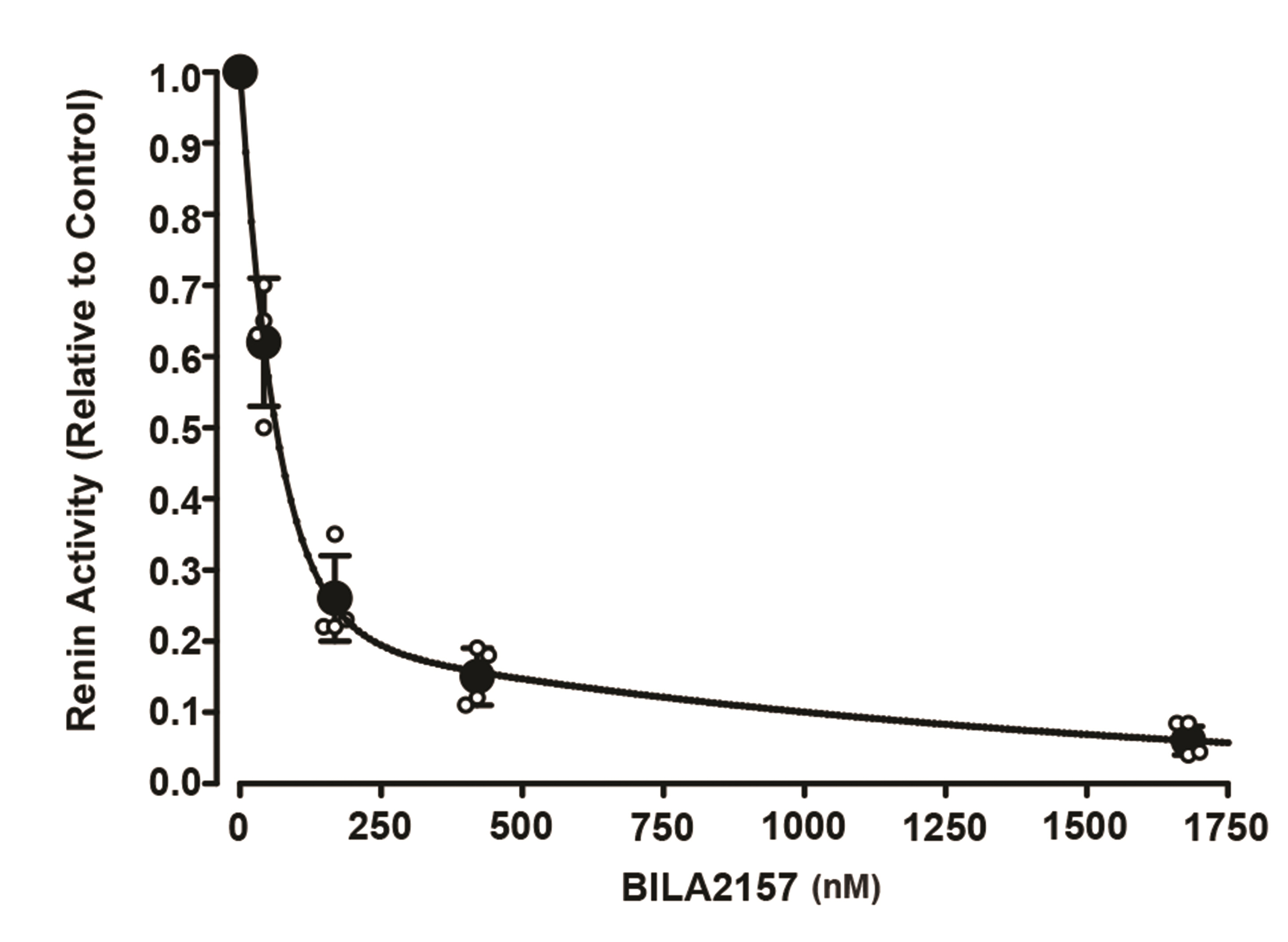
Fig. 2 BILA 2157 BS inhibits renin activity. Incubation media from postconfluent cells with a renin activity of 268.3 ± 51 ng ANG I/ml/h (relative value 1.0) was incubated with varying concentrations of BILA 2157 BS for 1 h. Active renin activity was determined by radioimmunoassay for ANG I. Each data point represents the mean ± SD of three samples. ANG I, angiotensin I.

Fig. 3 Effects of Ca2+, calmidazolium, ML-7, isoproterenol and forskolin on active renin secretion in postconfluent As4.1 cells. Cells on day 2 postconfluence were incubated in Ca2+-free DMEM containing 1 mM EGTA (–Ca2+ DMEM) (A, left panel) and then in 2 mM Ca2+-containing DMEM and 10% FBS (+Ca2+ DMEM) successively for 1 h each or in the reverse order (A, right panel, p < 0.001, n = 6). In the next series of experiments, cells were incubated in +Ca2+ DMEM for 1 h (control) and then another 1 h in +Ca2+ DMEM + 10 % FBS containing either calmidazolium (3 × 10⁻⁵ M) (B, n = 6), ML-7 (6 × 10⁻⁵M) (C, n = 6), isoproterenol (10−8–10−5 M) (D, n = 6) or forskolin (3 × 10⁻⁵ M) plus IBMX (10−4 M) (E, n = 6). The rate of active renin secretion was determined by radioimmunoassay for ANG I in (A) and (B) or by ELISA in (C), (D), and (E). ANG I, angiotensin I; DMEM, Dulbecco’s modified eagle medium; FBS, fetal bovine serum. Samples within groups were compared using paired Student’s t-tests and between groups using unpaired Student’s t-tests (A–C, E) or ANOVA (D), *p < 0.001 vs. control, except between samples at the two highest concentrations, which were compared by unpaired Student’s t-tests. †p < 0.001.
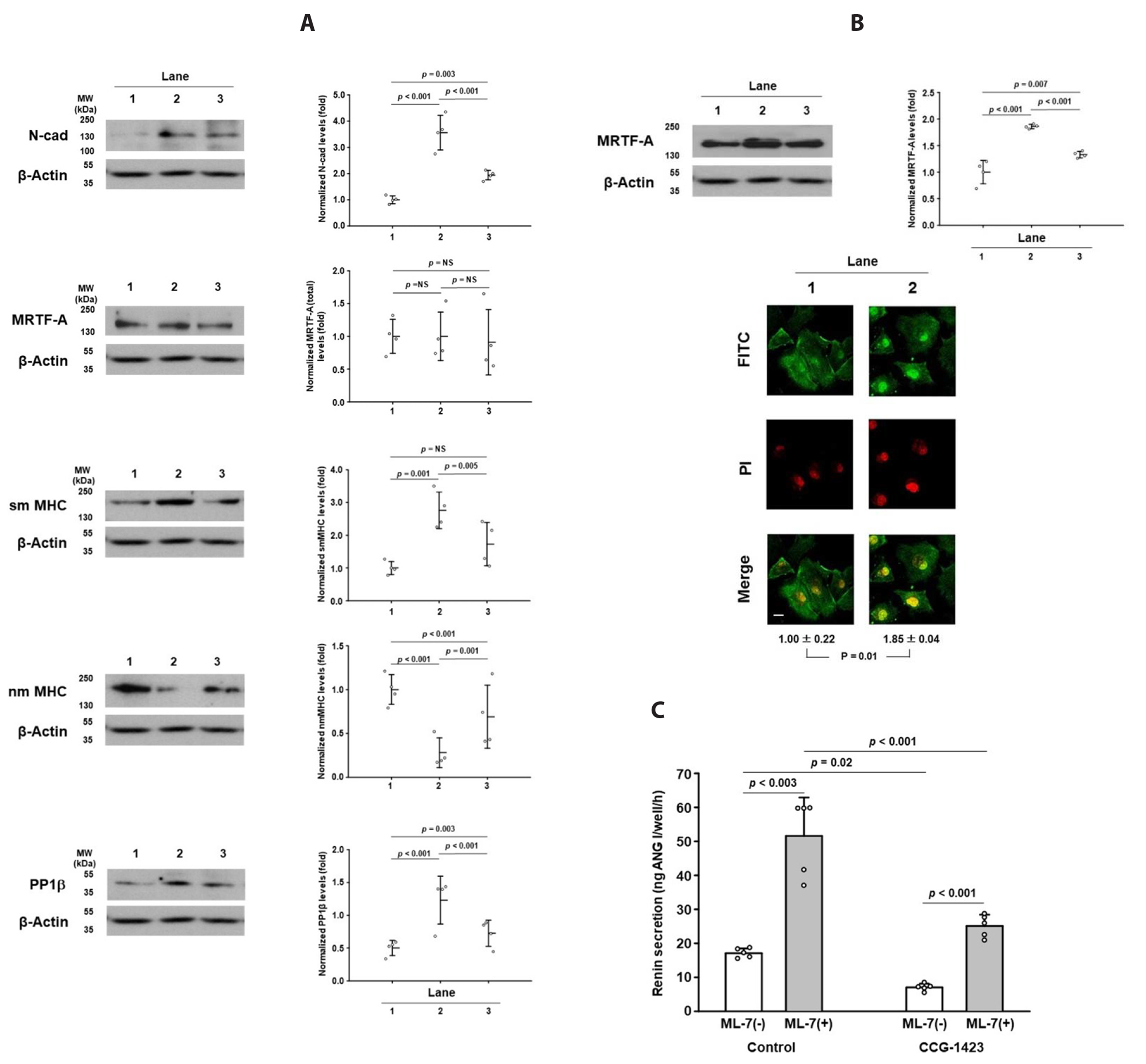
Fig. 4 Expression of N-cadherin, MRTF-A, sm MHC, nm MHC and PP1β changes to As4.1 cells before and after confluence. Cell lysates from cells grown to 70%–80% confluence in the presence of 10% FBS (A, lane 1), cells at day 2 postconfluence in the continuous presence of 10% FBS (A, lane 2) or in the absence of 10% FBS for two days after achieving 100% confluence (A, lane 3). After the aspiration of the incubation media, cells were lysed and centrifuged. Supernatant protein (60 μg) was resolved by SDS-PAGE on 7% or 15% acrylamide slab gels followed by Western blotting for N-cad, MRTF-A, sm MHC, nm MHC, and PP1β and then analyzed by densitometry. In the case of MRTF-A, a nuclear fraction was prepared, and 100 μg was resolved (B, upper panel). p-values were obtained by ANOVA. To assess nuclear localization of MRTF-A, cells were plated onto coverslips and fixed and permeabilized as described in the Methods section. Cells were then immunostained with primary MRTF-A antibody (1:100) and then with FITC-labelled goat anti-mouse secondary antibody to MRTF-A (1:1,000). Nuclei were stained with propidium iodide (DAPI) (B, lower panel). The density of preconfluent cells was arbitrarily set to 1. Fluorescence was analyzed by an image analyzer. Lane 1, preconfluent cells; lane 2, postconfluent cells in the presence of 10% FBS. p-values were obtained by unpaired Student’s t-tests (n = 4). Scale bar: 10 μM. (C) Postconfluent cells were incubated in DMEM + 10% FBS with or without CCG-1423 (5 × 10⁻⁶ M), an inhibitor of nuclear translocation of MRTF-A, for 1 h and then with ML-7 (6 × 10⁻⁵ M) for another 1 h. Secreted renin was measured by ELISA (C, n = 5). MRTF-A, myocardin related transcription factor-A; sm MHC, smooth muscle myosin heavy chain; nm MHC, nonmuscle myosin heavy chain; PP1β, protein phosphatase 1β; FBS, fetal bovine serum. NS, no significant difference. p-values within groups were obtained by paired Student’s t-tests, and those between groups were obtained by ANOVA.
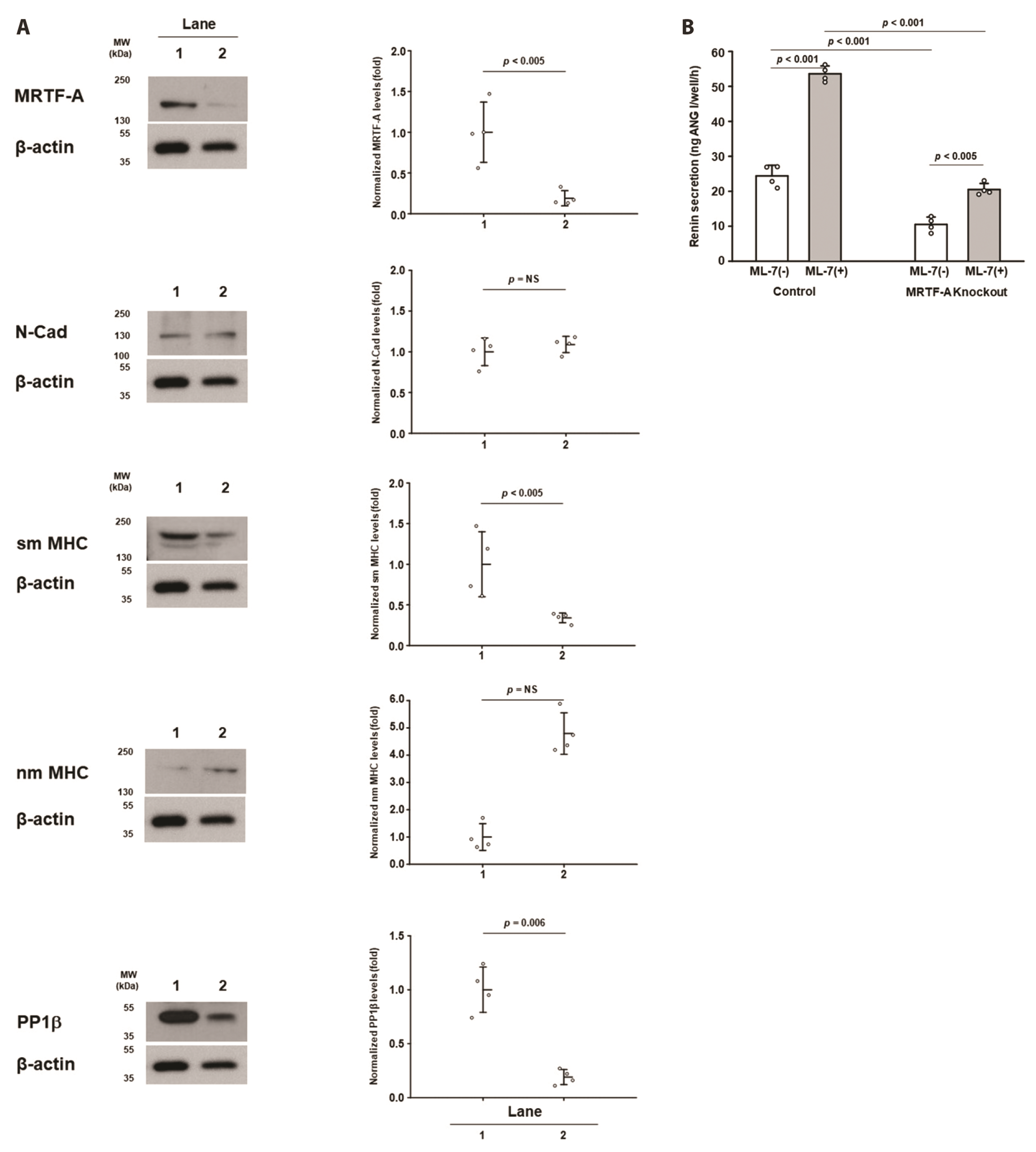
Fig. 5 Effects of MRTF-A knockout on phenotypic changes. Cells at ~70% confluence were transfected with control plasmids (A, lane 1) or the MRTF-A gene was knocked out using MRTF-A HDR CRISPR-associated 9 (Cas9) (A, lane 2) for three days. On day 2 postconfluence, the expression of MRTF-A, N-cad, sm MHC, nm MHC, and PP1β in the supernatant fraction (80 μg) was determined as described in the legend of . p-values were obtained by unpaired Student’s t-tests. (B) Control and knockout cells were incubated before and after stimulation with ML-7 (6 × 10⁻⁵ M) for 1 h each, and secreted active renin was measured by ELISA (B, n = 4). MRTF-A, myocardin related transcription factor-A; HDR, homology directed repair; CRISPR, clustered regularly interspaced short palindromic repeats; sm MHC, smooth muscle myosin heavy chain; nm MHC, nonmuscle myosin heavy chain; PP1β, protein phosphatase 1β. NS, no significant difference. p-values were obtained either by paired Student’s t-tests (within groups) or by ANOVA (between groups).
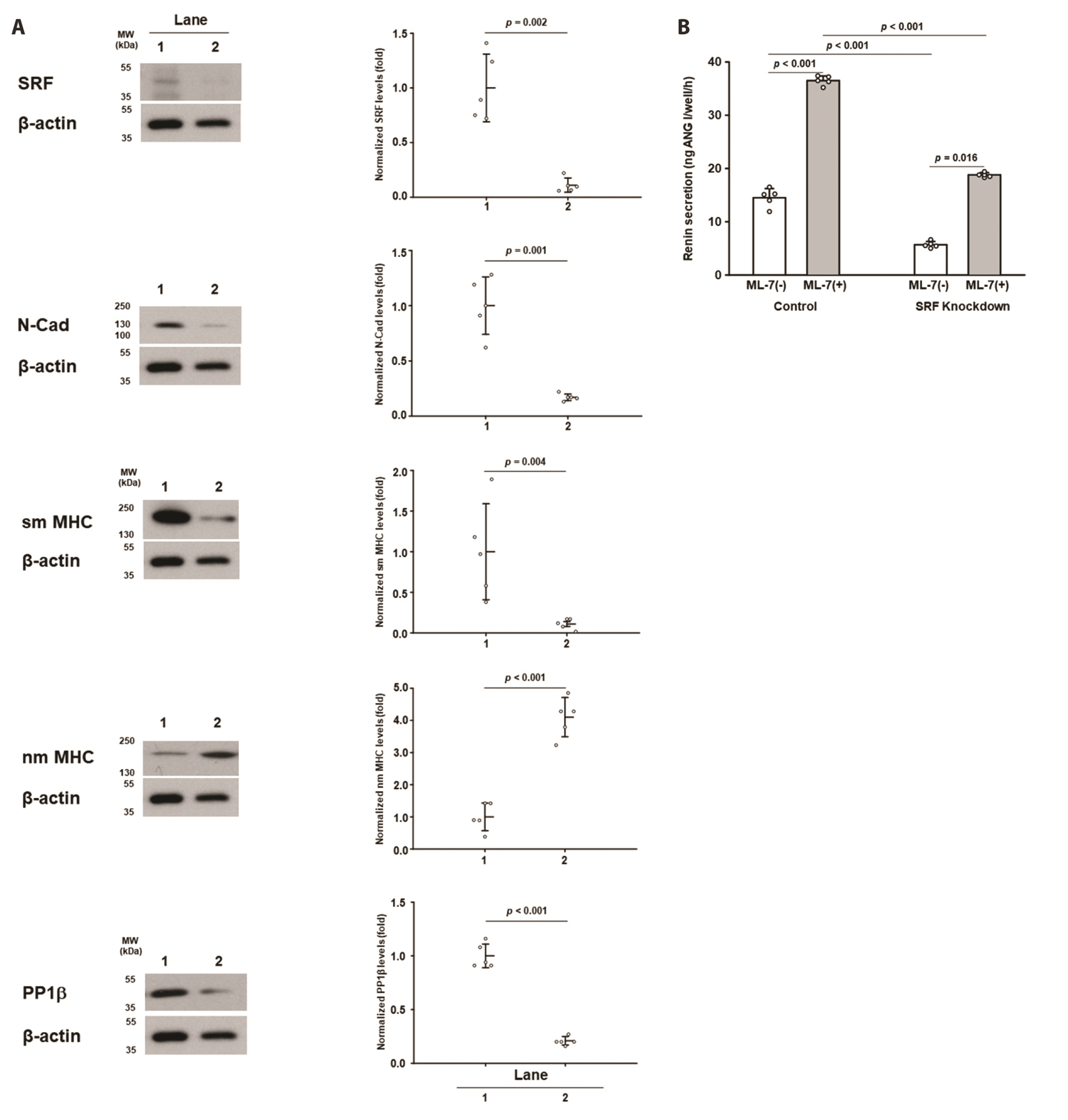
Fig. 6 Effects of SRF knockout on phenotypic changes. Cells were transfected with control plasmids (A, lane 1) or SRF HDR CRISPR/Cas9 plasmids (A, lane 2) as described in the legend of . Then, the expression of each protein was determined in the supernatant fraction (80 μg). Both control and knockout cells were incubated with ML-7 (6 × 10⁻⁵ M) before and after stimulation for 1 h each, and secreted active renin was measured by ELISA (B, n = 5). SRF, serum response factor; HDR, homology directed repair; CRISPR, clustered regularly interspaced short palindromic repeats. p-values were obtained either by paired Student’s t-tests (within groups) or by ANOVA (between groups).

Fig. 7 Effects of N-cad expression on pMLC20 and PP1β. Cells were cultured to 100% confluence and maintained for two more days in the presence of 10% FBS (lane 1) or in the presence of both FBS and IGF-1 antibody (10 μg/ml) (lane 2). Proteins in the supernatant (40 μg) were resolved by Western blotting as described above (n = 4). pMLC20, phosphorylated 20 kDa myosin light chain; PP1β, protein phosphatase 1β; FBS, fetal bovine serum; IGF-I, insulin-like growth factor-I. p-values were obtained by unpaired Student’s t-tests.
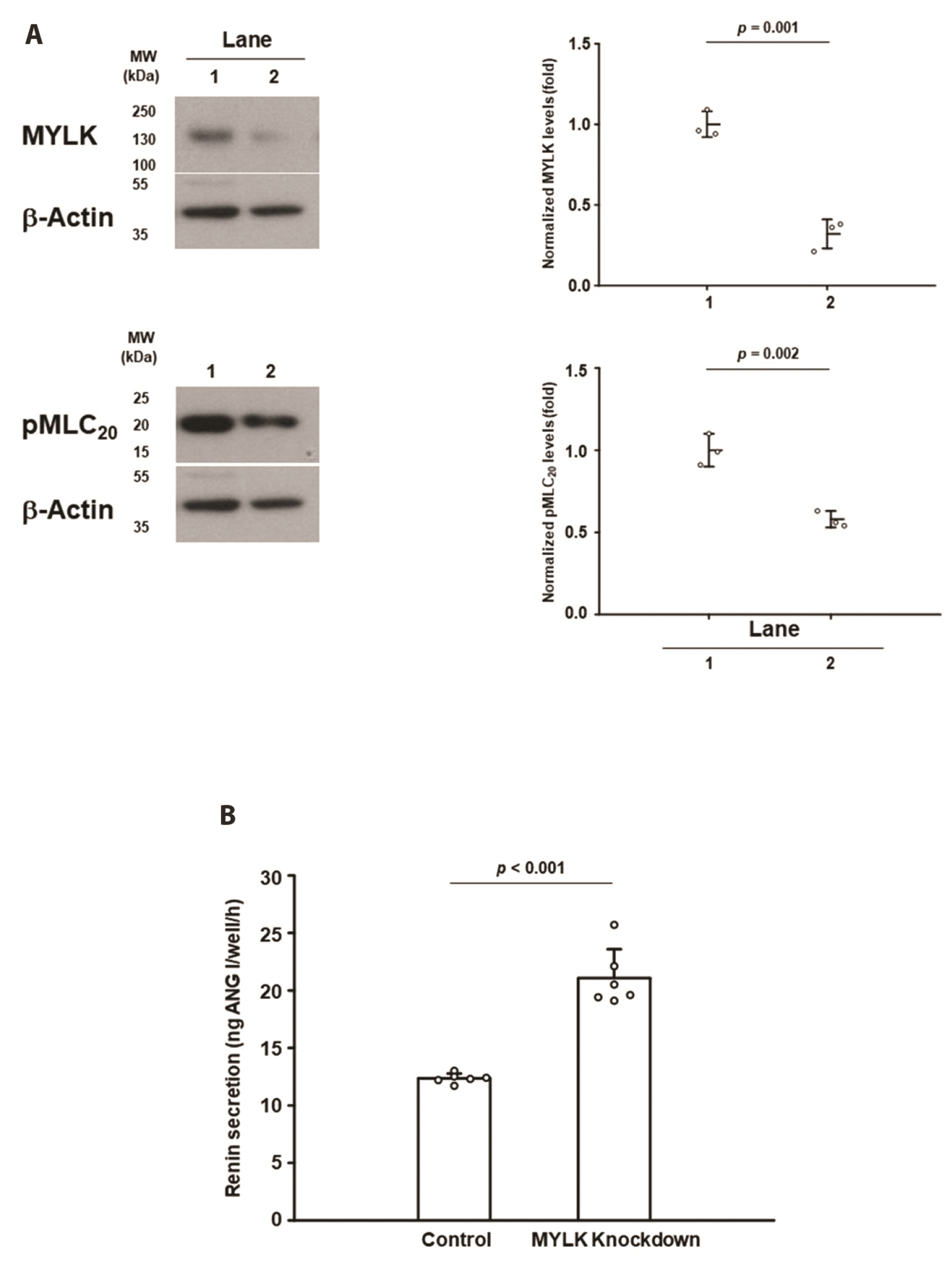
Fig. 8 Effects of MYLK knockdown on phenotypic changes. Cells were transfected with control or siRNA of MYLK plasmids as described in the legend of . Expression of MYLK and the cytosolic pMLC20 level (A) were determined using supernatant protein (60 μg). p-values were obtained by unpaired Student’s t-tests (n = 3). Active renin secretion from control and knockdown cells was determined by incubating cells in DMEM + 10% FBS for 1 h (B, n =6). pMLC20, phosphorylated 20 kDa myosin light chain; DMEM, Dulbecco’s modified eagle medium; FBS, fetal bovine serum. p-values by unpaired Student’s t-tests.
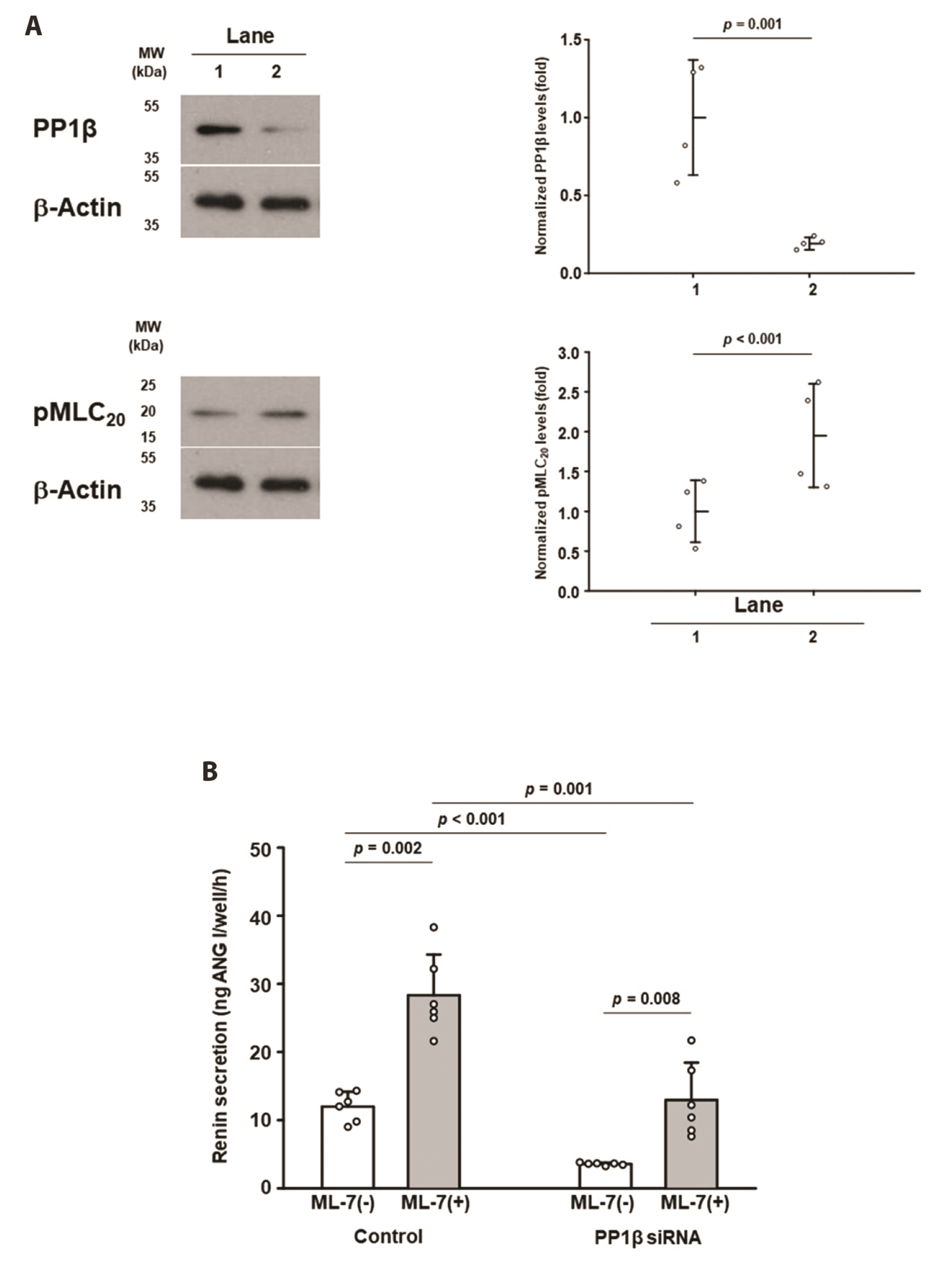
Fig. 9 Effects of PP1β knockdown on phenotypic changes. Cells were transfected with control (lane 1) or siRNA of PP1β plasmids as described in the legend for . The expression of PP1β (A, upper panel) and cytosolic pMLC20 level (A, lower panel) were determined using supernatant protein (60 μg). p-values were obtained by unpaired Student’s t-tests (n = 4). Active renin secretion was determined before and after stimulation by ML-7 (6 × 10⁻⁵ M) for 1 h each, and secreted renin was measured by ELISA (B). PP1β, protein phosphatase 1β; pMLC20, phosphorylated 20 kDa myosin light chain. p-values were obtained either by paired Student’s t-tests (within groups) or by ANOVA (between groups) (n = 6).

Fig. 10 Effects of ML-7 and calyculin A on the phosphorylation of MLC20. On day 2, postconfluent cells were incubated in DMEM + 10% FBS (control) in the presence of ML-7 (6 × 10⁻⁵ M; ML-7) and in the presence of both calyculin A (2 × 10⁻⁷ M) and ML-7 (6 × 10⁻⁵ M; ML-7 + Caly) for 1 h. The level of pMLC20 was determined using supernatant protein (80 μg). MLC20, 20 kDa myosin light chain; DMEM, Dulbecco’s modified eagle medium; FBS, fetal bovine serum. p-values were obtained by ANOVA (n = 5).

Fig. 11 Imaging of exocytotic discharge of renin and neutral red by ML-7. Cells on postconfluent day 2 were plated on cover slips were permeabilized and stained with antibody to renin. Many cytoplasmic stained granules are seen (A, control). When cells were pretreated with ML-7 (6 × 10⁻⁵ M) for 2 min and stained with renin antibody, no granules were observed (ML-7). Scale bar: 10 μm. Cells were incubated with 100 μM neutral red for 3 h, washed with fresh DMEM, and photographed with a light microscope. Numerous neutral red stained pink granules are seen (B). N denotes nucleus. Scale bar: 3.33 μm. (C, D) When neutral red-loaded granules were viewed at the isosbestic absorbent light at 470 nm, the granules observed were black. White granules are likely discharged granules prior to imaging. When cells were perfused with ML-7 (6 × 10⁻⁵ M), #1, #2, and #3 neutral red-loaded granules were discharged over 200 msec. (D) The time-lapsed movie shows the discharge of neutral red-loaded secretory granules (Supplementary Video). DMEM, Dulbecco’s modified eagle medium.
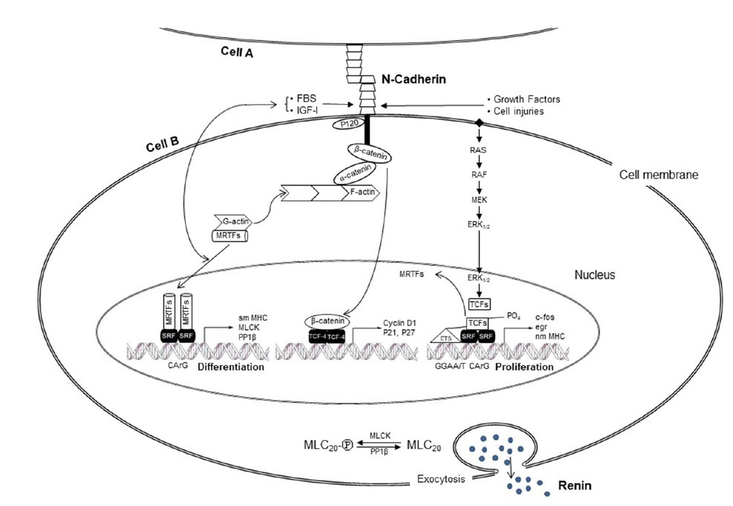
Fig. 12 Schematic summary of the transcriptional cascades regulating expression of proteins associated with renin secretory phenotypes in As4.1 cells. N-cad expression at the plasma membrane upon cell-cell contact triggers MRTF-A-SRF-CArG box, which activates transcription of smooth muscle-specific genes encoding contractile proteins in association with the regulatory phenotype of active renin secretion (left side). Without N-cad expression, β-catenin (signaling cascade in the middle) and growth factors (signaling cascade of right side) trigger a signaling cascade of growth factors of cell proliferation. MRTF-A, myocardin related transcription factor-A; SRF, serum response factor; MLC20, 20 kDa myosin light chain; PP1β, protein phosphatase 1β; FBS, fetal bovine serum; IGF-I, insulin-like growth factor-I; sm MHC, smooth muscle myosin heavy chain; nm MHC, nonmuscle myosin heavy chain.
Reference
-
1. Halban PA, Wollheim CB, Blondel B, Meda P, Niesor EN, Mintz DH. 1982; The possible importance of contact between pancreatic islet cells for the control of insulin release. Endocrinology. 111:86–94. DOI: 10.1210/endo-111-1-86. PMID: 6123433. PMID: https://www.scopus.com/inward/record.uri?partnerID=HzOxMe3b&scp=0020164058&origin=inward.
Article2. Hauge-Evans AC, Squires PE, Persaud SJ, Jones PM. 1999; Pancreatic beta-cell-to-beta-cell interactions are required for integrated responses to nutrient stimuli: enhanced Ca2+ and insulin secretory responses of MIN6 pseudoislets. Diabetes. 48:1402–1408. DOI: 10.2337/diabetes.48.7.1402. PMID: 10389845. PMID: https://www.scopus.com/inward/record.uri?partnerID=HzOxMe3b&scp=0032992378&origin=inward.
Article3. Lilla V, Webb G, Rickenbach K, Maturana A, Steiner DF, Halban PA, Irminger JC. 2003; Differential gene expression in well-regulated and dysregulated pancreatic beta-cell (MIN6) sublines. Endocrinology. 144:1368–1379. DOI: 10.1210/en.2002-220916. PMID: 12639920. PMID: https://www.scopus.com/inward/record.uri?partnerID=HzOxMe3b&scp=0037390423&origin=inward.
Article4. Hackenthal E, Paul M, Ganten D, Taugner R. 1990; Morphology, physiology, and molecular biology of renin secretion. Physiol Rev. 70:1067–1116. DOI: 10.1152/physrev.1990.70.4.1067. PMID: 2217555. PMID: https://www.scopus.com/inward/record.uri?partnerID=HzOxMe3b&scp=0025005906&origin=inward.
Article5. Cabandugama PK, Gardner MJ, Sowers JR. 2017; The renin angiotensin aldosterone system in obesity and hypertension: roles in the cardiorenal metabolic syndrome. Med Clin North Am. 101:129–137. DOI: 10.1016/j.mcna.2016.08.009. PMID: 27884224. PMCID: PMC5125542. PMID: https://www.scopus.com/inward/record.uri?partnerID=HzOxMe3b&scp=84996743333&origin=inward.6. Brunskill EW, Sequeira-Lopez ML, Pentz ES, Lin E, Yu J, Aronow BJ, Potter SS, Gomez RA. 2011; Genes that confer the identity of the renin cell. J Am Soc Nephrol. 22:2213–2225. DOI: 10.1681/ASN.2011040401. PMID: 22034642. PMCID: PMC3279933. PMID: https://www.scopus.com/inward/record.uri?partnerID=HzOxMe3b&scp=82655173696&origin=inward.
Article7. Della Bruna R, Kurtz A. 1995; Juxtaglomerular cells in culture. Exp Nephrol. 3:219–222. PMID: 8590034. PMID: https://www.scopus.com/inward/record.uri?partnerID=HzOxMe3b&scp=0029101966&origin=inward.8. Galen FX, Devaux C, Houot AM, Menard J, Corvol P, Corvol MT, Gubler MC, Mounier F, Camilleri JP. 1984; Renin biosynthesis by human tumoral juxtaglomerular cells. Evidences for a renin precursor. J Clin Invest. 73:1144–1155. DOI: 10.1172/JCI111300. PMID: 6323535. PMCID: PMC425128. PMID: https://www.scopus.com/inward/record.uri?partnerID=HzOxMe3b&scp=0021270652&origin=inward.
Article9. Pinet F, Corvol MT, Dench F, Bourguignon J, Feunteun J, Menard J, Corvol P. 1985; Isolation of renin-producing human cells by transfection with three simian virus 40 mutants. Proc Natl Acad Sci U S A. 82:8503–8507. DOI: 10.1073/pnas.82.24.8503. PMID: 3001706. PMCID: PMC390944. PMID: https://www.scopus.com/inward/record.uri?partnerID=HzOxMe3b&scp=0022371679&origin=inward.
Article10. Sigmund CD, Okuyama K, Ingelfinger J, Jones CA, Mullins JJ, Kane C, Kim U, Wu CZ, Kenny L, Rustum Y, Dzau VJ, Gross KW. 1990; Isolation and characterization of renin-expressing cell lines from transgenic mice containing a renin-promoter viral oncogene fusion construct. J Biol Chem. 265:19916–19922. DOI: 10.1016/S0021-9258(17)45460-3. PMID: 2174057.
Article11. Grünberger C, Obermayer B, Klar J, Kurtz A, Schweda F. 2006; The calcium paradoxon of renin release: calcium suppresses renin exocytosis by inhibition of calcium-dependent adenylate cyclases AC5 and AC6. Circ Res. 99:1197–1206. DOI: 10.1161/01.RES.0000251057.35537.d3. PMID: 17068292. PMID: https://www.scopus.com/inward/record.uri?partnerID=HzOxMe3b&scp=33751335626&origin=inward.12. Jensen BL, Lehle U, Müller M, Wagner C, Kurtz A. 1998; Interleukin-1 inhibits renin gene expression in As4.1 cells but not in native juxtaglomerular cells. Pflugers Arch. 436:673–678. DOI: 10.1007/s004240050688. PMID: 9716699. PMID: https://www.scopus.com/inward/record.uri?partnerID=HzOxMe3b&scp=0031693541&origin=inward.
Article13. Jones CA, Petrovic N, Novak EK, Swank RT, Sigmund CD, Gross KW. 1997; Biosynthesis of renin in mouse kidney tumor As4.1 cells. Eur J Biochem. 243:181–190. DOI: 10.1111/j.1432-1033.1997.0181a.x. PMID: 9030738. PMID: https://www.scopus.com/inward/record.uri?partnerID=HzOxMe3b&scp=0031019662&origin=inward.
Article14. Klar J, Sandner P, Müller MW, Kurtz A. 2002; Cyclic AMP stimulates renin gene transcription in juxtaglomerular cells. Pflugers Arch. 444:335–344. DOI: 10.1007/s00424-002-0818-9. PMID: 12111241. PMID: https://www.scopus.com/inward/record.uri?partnerID=HzOxMe3b&scp=0036448341&origin=inward.
Article15. Laframboise M, Reudelhuber TL, Jutras I, Brechler V, Seidah NG, Day R, Gross KW, Deschepper CF. 1997; Prorenin activation and prohormone convertases in the mouse As4.1 cell line. Kidney Int. 51:104–109. DOI: 10.1038/ki.1997.13. PMID: 8995723. PMID: https://www.scopus.com/inward/record.uri?partnerID=HzOxMe3b&scp=0031042265&origin=inward.
Article16. Peti-Peterdi J, Fintha A, Fuson AL, Tousson A, Chow RH. 2004; Real-time imaging of renin release in vitro. Am J Physiol Renal Physiol. 287:F329–F335. DOI: 10.1152/ajprenal.00420.2003. PMID: 15082450. PMID: https://www.scopus.com/inward/record.uri?partnerID=HzOxMe3b&scp=3242716881&origin=inward.
Article17. Beierwaltes WH. 2012; Hydrogen sulfide, renin, and regulating the second messenger cAMP. Focus on "Hydrogen sulfide regulates cAMP homeostasis and renin degranulation in As4.1 and rat renin-rich kidney cell.". Am J Physiol Cell Physiol. 302:C21–C23. DOI: 10.1152/ajpcell.00375.2011. PMID: 21998138. PMCID: PMC3328909. PMID: https://www.scopus.com/inward/record.uri?partnerID=HzOxMe3b&scp=83455223699&origin=inward.18. Chamley-Campbell JH, Campbell GR. 1981; What controls smooth muscle phenotype? Atherosclerosis. 40:347–357. https://doi.org/10.1016/0021-9150(81)90145-3. DOI: 10.1016/0021-9150(81)90145-3. PMID: https://www.scopus.com/inward/record.uri?partnerID=HzOxMe3b&scp=0019458817&origin=inward.
Article19. Owens GK, Kumar MS, Wamhoff BR. 2004; Molecular regulation of vascular smooth muscle cell differentiation in development and disease. Physiol Rev. 84:767–801. DOI: 10.1152/physrev.00041.2003. PMID: 15269336. PMID: https://www.scopus.com/inward/record.uri?partnerID=HzOxMe3b&scp=3042588831&origin=inward.
Article20. Kawamoto S, Adelstein RS. 1987; Characterization of myosin heavy chains in cultured aorta smooth muscle cells. A comparative study. J Biol Chem. 262:7282–7288. DOI: 10.1016/S0021-9258(18)48234-8. PMID: 2438275.
Article21. Martin KA, Rzucidlo EM, Merenick BL, Fingar DC, Brown DJ, Wagner RJ, Powell RJ. 2004; The mTOR/p70 S6K1 pathway regulates vascular smooth muscle cell differentiation. Am J Physiol Cell Physiol. 286:C507–C517. DOI: 10.1152/ajpcell.00201.2003. PMID: 14592809. PMID: https://www.scopus.com/inward/record.uri?partnerID=HzOxMe3b&scp=1442325390&origin=inward.
Article22. George SJ, Beeching CA. 2006; Cadherin:catenin complex: a novel regulator of vascular smooth muscle cell behaviour. Atherosclerosis. 188:1–11. DOI: 10.1016/j.atherosclerosis.2005.12.017. PMID: 16438974. PMID: https://www.scopus.com/inward/record.uri?partnerID=HzOxMe3b&scp=33746478732&origin=inward.
Article23. Quasnichka H, Slater SC, Beeching CA, Boehm M, Sala-Newby GB, George SJ. 2006; Regulation of smooth muscle cell proliferation by beta-catenin/T-cell factor signaling involves modulation of cyclin D1 and p21 expression. Circ Res. 99:1329–1337. DOI: 10.1161/01.RES.0000253533.65446.33. PMID: 17122440. PMID: https://www.scopus.com/inward/record.uri?partnerID=HzOxMe3b&scp=33845620229&origin=inward.
Article24. Uglow EB, Slater S, Sala-Newby GB, Aguilera-Garcia CM, Angelini GD, Newby AC, George SJ. 2003; Dismantling of cadherin-mediated cell-cell contacts modulates smooth muscle cell proliferation. Circ Res. 92:1314–1321. DOI: 10.1161/01.RES.0000079027.44309.53. PMID: 12775583. PMID: https://www.scopus.com/inward/record.uri?partnerID=HzOxMe3b&scp=0038801626&origin=inward.
Article25. Gurney EG, Gurney T Jr. 1979; Density dependent inhibition of both growth and T-antigen expression in revertants isolated from simian virus 40-transformed mouse SVT2 cells. J Virol. 32:667–671. DOI: 10.1128/jvi.32.2.667-671.1979. PMID: 228083. PMCID: PMC353598. PMID: https://www.scopus.com/inward/record.uri?partnerID=HzOxMe3b&scp=0018538248&origin=inward.
Article26. Ganot N, Meker S, Reytman L, Tzubery A, Tshuva EY. 2013; Anticancer metal complexes: synthesis and cytotoxicity evaluation by the MTT assay. J Vis Exp. (81):e50767. DOI: 10.3791/50767. PMID: 24300943. PMCID: PMC3989495. PMID: https://www.scopus.com/inward/record.uri?partnerID=HzOxMe3b&scp=84903080854&origin=inward.
Article27. Park CS, Lee HS, Chang SH, Honeyman TW, Hong CD. 1996; Inhibitory effect of Ca2+ on renin secretion elicited by chemiosmotic stimuli through actomyosin mediation. Am J Physiol. 271(1 Pt 1):C248–C254. DOI: 10.1152/ajpcell.1996.271.1.C248. PMID: 8760053. PMID: https://www.scopus.com/inward/record.uri?partnerID=HzOxMe3b&scp=0030192099&origin=inward.28. Ludowyke RI, Peleg I, Beaven MA, Adelstein RS. 1989; Antigen-induced secretion of histamine and the phosphorylation of myosin by protein kinase C in rat basophilic leukemia cells. J Biol Chem. 264:12492–12501. DOI: 10.1016/S0021-9258(18)63885-2. PMID: 2473073.
Article29. Bradford MM. 1976; A rapid and sensitive method for the quantitation of microgram quantities of protein utilizing the principle of protein-dye binding. Anal Biochem. 72:248–254. DOI: 10.1016/0003-2697(76)90527-3. PMID: 942051. PMID: https://www.scopus.com/inward/record.uri?partnerID=HzOxMe3b&scp=0017184389&origin=inward.
Article30. Harada K. 1954; Histochemical studies of the juxta glomerular apparatus. Rev Belg Pathol Med Exp. 23:311–320. PMID: 14372543. PMID: https://www.scopus.com/inward/record.uri?partnerID=HzOxMe3b&scp=77049215216&origin=inward.31. Knudsen KA, Frankowski C, Johnson KR, Wheelock MJ. 1998; A role for cadherins in cellular signaling and differentiation. J Cell Biochem. 72(S30-S31):168–176. DOI: 10.1002/(SICI)1097-4644(1998)72:30/31+<168::AID-JCB21>3.0.CO;2-V. PMID: 29345834.
Article32. Saitoh M, Ishikawa T, Matsushima S, Naka M, Hidaka H. 1987; Selective inhibition of catalytic activity of smooth muscle myosin light chain kinase. J Biol Chem. 262:7796–7801. DOI: 10.1016/S0021-9258(18)47638-7. PMID: 3108259.
Article33. Park CS, Kim MH, Leem CH, Jang YJ, Kim HW, Kim HS, et al. 1998; Inhibitory effect of calyculin A, a Ser/Thr protein phosphatase type I inhibitor, on renin secretion. Am J Physiol. 275:F664–F670. DOI: 10.1152/ajprenal.1998.275.5.F664. PMID: 9815125. PMID: https://www.scopus.com/inward/record.uri?partnerID=HzOxMe3b&scp=17344364413&origin=inward.
Article34. Martin KA, Merenick BL, Ding M, Fetalvero KM, Rzucidlo EM, Kozul CD, Brown DJ, Chiu HY, Shyu M, Drapeau BL, Wagner RJ, Powell RJ. 2007; Rapamycin promotes vascular smooth muscle cell differentiation through insulin receptor substrate-1/phosphatidylinositol 3-kinase/Akt2 feedback signaling. J Biol Chem. 282:36112–36120. DOI: 10.1074/jbc.M703914200. PMID: 17908691. PMID: https://www.scopus.com/inward/record.uri?partnerID=HzOxMe3b&scp=37249046846&origin=inward.
Article35. Park CS, Chang SH, Lee HS, Kim SH, Chang JW, Hong CD. 1996; Inhibition of renin secretion by Ca2+ through activation of myosin light chain kinase. Am J Physiol. 271(1 Pt 1):C242–C247. DOI: 10.1152/ajpcell.1996.271.1.C242. PMID: 8760052. PMID: https://www.scopus.com/inward/record.uri?partnerID=HzOxMe3b&scp=0043062950&origin=inward.36. Park CS, Sigmon DH, Han DS, Honeyman TW, Fray JC. 1986; Control of renin secretion by Ca2+ and cyclic AMP through two parallel mechanisms. Am J Physiol. 251(3 Pt 2):R531–R536. DOI: 10.1152/ajpregu.1986.251.3.R531. PMID: 3019164. PMID: https://www.scopus.com/inward/record.uri?partnerID=HzOxMe3b&scp=0023019413&origin=inward.37. Ishihara H, Martin BL, Brautigan DL, Karaki H, Ozaki H, Kato Y, Fusetani N, Watabe S, Hashimoto K, Uemura D, Hartshorne DJ. 1989; Calyculin A and okadaic acid: inhibitors of protein phosphatase activity. Biochem Biophys Res Commun. 159:871–877. DOI: 10.1016/0006-291X(89)92189-X. PMID: 2539153. PMID: https://www.scopus.com/inward/record.uri?partnerID=HzOxMe3b&scp=0024517280&origin=inward.
Article38. Kim MH, Kim SH, Kim HS, Chang JW, Hong YS, Kim HW, Park CS. 1998; Regulation of renin secretion through reversible phosphorylation of myosin by myosin light chain kinase and protein phosphatase type 1. J Pharmacol Exp Ther. 285:968–974. PMID: 9618396. PMID: https://www.scopus.com/inward/record.uri?partnerID=HzOxMe3b&scp=7144261705&origin=inward.39. Haber E, Koerner T, Page LB, Kliman B, Purnode A. 1969; Application of a radioimmunoassay for angiotensin I to the physiologic measurements of plasma renin activity in normal human subjects. J Clin Endocrinol Metab. 29:1349–1355. DOI: 10.1210/jcem-29-10-1349. PMID: 4311082. PMID: https://www.scopus.com/inward/record.uri?partnerID=HzOxMe3b&scp=0014584257&origin=inward.
Article40. Taugner R, Whalley A, Angermüller S, Bührle CP, Hackenthal E. 1985; Are the renin-containing granules of juxtaglomerular epithelioid cells modified lysosomes? Cell Tissue Res. 239:575–587. DOI: 10.1007/BF00219236. PMID: 3886148. PMID: https://www.scopus.com/inward/record.uri?partnerID=HzOxMe3b&scp=0021999739&origin=inward.
Article41. Faust PL, Kornfeld S, Chirgwin JM. 1985; Cloning and sequence analysis of cDNA for human cathepsin D. Proc Natl Acad Sci U S A. 82:4910–4914. DOI: 10.1073/pnas.82.15.4910. PMID: 3927292. PMCID: PMC390467. PMID: https://www.scopus.com/inward/record.uri?partnerID=HzOxMe3b&scp=0011288815&origin=inward.
Article42. Beaulieu PL, Gillard J, Bailey M, Beaulieu C, Duceppe JS, Lavallée P, Wernic D. 1999; Practical synthesis of BILA 2157 BS, a potent and orally active renin inhibitor: use of an enzyme-catalyzed hydrolysis for the preparation of homochiral succinic acid derivatives. J Org Chem. 64:6622–6634. DOI: 10.1021/jo990321x. PMID: 11674665. PMID: https://www.scopus.com/inward/record.uri?partnerID=HzOxMe3b&scp=0032885570&origin=inward.
Article43. Park CS, Honeyman TW, Chung ES, Lee JS, Sigmon DH, Fray JC. 1986; Involvement of calmodulin in mediating inhibitory action of intracellular Ca2+ on renin secretion. Am J Physiol. 251(6 Pt 2):F1055–F1062. DOI: 10.1152/ajprenal.1986.251.6.F1055. PMID: 3538904. PMID: https://www.scopus.com/inward/record.uri?partnerID=HzOxMe3b&scp=0023007879&origin=inward.44. Park CS, Malvin RL. 1978; Calcium in the control of renin release. Am J Physiol. 235:F22–F25. DOI: 10.1152/ajprenal.1978.235.1.F22. PMID: 98060. PMID: https://www.scopus.com/inward/record.uri?partnerID=HzOxMe3b&scp=18144444810&origin=inward.
Article45. Park CS, Hong CD, Honeyman TW. 1992; Calcium-dependent inhibitory step in control of renin secretion. Am J Physiol. 262(5 Pt 2):F793–F798. DOI: 10.1152/ajprenal.1992.262.5.F793. PMID: 1590424.
Article46. Van Belle H. 1981; R 24 571: a potent inhibitor of calmodulin-activated enzymes. Cell Calcium. 2:483–494. https://doi.org/10.1016/0143-4160(81)90007-5. DOI: 10.1016/0143-4160(81)90007-5. PMID: https://www.scopus.com/inward/record.uri?partnerID=HzOxMe3b&scp=0019718498&origin=inward.
Article47. Seamon KB, Daly JW. 1981; Forskolin: a unique diterpene activator of cyclic AMP-generating systems. J Cyclic Nucleotide Res. 7:201–224. PMID: 6278005. PMID: https://www.scopus.com/inward/record.uri?partnerID=HzOxMe3b&scp=0019811368&origin=inward.48. Miralles F, Posern G, Zaromytidou AI, Treisman R. 2003; Actin dynamics control SRF activity by regulation of its coactivator MAL. Cell. 113:329–342. DOI: 10.1016/S0092-8674(03)00278-2. PMID: 12732141. PMID: https://www.scopus.com/inward/record.uri?partnerID=HzOxMe3b&scp=0038737042&origin=inward.
Article49. Wang Z, Wang DZ, Pipes GC, Olson EN. 2003; Myocardin is a master regulator of smooth muscle gene expression. Proc Natl Acad Sci U S A. 100:7129–7134. DOI: 10.1073/pnas.1232341100. PMID: 12756293. PMCID: PMC165841. PMID: https://www.scopus.com/inward/record.uri?partnerID=HzOxMe3b&scp=0037795465&origin=inward.
Article50. Evelyn CR, Wade SM, Wang Q, Wu M, Iñiguez-Lluhí JA, Merajver SD, Neubig RR. 2007; CCG-1423: a small-molecule inhibitor of RhoA transcriptional signaling. Mol Cancer Ther. 6:2249–2260. DOI: 10.1158/1535-7163.MCT-06-0782. PMID: 17699722. PMID: https://www.scopus.com/inward/record.uri?partnerID=HzOxMe3b&scp=34548066554&origin=inward.
Article51. Cong L, Ran FA, Cox D, Lin S, Barretto R, Habib N, Hsu PD, Wu X, Jiang W, Marraffini LA, Zhang F. 2013; Multiplex genome engineering using CRISPR/Cas systems. Science. 339:819–823. DOI: 10.1126/science.1231143. PMID: 23287718. PMCID: PMC3795411. PMID: https://www.scopus.com/inward/record.uri?partnerID=HzOxMe3b&scp=84873729095&origin=inward.
Article52. Miano JM, Long X, Fujiwara K. 2007; Serum response factor: master regulator of the actin cytoskeleton and contractile apparatus. Am J Physiol Cell Physiol. 292:C70–C81. DOI: 10.1152/ajpcell.00386.2006. PMID: 16928770. PMID: https://www.scopus.com/inward/record.uri?partnerID=HzOxMe3b&scp=33846301667&origin=inward.
Article53. Heemskerk JW, Farndale RW, Sage SO. 1997; Effects of U73122 and U73343 on human platelet calcium signalling and protein tyrosine phosphorylation. Biochim Biophys Acta. 1355:81–88. DOI: 10.1016/S0167-4889(96)00113-9. PMID: 9030204. PMID: https://www.scopus.com/inward/record.uri?partnerID=HzOxMe3b&scp=0031037686&origin=inward.
Article54. Aldehni F, Tang T, Madsen K, Plattner M, Schreiber A, Friis UG, Hammond HK, Han PL, Schweda F. 2011; Stimulation of renin secretion by catecholamines is dependent on adenylyl cyclases 5 and 6. Hypertension. 57:460–468. DOI: 10.1161/HYPERTENSIONAHA.110.167130. PMID: 21282557. PMCID: PMC3106204. PMID: https://www.scopus.com/inward/record.uri?partnerID=HzOxMe3b&scp=79953241859&origin=inward.
Article55. Luetscher JA, Kraemer FB, Wilson DM, Schwartz HC, Bryer-Ash M. 1985; Increased plasma inactive renin in diabetes mellitus. A marker of microvascular complications. N Engl J Med. 312:1412–1417. DOI: 10.1056/NEJM198505303122202. PMID: 3887168. PMID: https://www.scopus.com/inward/record.uri?partnerID=HzOxMe3b&scp=0021889169&origin=inward.
Article56. Todaro GJ, Lazar GK, Green H. 1965; The initiation of cell division in a contact-inhibited mammalian cell line. J Cell Physiol. 66:325–333. DOI: 10.1002/jcp.1030660310. PMID: 5884360. PMID: https://www.scopus.com/inward/record.uri?partnerID=HzOxMe3b&scp=0013831072&origin=inward.
Article57. Nourse J, Firpo E, Flanagan WM, Coats S, Polyak K, Lee MH, Massague J, Crabtree GR, Roberts JM. 1994; Interleukin-2-mediated elimination of the p27Kip1 cyclin-dependent kinase inhibitor prevented by rapamycin. Nature. 372:570–573. DOI: 10.1038/372570a0. PMID: 7990932. PMID: https://www.scopus.com/inward/record.uri?partnerID=HzOxMe3b&scp=0028172867&origin=inward.
Article58. Posern G, Treisman R. 2006; Actin' together: serum response factor, its cofactors and the link to signal transduction. Trends Cell Biol. 16:588–596. DOI: 10.1016/j.tcb.2006.09.008. PMID: 17035020. PMID: https://www.scopus.com/inward/record.uri?partnerID=HzOxMe3b&scp=33750343214&origin=inward.
Article59. Wang D, Chang PS, Wang Z, Sutherland L, Richardson JA, Small E, Krieg PA, Olson EN. 2001; Activation of cardiac gene expression by myocardin, a transcriptional cofactor for serum response factor. Cell. 105:851–862. DOI: 10.1016/S0092-8674(01)00404-4. PMID: 11439182. PMID: https://www.scopus.com/inward/record.uri?partnerID=HzOxMe3b&scp=0035967868&origin=inward.
Article60. Wang Z, Wang DZ, Hockemeyer D, McAnally J, Nordheim A, Olson EN. 2004; Myocardin and ternary complex factors compete for SRF to control smooth muscle gene expression. Nature. 428:185–189. DOI: 10.1038/nature02382. PMID: 15014501. PMID: https://www.scopus.com/inward/record.uri?partnerID=HzOxMe3b&scp=1642297200&origin=inward.
Article61. Simons M, Rosenberg RD. 1992; Antisense nonmuscle myosin heavy chain and c-myb oligonucleotides suppress smooth muscle cell proliferation in vitro. Circ Res. 70:835–843. DOI: 10.1161/01.RES.70.4.835. PMID: 1551207. PMID: https://www.scopus.com/inward/record.uri?partnerID=HzOxMe3b&scp=0026536410&origin=inward.
Article
- Full Text Links
-
- Actions
-
Cited
- CITED
-
- Close
- Share
-
- Similar articles
-
- E-Cadherin Expression and DNA Ploidy Analysis in Invasive Squamous Cell Carcinoma of the Uterine Cervix Comparison with those of CIN
- Cyclooxygenase-2 Expression Is Related to the Epithelial-to-Mesenchymal Transition in Human Colon Cancers
- Regulatory T Cells in B Cell Follicles
- Changes in the Rate of Renin Secretion During Cell Cycle of As 4.1 Cells
- Role of Basal Cell and Secretory Cell in Benign Prostatic Hyperplasia and Prostatic Cancer
