Involvement of adaptor protein, phosphotyrosine interacting with PH domain and leucine zipper 1 in diallyl trisulfide-induced cytotoxicity in hepatocellular carcinoma cells
- Affiliations
-
- 1Department of Pathology, Renmin Hospital of Wuhan University, Wuhan 430060, China
- 2Department of Hepatobiliary & Laparascopic Surgery, Renmin Hospital of Wuhan University, Wuhan 430060, China
- 3Tongji Medical College Huazhong University of Science and Technology, School of Nursing, Wuhan 430030, China
- 4Department of Pathology and Pathophysiology, Wuhan University Taikang Medical School (School of Basic Medical Sciences), Wuhan 430071, China
- 5Department of Breast and Thyroid Surgery, Zhongnan Hospital of Wuhan University, Wuhan 430071, China
- KMID: 2534522
- DOI: http://doi.org/10.4196/kjpp.2022.26.6.457
Abstract
- It has been demonstrated that APPL1 (adaptor protein, phosphotyrosine interacting with PH domain and leucine zipper 1) is involved in the regulation of several growth-related signaling pathways and thus closely associated with the development and progression of some cancers. Diallyl trisulfide (DAT), a garlic-derived bioactive compound, exerts selective cytotoxicity to various human cancer cells through interfering with pro-survival signaling pathways. However, whether and how DAT affects survival of human hepatocellular carcinoma (HCC) cells remain unclear. Herein, we tested the hypothesis of the involvement of APPL1 in DAT-induced cytotoxicity in HCC HepG2 cells. We found that Lys 63 (K63)-linked polyubiquitination of APPL1 was significantly decreased whereas phosphorylation of APPL1 at serine residues remained unchanged in DAT-treated HepG2 cells. Compared with wild-type APPL1, overexpression of APPL1 K63R mutant dramatically increased cell apoptosis and mitigated cell survival, along with a reduction of phosphorylation of STAT3, Akt, and Erk1/2. In addition, DAT administration markedly reduced protein levels of intracellular TNF receptor-associated factor 6 (TRAF6). Genetic inhibition of TRAF6 decreased K63-linked polyubiquitination of APPL1. Moreover, the cytotoxicity impacts of DAT on HepG2 cells were greatly attenuated by overexpression of wild-type APPL1. Taken together, these results suggest that APPL1 polyubiquitination probably mediates the inhibitory effects of DAT on survival of HepG2 cells by modulating STAT3, Akt, and Erk1/2 pathways.
Figure
-
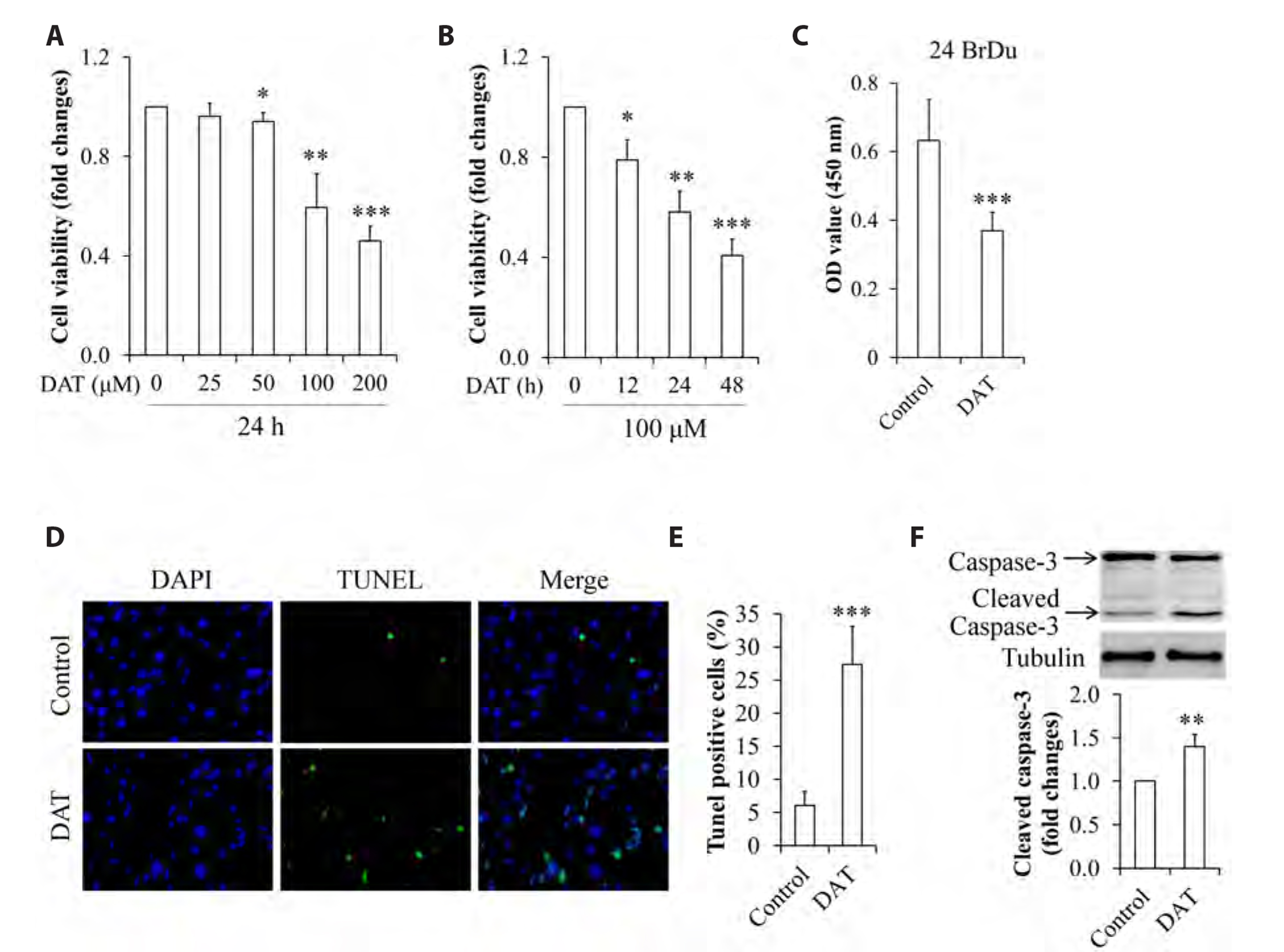
Fig. 1 Effect of DAT on cell survival and proliferation of HepG2 cells. (A) Dose-response of DAT on cell viability. (B) Time-response of DAT on cell viability. (C) Effect of DAT on cell proliferation. (D) Representative images of cell apoptosis by DAT (×400). (E) Quantitative analysis of cell apoptosis in (D). (F) Impact of DAT on cleaved caspase-3 levels. DAT, diallyl trisulfide. *p < 0.05, **p < 0.01, ***p < 0.001 vs. control group.
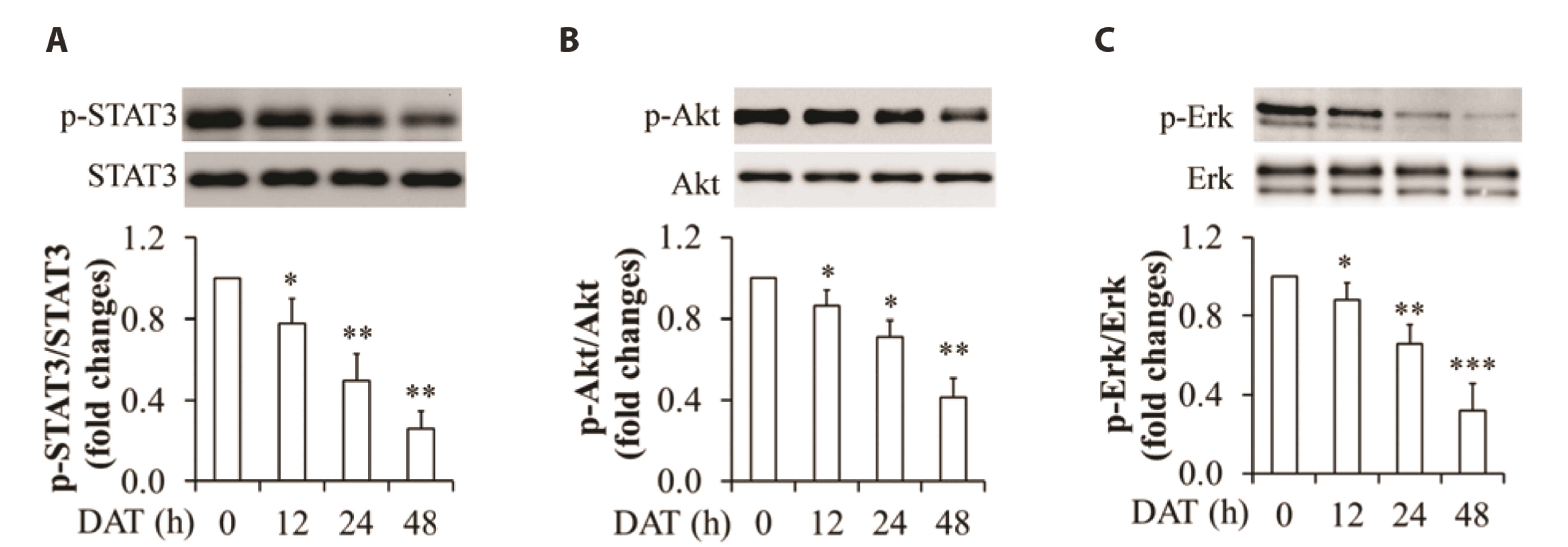
Fig. 2 Effect of DAT on phosphorylation of STAT3, Akt, and Erk1/2 in HepG2 cells. (A) Impact of DAT on STAT3 phosphorylation. (B) Impact of DAT on Akt phosphorylation. (C) Impact of DAT on Erk1/2 phosphorylation. Upper panels are representative Western blot images and bottom panels are quantitative analysis of the ratio of phosphorylated proteins to the related-proteins. DAT, diallyl trisulfide. *p < 0.05, **p < 0.01, ***p < 0.01 vs. control group.

Fig. 3 Effect of DAT on phosphorylation and polyubiquitination of APPL1 in HepG2 cells. (A) Impact of DAT on the levels of intracellular APPL1 protein. (B) Impact of DAT on serine phosphorylation of APPL1. APPL1 was immunoprecipitated by specific antibody and serine phosphorylation was then detected by western blot using a specific anti-serine antibody. (C) Impact of DAT on K63-linked polyubiquitination of APPL1. APPL1 was immunoprecipitated by specific antibody and polyubiquitination was detected by Western blot using a specific antibody against K63-linked polyubiquitination. (D) Quantitative analysis of K63-linked polyubiquitination of APPL1 in (C). (E) Impact of DAT on the levels of intracellular Nedd4 and TRAF6 proteins. (F) Quantitative analysis of TRAF6 levels in (E). (G) Impact of TRAF6 knockdown on K63-linked polyubiquitination of APPL1. DAT, diallyl trisulfide; APPL1, adaptor protein, phosphotyrosine interacting with PH domain and leucine zipper 1; K63, Lys 63; Nedd4, neural precursor cell expressed, developmentally down-regulated protein 4; TRAF6, TNF receptor-associated factor 6; IP, immunoprecipitation; Ub, ubiquitin; polyub, APPL1 polyubiquitination. *p < 0.05, **p < 0.01 vs. control group.
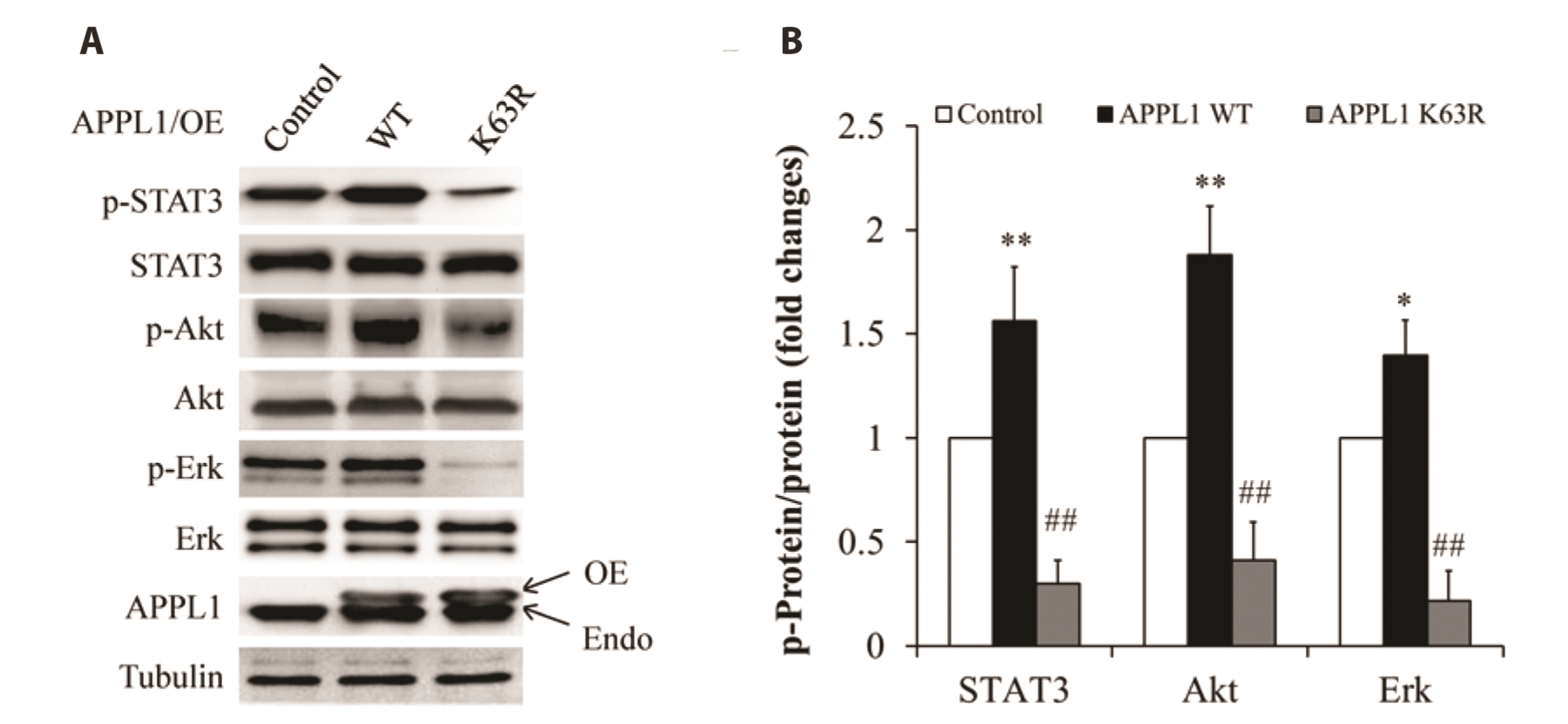
Fig. 4 Effect of APPL1 K63R mutant on phosphorylation of STAT3, Akt, and Erk1/2 in HepG2 cells. (A) Representative Western blot images. (B). Quantitative analysis of the ratio of phosphorylated proteins to the related-proteins. APPL1, adaptor protein, phosphotyrosine interacting with PH domain and leucine zipper 1; K63, Lys 63; OE, overexpression; Endo, endogenous; WT, wild type. *p < 0.05, **p < 0.01 vs. control group; ##p < 0.01 vs. APPL1 WT group.
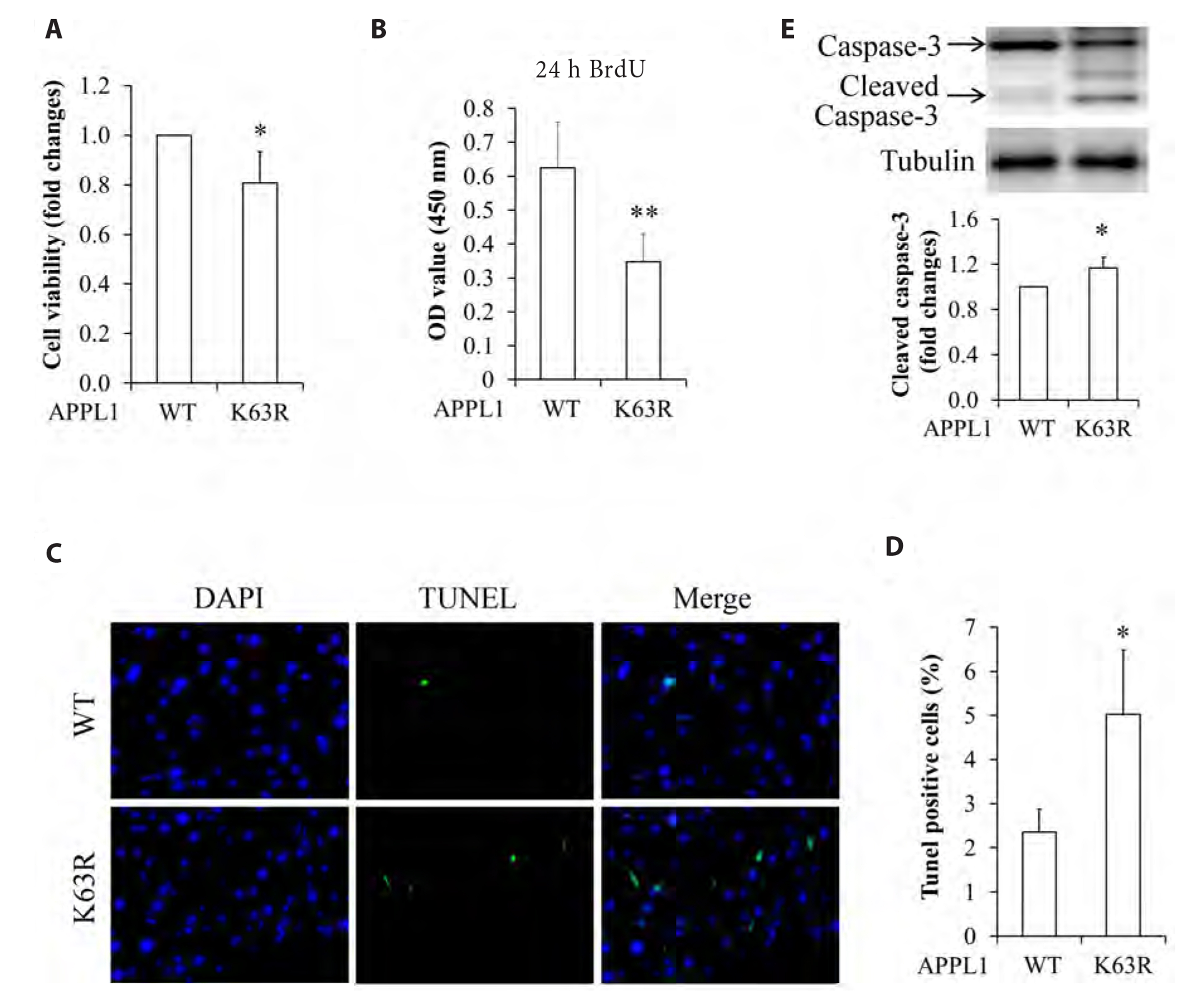
Fig. 5 Effect of APPL1 K63R mutant on survival and proliferation of HepG2 cells. (A) Impact of K63R mutation of APPL1 on cell viability. (B) Impact of K63 mutation of APPL1 on cell proliferation. (C) Representative images of cell apoptosis by APPL1 K63R mutation (×400). (D) Quantitative analysis of cell apoptosis in (C). (E) Impact of APPL1 K63R mutation on cleaved caspase-3 levels. APPL1, adaptor protein, phosphotyrosine interacting with PH domain and leucine zipper 1; K63, Lys 63; WT, wild type. *p < 0.05, **p < 0.01 vs. control group.

Fig. 6 Effect of overexpression of wild-type APPL1 on phosphorylation of STAT3, Akt, and Erk1/2 in DAT-treated HepG2 cells. (A) Representative Western blot images. (B) Quantitative analysis of the ratio of phosphorylated proteins to the related-proteins. APPL1, adaptor protein, phosphotyrosine interacting with PH domain and leucine zipper 1; DAT, diallyl trisulfide; OE, overexpression; Endo, endogenous; WT, wild type. **p < 0.01 vs. control group; #p < 0.05, ##p < 0.01 vs. DAT-treated control group.

Fig. 7 Effect of overexpression of wild-type APPL1 on cell survival and proliferation in DAT-treated HepG2 cells. (A) Overexpression of wild-type APPL1 on cell viability. (B) Overexpression of wild-type APPL1 on cell proliferation. (C) Representative images of cell apoptosis by overexpression of wild-type APPL1 (×400). (D) Quantitative analysis of cell apoptosis in (C). (E) Impact of overexpression of wild-type APPL1 on cleaved caspase-3 levels. APPL1, adaptor protein, phosphotyrosine interacting with PH domain and leucine zipper 1; DAT, diallyl trisulfide. **p < 0.01 vs. control group; #p < 0.05, ##p < 0.01 vs. DAT-treated control group.
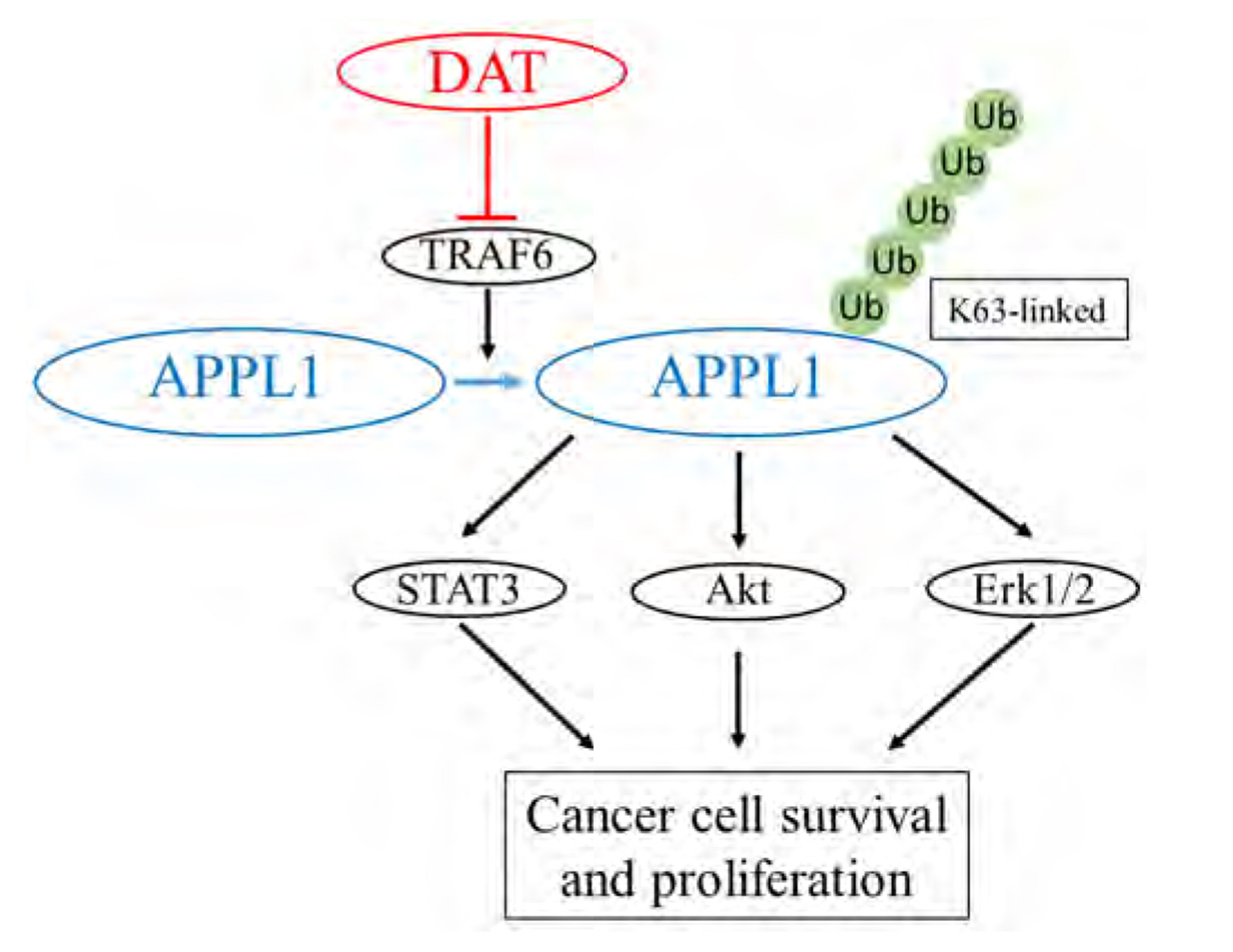
Fig. 8 A schematic diagram of mechanism underlying DAT action on HepG2 cell survival. TRAF6 functions as an E3 ligase. In the present study, TRAF6-modulated K63-linked polyubiquitination of APPL1 mediates the inhibitory effects of DAT on the survival of HepG2 cells by downregulating pro-survival signaling pathways such as STAT3, Akt, and Erk1/2 pathways. DAT, diallyl trisulfide; TRAF6, TNF receptor-associated factor 6; K63, Lys 63; APPL1, adaptor protein, phosphotyrosine interacting with PH domain and leucine zipper 1; Ub, ubiquitin.
Reference
-
1. Balogh J, Victor D 3rd, Asham EH, Burroughs SG, Boktour M, Saharia A, Li X, Ghobrial RM, Monsour HP Jr. 2016; Hepatocellular carcinoma: a review. J Hepatocell Carcinoma. 3:41–53. DOI: 10.2147/JHC.S61146. PMID: 27785449. PMCID: PMC5063561.
Article2. Chacko S, Samanta S. 2016; "Hepatocellular carcinoma: a life-threatening disease". Biomed Pharmacother. 84:1679–1688. DOI: 10.1016/j.biopha.2016.10.078. PMID: 27823920. PMID: https://www.scopus.com/inward/record.uri?partnerID=HzOxMe3b&scp=85001969730&origin=inward.
Article3. Best J, Schotten C, Theysohn JM, Wetter A, Müller S, Radünz S, Schulze M, Canbay A, Dechêne A, Gerken G. 2017; Novel implications in the treatment of hepatocellular carcinoma. Ann Gastroenterol. 30:23–32. DOI: 10.20524/aog.2016.0092. PMID: 28042235. PMCID: PMC5198244. PMID: https://www.scopus.com/inward/record.uri?partnerID=HzOxMe3b&scp=85008180773&origin=inward.4. Dutta R, Mahato RI. 2017; Recent advances in hepatocellular carcinoma therapy. Pharmacol Ther. 173:106–117. DOI: 10.1016/j.pharmthera.2017.02.010. PMID: 28174094. PMCID: PMC5777523. PMID: https://www.scopus.com/inward/record.uri?partnerID=HzOxMe3b&scp=85012930297&origin=inward.
Article5. Sapisochin G, Bruix J. 2017; Liver transplantation for hepatocellular carcinoma: outcomes and novel surgical approaches. Nat Rev Gastroenterol Hepatol. 14:203–217. DOI: 10.1038/nrgastro.2016.193. PMID: 28053342. PMID: https://www.scopus.com/inward/record.uri?partnerID=HzOxMe3b&scp=85008330472&origin=inward.
Article6. Adaki S, Adaki R, Shah K, Karagir A. 2014; Garlic: review of literature. Indian J Cancer. 51:577–581. DOI: 10.4103/0019-509X.175383. PMID: 26842201. PMID: https://www.scopus.com/inward/record.uri?partnerID=HzOxMe3b&scp=84958178966&origin=inward.
Article7. Yu L, Li S, Tang X, Li Z, Zhang J, Xue X, Han J, Liu Y, Zhang Y, Zhang Y, Xu Y, Yang Y, Wang H. 2017; Diallyl trisulfide ameliorates myocardial ischemia-reperfusion injury by reducing oxidative stress and endoplasmic reticulum stress-mediated apoptosis in type 1 diabetic rats: role of SIRT1 activation. Apoptosis. 22:942–954. DOI: 10.1007/s10495-017-1378-y. PMID: 28455824. PMID: https://www.scopus.com/inward/record.uri?partnerID=HzOxMe3b&scp=85018294902&origin=inward.
Article8. Zhu X, Zhang F, Zhou L, Kong D, Chen L, Lu Y, Zheng S. 2014; Diallyl trisulfide attenuates carbon tetrachloride-caused liver injury and fibrogenesis and reduces hepatic oxidative stress in rats. Naunyn Schmiedebergs Arch Pharmacol. 387:445–455. DOI: 10.1007/s00210-014-0959-3. PMID: 24557053. PMID: https://www.scopus.com/inward/record.uri?partnerID=HzOxMe3b&scp=84899965294&origin=inward.
Article9. Tsai CY, Wen SY, Shibu MA, Yang YC, Peng H, Wang B, Wei YM, Chang HY, Lee CY, Huang CY, Kuo WW. 2015; Diallyl trisulfide protects against high glucose-induced cardiac apoptosis by stimulating the production of cystathionine gamma-lyase-derived hydrogen sulfide. Int J Cardiol. 195:300–310. DOI: 10.1016/j.ijcard.2015.05.111. PMID: 26056963. PMID: https://www.scopus.com/inward/record.uri?partnerID=HzOxMe3b&scp=84937611169&origin=inward.
Article10. Hayashida R, Kondo K, Morita S, Unno K, Shintani S, Shimizu Y, Calvert JW, Shibata R, Murohara T. 2017; Diallyl trisulfide augments ischemia-induced angiogenesis via an endothelial nitric oxide synthase-dependent mechanism. Circ J. 81:870–878. DOI: 10.1253/circj.CJ-16-1097. PMID: 28216514. PMID: https://www.scopus.com/inward/record.uri?partnerID=HzOxMe3b&scp=85019773175&origin=inward.
Article11. Chen LY, Chen Q, Cheng YF, Jin HH, Kong DS, Zhang F, Wu L, Shao JJ, Zheng SZ. 2016; Diallyl trisulfide attenuates ethanol-induced hepatic steatosis by inhibiting oxidative stress and apoptosis. Biomed Pharmacother. 79:35–43. DOI: 10.1016/j.biopha.2016.01.009. PMID: 27044810. PMID: https://www.scopus.com/inward/record.uri?partnerID=HzOxMe3b&scp=84961894562&origin=inward.
Article12. Wang S, Li M, Wang X, Li X, Yin H, Jiang L, Han W, Irving G, Zeng T, Xie K. 2017; Diallyl trisulfide attenuated n-hexane induced neurotoxicity in rats by modulating P450 enzymes. Chem Biol Interact. 265:1–7. DOI: 10.1016/j.cbi.2017.01.013. PMID: 28115069. PMID: https://www.scopus.com/inward/record.uri?partnerID=HzOxMe3b&scp=85010399976&origin=inward.
Article13. Lii CK, Huang CY, Chen HW, Chow MY, Lin YR, Huang CS, Tsai CW. 2012; Diallyl trisulfide suppresses the adipogenesis of 3T3-L1 preadipocytes through ERK activation. Food Chem Toxicol. 50:478–484. DOI: 10.1016/j.fct.2011.11.020. PMID: 22137902. PMID: https://www.scopus.com/inward/record.uri?partnerID=HzOxMe3b&scp=84863298355&origin=inward.
Article14. Zhang F, Zhang Y, Wang K, Liu G, Yang M, Zhao Z, Li S, Cai J, Cao J. 2016; Protective effect of diallyl trisulfide against naphthalene-induced oxidative stress and inflammatory damage in mice. Int J Immunopathol Pharmacol. 29:205–216. DOI: 10.1177/0394632015627160. PMID: 26813860. PMCID: PMC5806724. PMID: https://www.scopus.com/inward/record.uri?partnerID=HzOxMe3b&scp=84966701534&origin=inward.
Article15. Kiesel VA, Stan SD. 2017; Diallyl trisulfide, a chemopreventive agent from Allium vegetables, inhibits alpha-secretases in breast cancer cells. Biochem Biophys Res Commun. 484:833–838. DOI: 10.1016/j.bbrc.2017.01.184. PMID: 28161636. PMID: https://www.scopus.com/inward/record.uri?partnerID=HzOxMe3b&scp=85011878196&origin=inward.
Article16. Jiang X, Zhu X, Liu N, Xu H, Zhao Z, Li S, Li S, Cai J, Cao J. 2017; Diallyl trisulfide inhibits growth of NCI-H460 in vitro and in vivo, and ameliorates cisplatin-induced oxidative injury in the treatment of lung carcinoma in xenograft mice. Int J Biol Sci. 13:167–178. DOI: 10.7150/ijbs.16828. PMID: 28255269. PMCID: PMC5332871. PMID: https://www.scopus.com/inward/record.uri?partnerID=HzOxMe3b&scp=85011831927&origin=inward.
Article17. Jiang XY, Zhu XS, Xu HY, Zhao ZX, Li SY, Li SZ, Cai JH, Cao JM. 2017; Diallyl trisulfide suppresses tumor growth through the attenuation of Nrf2/Akt and activation of p38/JNK and potentiates cisplatin efficacy in gastric cancer treatment. Acta Pharmacol Sin. 38:1048–1058. DOI: 10.1038/aps.2016.176. PMID: 28344324. PMCID: PMC5519247. PMID: https://www.scopus.com/inward/record.uri?partnerID=HzOxMe3b&scp=85021820280&origin=inward.
Article18. Hosono T, Fukao T, Ogihara J, Ito Y, Shiba H, Seki T, Ariga T. 2005; Diallyl trisulfide suppresses the proliferation and induces apoptosis of human colon cancer cells through oxidative modification of beta-tubulin. J Biol Chem. 280:41487–41493. DOI: 10.1074/jbc.M507127200. PMID: 16219763. PMID: https://www.scopus.com/inward/record.uri?partnerID=HzOxMe3b&scp=29244484434&origin=inward.
Article19. Lai KC, Hsu SC, Yang JS, Yu CC, Lein JC, Chung JG. 2015; Diallyl trisulfide inhibits migration, invasion and angiogenesis of human colon cancer HT-29 cells and umbilical vein endothelial cells, and suppresses murine xenograft tumour growth. J Cell Mol Med. 19:474–484. DOI: 10.1111/jcmm.12486. PMID: 25403643. PMCID: PMC4407594. PMID: https://www.scopus.com/inward/record.uri?partnerID=HzOxMe3b&scp=84921786219&origin=inward.
Article20. Xiao D, Choi S, Johnson DE, Vogel VG, Johnson CS, Trump DL, Lee YJ, Singh SV. 2004; Diallyl trisulfide-induced apoptosis in human prostate cancer cells involves c-Jun N-terminal kinase and extracellular-signal regulated kinase-mediated phosphorylation of Bcl-2. Oncogene. 23:5594–5606. DOI: 10.1038/sj.onc.1207747. PMID: 15184882. PMID: https://www.scopus.com/inward/record.uri?partnerID=HzOxMe3b&scp=3342895063&origin=inward.
Article21. Xiao D, Lew KL, Kim YA, Zeng Y, Hahm ER, Dhir R, Singh SV. 2006; Diallyl trisulfide suppresses growth of PC-3 human prostate cancer xenograft in vivo in association with Bax and Bak induction. Clin Cancer Res. 12:6836–6843. DOI: 10.1158/1078-0432.CCR-06-1273. PMID: 17121905. PMID: https://www.scopus.com/inward/record.uri?partnerID=HzOxMe3b&scp=33845299153&origin=inward.
Article22. Li Y, Zhang J, Zhang L, Si M, Yin H, Li J. 2013; Diallyl trisulfide inhibits proliferation, invasion and angiogenesis of osteosarcoma cells by switching on suppressor microRNAs and inactivating of Notch-1 signaling. Carcinogenesis. 34:1601–1610. DOI: 10.1093/carcin/bgt065. PMID: 23430952. PMID: https://www.scopus.com/inward/record.uri?partnerID=HzOxMe3b&scp=84880320805&origin=inward.
Article23. Zhang ZM, Yang XY, Deng SH, Xu W, Gao HQ. 2007; Anti-tumor effects of polybutylcyanoacrylate nanoparticles of diallyl trisulfide on orthotopic transplantation tumor model of hepatocellular carcinoma in BALB/c nude mice. Chin Med J (Engl). 120:1336–1342. DOI: 10.1097/00029330-200708010-00008. PMID: 17711740. PMID: https://www.scopus.com/inward/record.uri?partnerID=HzOxMe3b&scp=34548296990&origin=inward.
Article24. Deepa SS, Dong LQ. 2009; APPL1: role in adiponectin signaling and beyond. Am J Physiol Endocrinol Metab. 296:E22–E36. DOI: 10.1152/ajpendo.90731.2008. PMID: 18854421. PMCID: PMC2636986. PMID: https://www.scopus.com/inward/record.uri?partnerID=HzOxMe3b&scp=58249105213&origin=inward.
Article25. Liu Z, Xiao T, Peng X, Li G, Hu F. 2017; APPLs: more than just adiponectin receptor binding proteins. Cell Signal. 32:76–84. DOI: 10.1016/j.cellsig.2017.01.018. PMID: 28108259. PMID: https://www.scopus.com/inward/record.uri?partnerID=HzOxMe3b&scp=85010297595&origin=inward.
Article26. Johnson IR, Parkinson-Lawrence EJ, Keegan H, Spillane CD, Barry-O'Crowley J, Watson WR, Selemidis S, Butler LM, O'Leary JJ, Brooks DA. 2015; Endosomal gene expression: a new indicator for prostate cancer patient prognosis? Oncotarget. 6:37919–37929. DOI: 10.18632/oncotarget.6114. PMID: 26473288. PMCID: PMC4741974. PMID: https://www.scopus.com/inward/record.uri?partnerID=HzOxMe3b&scp=84947786217&origin=inward.
Article27. Song J, Mu Y, Li C, Bergh A, Miaczynska M, Heldin CH, Landström M. 2016; APPL proteins promote TGFβ-induced nuclear transport of the TGFβ type I receptor intracellular domain. Oncotarget. 7:279–292. DOI: 10.18632/oncotarget.6346. PMID: 26583432. PMCID: PMC4807998. PMID: https://www.scopus.com/inward/record.uri?partnerID=HzOxMe3b&scp=85020883234&origin=inward.
Article28. Ding Y, Cao Y, Wang B, Wang L, Zhang Y, Zhang D, Chen X, Li M, Wang C. 2016; APPL1-mediating leptin signaling contributes to proliferation and migration of cancer cells. PLoS One. 11:e0166172. DOI: 10.1371/journal.pone.0166172. PMID: 27820851. PMCID: PMC5098739. PMID: https://www.scopus.com/inward/record.uri?partnerID=HzOxMe3b&scp=84994589804&origin=inward.
Article29. Hennig J, McShane MP, Cordes N, Eke I. 2014; APPL proteins modulate DNA repair and radiation survival of pancreatic carcinoma cells by regulating ATM. Cell Death Dis. 5:e1199. DOI: 10.1038/cddis.2014.167. PMID: 24763056. PMCID: PMC4001316. PMID: https://www.scopus.com/inward/record.uri?partnerID=HzOxMe3b&scp=84901045038&origin=inward.
Article30. Kunkel TA, Roberts JD, Zakour RA. 1987; Rapid and efficient site-specific mutagenesis without phenotypic selection. Methods Enzymol. 154:367–382. DOI: 10.1016/0076-6879(87)54085-X. PMID: 3323813. PMID: https://www.scopus.com/inward/record.uri?partnerID=HzOxMe3b&scp=0023613953&origin=inward.
Article31. Cheng KK, Lam KS, Wang Y, Wu D, Zhang M, Wang B, Li X, Hoo RL, Huang Z, Sweeney G, Xu A. 2013; TRAF6-mediated ubiquitination of APPL1 enhances hepatic actions of insulin by promoting the membrane translocation of Akt. Biochem J. 455:207–216. DOI: 10.1042/BJ20130760. PMID: 23909487. PMID: https://www.scopus.com/inward/record.uri?partnerID=HzOxMe3b&scp=84884787995&origin=inward.
Article32. Wang C, Xin X, Xiang R, Ramos FJ, Liu M, Lee HJ, Chen H, Mao X, Kikani CK, Liu F, Dong LQ. 2009; Yin-Yang regulation of adiponectin signaling by APPL isoforms in muscle cells. J Biol Chem. 284:31608–31615. DOI: 10.1074/jbc.M109.010355. PMID: 19661063. PMCID: PMC2797231. PMID: https://www.scopus.com/inward/record.uri?partnerID=HzOxMe3b&scp=70450265225&origin=inward.
Article33. Ding Y, Zhang D, Wang B, Zhang Y, Wang L, Chen X, Li M, Tang Z, Wang C. 2016; APPL1-mediated activation of STAT3 contributes to inhibitory effect of adiponectin on hepatic gluconeogenesis. Mol Cell Endocrinol. 433:12–19. DOI: 10.1016/j.mce.2016.05.021. PMID: 27246173. PMID: https://www.scopus.com/inward/record.uri?partnerID=HzOxMe3b&scp=84973326543&origin=inward.
Article34. Guan F, Ding Y, Zhang Y, Zhou Y, Li M, Wang C. 2016; Curcumin suppresses proliferation and migration of MDA-MB-231 breast cancer cells through autophagy-dependent Akt degradation. PLoS One. 11:e0146553. DOI: 10.1371/journal.pone.0146553. PMID: 26752181. PMCID: PMC4708990. PMID: https://www.scopus.com/inward/record.uri?partnerID=HzOxMe3b&scp=84954567599&origin=inward.
Article35. Ding Y, Wang B, Chen X, Zhou Y, Ge J. 2017; Staurosporine suppresses survival of HepG2 cancer cells through Omi/HtrA2-mediated inhibition of PI3K/Akt signaling pathway. Tumour Biol. 39:1010428317694317. DOI: 10.1177/1010428317694317. PMID: 28349827. PMID: 26a7cdaef35d4ddb81d28370ec20d244. PMID: https://www.scopus.com/inward/record.uri?partnerID=HzOxMe3b&scp=85016925370&origin=inward.
Article36. Bai J, Cederbaum AI. 2006; Cycloheximide protects HepG2 cells from serum withdrawal-induced apoptosis by decreasing p53 and phosphorylated p53 levels. J Pharmacol Exp Ther. 319:1435–1443. DOI: 10.1124/jpet.106.110007. PMID: 16971506. PMID: https://www.scopus.com/inward/record.uri?partnerID=HzOxMe3b&scp=33751188946&origin=inward.37. Pilecka I, Sadowski L, Kalaidzidis Y, Miaczynska M. 2011; Recruitment of APPL1 to ubiquitin-rich aggresomes in response to proteasomal impairment. Exp Cell Res. 317:1093–1107. DOI: 10.1016/j.yexcr.2011.02.002. PMID: 21320486. PMCID: PMC3072527. PMID: https://www.scopus.com/inward/record.uri?partnerID=HzOxMe3b&scp=79953069853&origin=inward.
Article38. Xiao D, Singh SV. 2006; Diallyl trisulfide, a constituent of processed garlic, inactivates Akt to trigger mitochondrial translocation of BAD and caspase-mediated apoptosis in human prostate cancer cells. Carcinogenesis. 27:533–540. DOI: 10.1093/carcin/bgi228. PMID: 16169930. PMID: https://www.scopus.com/inward/record.uri?partnerID=HzOxMe3b&scp=33644869135&origin=inward.
Article39. Shrotriya S, Kundu JK, Na HK, Surh YJ. 2010; Diallyl trisulfide inhibits phorbol ester-induced tumor promotion, activation of AP-1, and expression of COX-2 in mouse skin by blocking JNK and Akt signaling. Cancer Res. 70:1932–1940. Erratum in: Cancer Res. 2010;70:3414. DOI: 10.1158/0008-5472.CAN-09-3501. PMID: 20179211. PMID: https://www.scopus.com/inward/record.uri?partnerID=HzOxMe3b&scp=77950280661&origin=inward.
Article40. Chandra-Kuntal K, Singh SV. 2010; Diallyl trisulfide inhibits activation of signal transducer and activator of transcription 3 in prostate cancer cells in culture and in vivo. Cancer Prev Res (Phila). 3:1473–1483. DOI: 10.1158/1940-6207.CAPR-10-0123. PMID: 20959517. PMCID: PMC2988081. PMID: https://www.scopus.com/inward/record.uri?partnerID=HzOxMe3b&scp=78649238180&origin=inward.
Article41. Wang H, Sun N, Li X, Li K, Tian J, Li J. 2016; Diallyl trisulfide induces osteosarcoma cell apoptosis through reactive oxygen species-mediated downregulation of the PI3K/Akt pathway. Oncol Rep. 35:3648–3658. DOI: 10.3892/or.2016.4722. PMID: 27035545. PMID: https://www.scopus.com/inward/record.uri?partnerID=HzOxMe3b&scp=84964910272&origin=inward.
Article42. Saxena NK, Sharma D, Ding X, Lin S, Marra F, Merlin D, Anania FA. 2007; Concomitant activation of the JAK/STAT, PI3K/AKT, and ERK signaling is involved in leptin-mediated promotion of invasion and migration of hepatocellular carcinoma cells. Cancer Res. 67:2497–2507. DOI: 10.1158/0008-5472.CAN-06-3075. PMID: 17363567. PMCID: PMC2925446. PMID: https://www.scopus.com/inward/record.uri?partnerID=HzOxMe3b&scp=34047250349&origin=inward.
Article43. Gant-Branum RL, Broussard JA, Mahsut A, Webb DJ, McLean JA. 2010; Identification of phosphorylation sites within the signaling adaptor APPL1 by mass spectrometry. J Proteome Res. 9:1541–1548. DOI: 10.1021/pr901043e. PMID: 20095645. PMCID: PMC2845304. PMID: https://www.scopus.com/inward/record.uri?partnerID=HzOxMe3b&scp=77949780036&origin=inward.
Article44. Holmes RM, Yi Z, De Filippis E, Berria R, Shahani S, Sathyanarayana P, Sherman V, Fujiwara K, Meyer C, Christ-Roberts C, Hwang H, Finlayson J, Dong LQ, Mandarino LJ, Bajaj M. 2011; Increased abundance of the adaptor protein containing pleckstrin homology domain, phosphotyrosine binding domain and leucine zipper motif (APPL1) in patients with obesity and type 2 diabetes: evidence for altered adiponectin signalling. Diabetologia. 54:2122–2131. DOI: 10.1007/s00125-011-2173-x. PMID: 21562756. PMCID: PMC3131511. PMID: https://www.scopus.com/inward/record.uri?partnerID=HzOxMe3b&scp=79960912411&origin=inward.
Article45. Liu M, Zhou L, Wei L, Villarreal R, Yang X, Hu D, Riojas RA, Holmes BM, Langlais PR, Lee H, Dong LQ. 2012; Phosphorylation of adaptor protein containing pleckstrin homology domain, phosphotyrosine binding domain, and leucine zipper motif 1 (APPL1) at Ser430 mediates endoplasmic reticulum (ER) stress-induced insulin resistance in hepatocytes. J Biol Chem. 287:26087–26093. DOI: 10.1074/jbc.M112.372292. PMID: 22685300. PMCID: PMC3406692. PMID: https://www.scopus.com/inward/record.uri?partnerID=HzOxMe3b&scp=84864390268&origin=inward.
Article46. Wang J, Lu W, Chen L, Zhang P, Qian T, Cao W, Luo J. 2016; Serine 707 of APPL1 is critical for the synaptic NMDA receptor-mediated Akt phosphorylation signaling pathway. Neurosci Bull. 32:323–330. DOI: 10.1007/s12264-016-0042-9. PMID: 27300007. PMCID: PMC5563782. PMID: https://www.scopus.com/inward/record.uri?partnerID=HzOxMe3b&scp=84974851680&origin=inward.
Article47. Komander D. 2009; The emerging complexity of protein ubiquitination. Biochem Soc Trans. 37(Pt 5):937–953. DOI: 10.1042/BST0370937. PMID: 19754430. PMID: https://www.scopus.com/inward/record.uri?partnerID=HzOxMe3b&scp=70350150000&origin=inward.
Article48. Xu P, Duong DM, Seyfried NT, Cheng D, Xie Y, Robert J, Rush J, Hochstrasser M, Finley D, Peng J. 2009; Quantitative proteomics reveals the function of unconventional ubiquitin chains in proteasomal degradation. Cell. 137:133–145. DOI: 10.1016/j.cell.2009.01.041. PMID: 19345192. PMCID: PMC2668214. PMID: https://www.scopus.com/inward/record.uri?partnerID=HzOxMe3b&scp=63049125531&origin=inward.
Article49. Deng L, Wang C, Spencer E, Yang L, Braun A, You J, Slaughter C, Pickart C, Chen ZJ. 2000; Activation of the IkappaB kinase complex by TRAF6 requires a dimeric ubiquitin-conjugating enzyme complex and a unique polyubiquitin chain. Cell. 103:351–361. DOI: 10.1016/S0092-8674(00)00126-4. PMID: 11057907. PMID: https://www.scopus.com/inward/record.uri?partnerID=HzOxMe3b&scp=0034644474&origin=inward.
Article50. Hoege C, Pfander B, Moldovan GL, Pyrowolakis G, Jentsch S. 2002; RAD6-dependent DNA repair is linked to modification of PCNA by ubiquitin and SUMO. Nature. 419:135–141. DOI: 10.1038/nature00991. PMID: 12226657. PMID: https://www.scopus.com/inward/record.uri?partnerID=HzOxMe3b&scp=0037068455&origin=inward.
Article51. Lauwers E, Jacob C, André B. 2009; K63-linked ubiquitin chains as a specific signal for protein sorting into the multivesicular body pathway. J Cell Biol. 185:493–502. DOI: 10.1083/jcb.200810114. PMID: 19398763. PMCID: PMC2700384. PMID: https://www.scopus.com/inward/record.uri?partnerID=HzOxMe3b&scp=65649128660&origin=inward.
Article52. Silva GM, Finley D, Vogel C. 2015; K63 polyubiquitination is a new modulator of the oxidative stress response. Nat Struct Mol Biol. 22:116–123. DOI: 10.1038/nsmb.2955. PMID: 25622294. PMCID: PMC4318705. PMID: https://www.scopus.com/inward/record.uri?partnerID=HzOxMe3b&scp=84926417515&origin=inward.
Article53. Zu Y, Yang Y, Zhu J, Bo X, Hou S, Zhang B, Qiu J, Zheng J. 2016; MiR-146a suppresses hepatocellular carcinoma by downregulating TRAF6. Am J Cancer Res. 6:2502–2513. PMID: 27904767. PMCID: PMC5126269.54. Li JJ, Luo J, Lu JN, Liang XN, Luo YH, Liu YR, Yang J, Ding H, Qin GH, Yang LH, Dang YW, Yang H, Chen G. 2016; Relationship between TRAF6 and deterioration of HCC: an immunohistochemical and in vitro study. Cancer Cell Int. 16:76. Erratum in: Cancer Cell Int. 2020;20:60. DOI: 10.1186/s12935-016-0352-z. PMID: 27708550. PMCID: PMC5041287. PMID: https://www.scopus.com/inward/record.uri?partnerID=HzOxMe3b&scp=84988930452&origin=inward.
Article55. Shigemi Z, Furukawa Y, Hosokawa K, Minami S, Matsuhiro J, Nakata S, Watanabe T, Kagawa H, Nakagawa K, Takeda H, Fujimuro M. 2016; Diallyl trisulfide induces apoptosis by suppressing NF-κB signaling through destabilization of TRAF6 in primary effusion lymphoma. Int J Oncol. 48:293–304. DOI: 10.3892/ijo.2015.3247. PMID: 26647777. PMID: https://www.scopus.com/inward/record.uri?partnerID=HzOxMe3b&scp=84953329084&origin=inward.
Article56. Schmitz KJ, Wohlschlaeger J, Lang H, Sotiropoulos GC, Malago M, Steveling K, Reis H, Cicinnati VR, Schmid KW, Baba HA. 2008; Activation of the ERK and AKT signalling pathway predicts poor prognosis in hepatocellular carcinoma and ERK activation in cancer tissue is associated with hepatitis C virus infection. J Hepatol. 48:83–90. DOI: 10.1016/j.jhep.2007.08.018. PMID: 17998146. PMID: https://www.scopus.com/inward/record.uri?partnerID=HzOxMe3b&scp=36549013178&origin=inward.
Article57. Yang J, Cai X, Lu W, Hu C, Xu X, Yu Q, Cao P. 2013; Evodiamine inhibits STAT3 signaling by inducing phosphatase shatterproof 1 in hepatocellular carcinoma cells. Cancer Lett. 328:243–251. DOI: 10.1016/j.canlet.2012.09.019. PMID: 23032719. PMID: https://www.scopus.com/inward/record.uri?partnerID=HzOxMe3b&scp=84870364791&origin=inward.
Article
- Full Text Links
-
- Actions
-
Cited
- CITED
-
- Close
- Share
-
- Similar articles
-
- The centrosomal localization of KM-HN-1 (MGC33607) depends on the leucine zipper motif and the C-terminal coiled-coil domain
- Association of Killer Cell Ig-like Receptor (KIR) with an Adaptor Protein Shc
- Exploring the role and mechanisms of diallyl trisulfide and diallyl disulfide in chronic constriction-induced neuropathic pain in rats
- Ubiquitylation of Fe65 adaptor protein by neuronal precursor cell expressed developmentally down regulated 4-2 (Nedd4-2) via the WW domain interaction with Fe65
- Gene Expression Changes by Diallyl Trisulfide Administration in Chemically-induced Mammary Tumors in Rats
