Ectopic Pancreas with Walled-off Necrosis Mimicking Malignant Submucosal Gastric Tumor
- Affiliations
-
- 1Departments of Medicine, Samsung Medical Center, Sungkyunkwan University School of Medicine, Seoul, Korea
- 2Departments of Pathology, Samsung Medical Center, Sungkyunkwan University School of Medicine, Seoul, Korea
- KMID: 2534469
- DOI: http://doi.org/10.4166/kjg.2022.078
Abstract
- An ectopic pancreas rarely transforms into a malignancy, and the symptoms vary from patient to patient. The most commonly observed site of an ectopic pancreas is the antrum of the stomach. A 59-year-old male patient with severe abdominal pain underwent CT. A 9.6 cm-sized well-defined exophytic huge mass with heterogenic density was located between the stomach distal antrum and duodenum. A malignant submucosal tumor was suspected because of the exophytic dirty huge mass. Initially, surgery was considered to confirm the histological evaluation. After 2 months, the abdominal pain disappeared, and the follow-up MRI scan showed a decrease in size, which contained a necrotic component inside. It was confirmed that the parenchymal tissue was the pancreas. The pathology through EUS-guided fine needle aspiration (EUS-FNA) was normal pancreatic acinar cells, smooth muscle fragments, squamous cyst, and some neutrophils (abscess). Walled-off necrosis occurs as a complication of acute pancreatitis with parenchymal tissues and surrounding tissues, but complications of ectopic pancreatitis occurred in this case. Abdominal pain due to ectopic pancreas leading to the formation of a giant abscess has been reported as a very rare case. Diagnosis through biopsy is most important when a malignant submucosal tumor is suspected. In addition, it is important to determine the clinical features, examination findings, such as EUS, CT, and MRI, and the changes according to the follow-up period. This paper reports a case of ectopic pancreas, resulting in necrotic tissue and walled-off necrosis, abdominal pain, and spontaneous improvement.
Figure
-
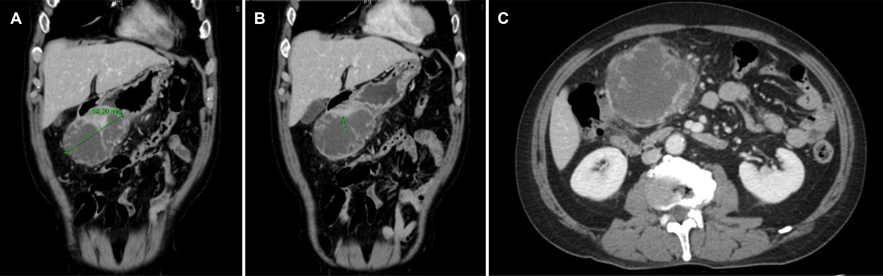
Fig. 1 Abdominal pelvis computed tomography. (A, B) Necrotic mass-like lesion abutting the stomach distal antrum and duodenum occupying omentum and mesentery was noticed in coronal view. (C) Severe necrosis and abscess changes were observed in the axial view.
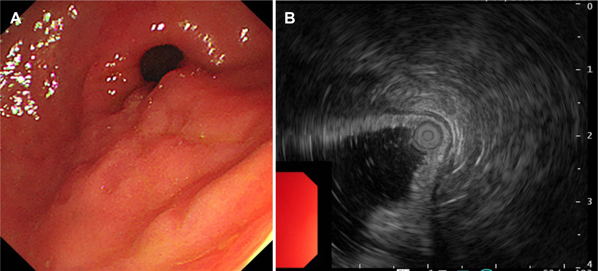
Fig. 2 (A) Esophagogastroduodenoscopy. The uneven surface without a loss of mucosa was observed on the antrum posterior wall. (B) Endoscopic ultrasonography. A huge hyperechoic lesion was noted.
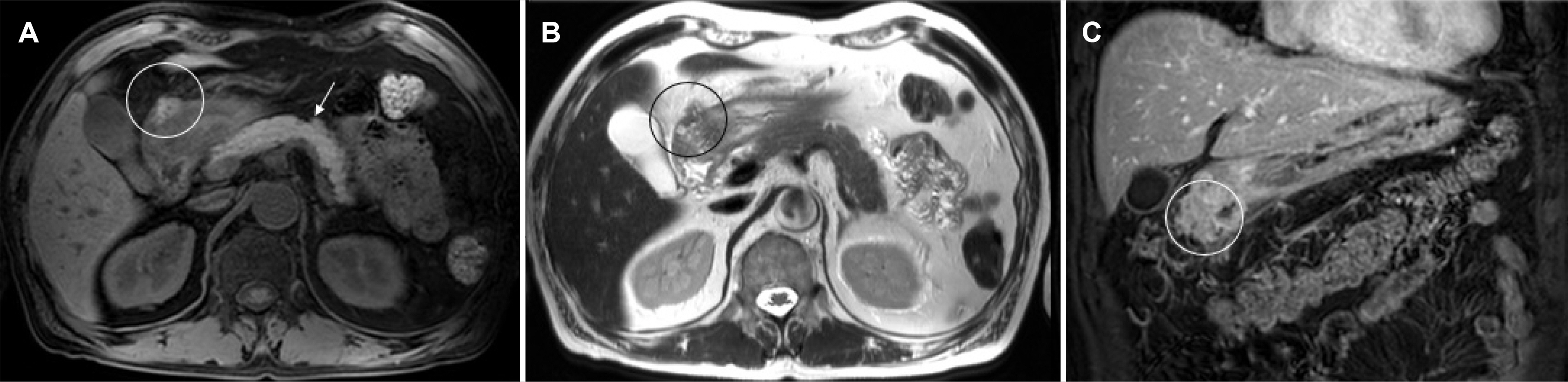
Fig. 3 Pancreas magnetic resonance imaging (pre- & post-contrast)+diffusion. Probable necrotizing pancreatitis with walled-off necrosis arising from ectopic pancreas rather than a malignant condition. (A) High signal intensity was observed in fat-suppressed pre-contrast T1-weighted image. (B) Soft tissue contains microcystic or microductular structure in the T2-weighted image. (C) High signal intensity was observed in the 3 min delayed-coronal view.
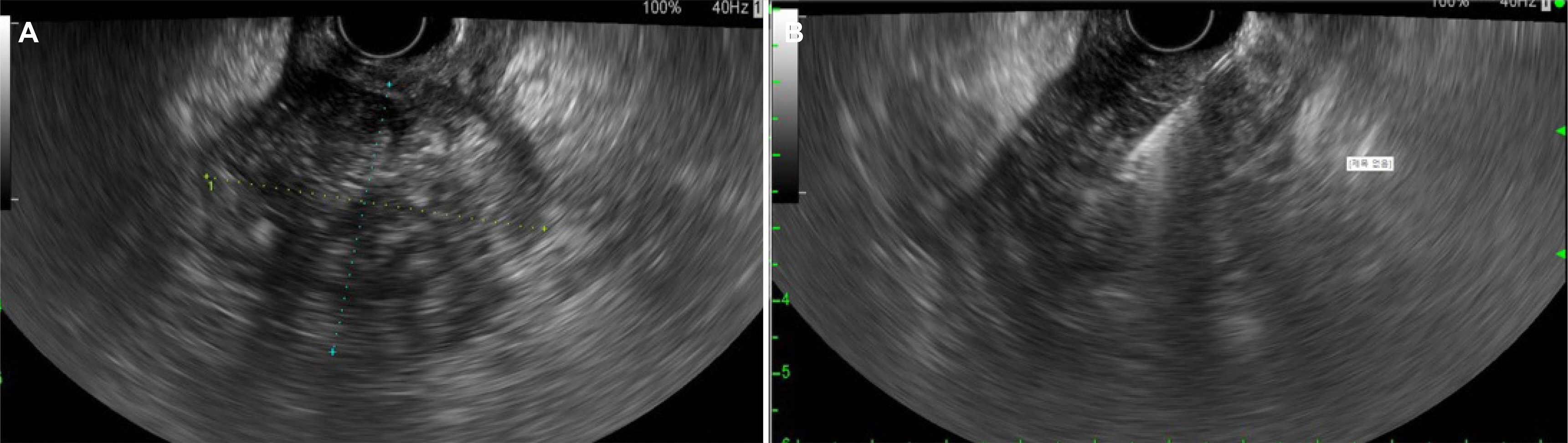
Fig. 4 (A) A 3×4 cm-sized heterogeneous echogenic mass observed in the antrum. (B) Endoscopic ultrasound-guided fine-needle aspiration. Cytology and biopsy were performed twice with a 19G needle.

Fig. 5 Histology examination. (A) normal pancreatic acinar cells. (B) Smooth muscle fragments. (C) Neutrophils (abscess). (D) Squamoid cyst.
Reference
-
1. Liu X, Wu X, Tuo B, Wu H. 2021; Ectopic pancreas appearing as a giant gastric cyst mimicking gastric lymphangioma: a case report and a brief review. BMC Gastroenterol. 21:151. DOI: 10.1186/s12876-021-01686-9. PMID: 33823798. PMCID: PMC8022400.2. Flores A, Papafragkakis C, Uberoi AS, Thaiudom S, Bhutani MS. 2018; EUS of an atypical ectopic pancreas. Endosc Ultrasound. 7:216–217. DOI: 10.4103/eus.eus_111_17. PMID: 29697071. PMCID: PMC6032704.3. Kung JW, Brown A, Kruskal JB, Goldsmith JD, Pedrosa I. 2010; Heterotopic pancreas: typical and atypical imaging findings. Clin Radiol. 65:403–407. DOI: 10.1016/j.crad.2010.01.005. PMID: 20380941.4. Cazacu IM, Luzuriaga Chavez AA, Nogueras Gonzalez GM, Saftoiu A, Bhutani MS. 2019; Malignant transformation of ectopic pancreas. Dig Dis Sci. 64:655–668. DOI: 10.1007/s10620-018-5366-z. PMID: 30415408.5. Alastal Y, Khalil B, Singh S, Almadani SB. 2018; Ectopic pancreas in the gastric antrum wall complicated by ectopic pancreatitis and persistent gastric abscess. ACG Case Rep J. 5:e34. DOI: 10.14309/crj.2018.34. PMID: 29774224. PMCID: PMC5948316.6. Kaneda M, Yano T, Yamamoto T, et al. 1989; Ectopic pancreas in the stomach presenting as an inflammatory abdominal mass. Am J Gastroenterol. 84:663–666.7. Manikkavasakar S, AlObaidy M, Busireddy KK, et al. 2014; Magnetic resonance imaging of pancreatitis: an update. World J Gastroenterol. 20:14760–14777. DOI: 10.3748/wjg.v20.i40.14760. PMID: 25356038. PMCID: PMC4209541.8. Pamuklar E, Semelka RC. 2005; MR imaging of the pancreas. Magn Reson Imaging Clin N Am. 13:313–330. DOI: 10.1016/j.mric.2005.03.012. PMID: 15935314.9. Heyn C, Sue-Chue-Lam D, Jhaveri K, Haider MA. 2012; MRI of the pancreas: problem solving tool. J Magn Reson Imaging. 36:1037–1051. DOI: 10.1002/jmri.23708. PMID: 23090915.
- Full Text Links
-
- Actions
-
Cited
- CITED
-
- Close
- Share
-
- Similar articles
-
- A case of asymptomatic gastric ectopic pancreas associated with elevated serum CA 19-9
- Perilesional Steatosis in Ectopic Pancreas Mimicking Exogastric Mass : A Case Report
- Malignant Solitary Fibrous Tumor of Retroperitoneum Mimicking Gastric Submucosal Tumor
- Endoscopic Findings of Ectopic Pancreas in the Stomach
- Synchronous Ectopic Pancreases in the Cardia and Antrum of the Stomach: A Case Report
