Single-operator Cholangioscopy Guided Lithotripsy
- Affiliations
-
- 1Division of Gastroenterology and Hepatology, Department of Internal Medicine, Kyung Hee University Hospital, Kyung Hee University College of Medicine, Seoul, Korea
- KMID: 2534464
- DOI: http://doi.org/10.4166/kjg.2022.117
Abstract
- ERCP is the standard treatment for common bile duct stones (CBD stones). On the other hand, in approximately 10% of patients with CBD stones, the complete removal of the stones cannot be achieved by conventional ERCP, which performs endoscopic sphincterotomy followed by stone extraction. Additional advanced and complex procedures are often necessary to remove these “difficult bile duct stones”, including endoscopic papillary large balloon dilatation or mechanical lithotripsy. Advances in cholangioscopy have made electrohydraulic or laser lithotripsy under direct endoscopic visualization possible during ERCP. Cholangioscopy-guided lithotripsy using the SpyGlass DS system could be a better treatment tool for removing difficult stones. The focus of this review was to describe single-operator cholangioscopy in the management of difficult CBD stones.
Keyword
Figure
-

Fig. 1 Single-operator cholangioscopy using the SpyGlass DS system. (A) The SpySope is inserted into the duodenoscope through the working channel. (B) A single endoscopist controls both the duodenoscope and SpyScope (the reason this is called “single-operator” cholangioscopy). (C) The SpyScope, which is the single-use cholangioscope of the SpyGlass DS System.
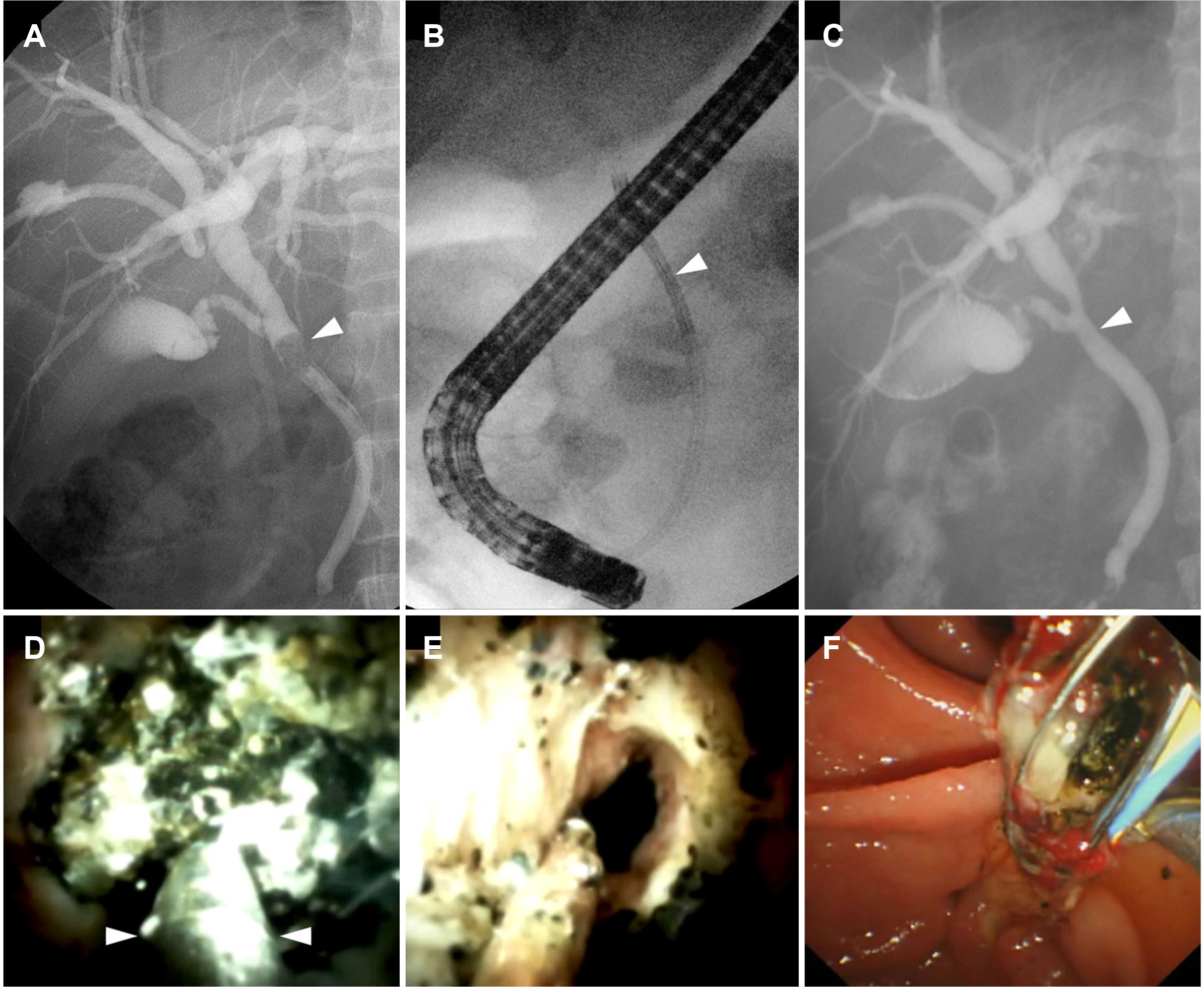
Fig. 2 Single-operator cholangioscopy-guided electrohydraulic lithotripsy (EHL). (A) Fluoroscopy image showing an impacted bile duct stone (arrowhead). (B) X-ray image showing SpyScope (arrowhead) inserted into the bile duct. (C) Final cholangiogram showing no residual filling defect. (arrowhead). (D) SpyGlass showing an impacted stone. EHL (arrowheads, EHL probe) is performed under direct visualization. (E) SpyGlass showing the cleared bile duct after EHL. (F) Fragmented bile duct stones are extracted using a stone basket after lithotripsy.
Reference
-
1. Collins C, Maguire D, Ireland A, Fitzgerald E, O'Sullivan GC. 2004; A prospective study of common bile duct calculi in patients undergoing laparoscopic cholecystectomy: natural history of choledocholithiasis revisited. Ann Surg. 239:28–33. DOI: 10.1097/01.sla.0000103069.00170.9c. PMID: 14685097. PMCID: PMC1356189.2. Freitas ML, Bell RL, Duffy AJ. 2006; Choledocholithiasis: evolving standards for diagnosis and management. World J Gastroenterol. 12:3162–3167. DOI: 10.3748/wjg.v12.i20.3162. PMID: 16718834. PMCID: PMC4087957.3. Yasuda I, Itoi T. 2013; Recent advances in endoscopic management of difficult bile duct stones. Dig Endosc. 25:376–385. DOI: 10.1111/den.12118. PMID: 23650878.4. Manes G, Paspatis G, Aabakken L, et al. 2019; Endoscopic management of common bile duct stones: European Society of Gastrointestinal Endoscopy (ESGE) guideline. Endoscopy. 51:472–491. DOI: 10.1055/a-0862-0346. PMID: 30943551.5. Lux G, Ell C, Hochberger J, Müller D, Demling L. 1986; The first successful endoscopic retrograde laser lithotripsy of common bile duct stones in man using a pulsed neodymium-YAG laser. Endoscopy. 18:144–145. DOI: 10.1055/s-2007-1018356. PMID: 2874019.6. Trikudanathan G, Navaneethan U, Parsi MA. 2013; Endoscopic management of difficult common bile duct stones. World J Gastroenterol. 19:165–173. DOI: 10.3748/wjg.v19.i2.165. PMID: 23345939. PMCID: PMC3547556.7. Patel SN, Rosenkranz L, Hooks B, et al. 2014; Holmium-yttrium aluminum garnet laser lithotripsy in the treatment of biliary calculi using single-operator cholangioscopy: a multicenter experience (with video). Gastrointest Endosc. 79:344–348. DOI: 10.1016/j.gie.2013.07.054. PMID: 24268531.8. Ishida Y, Itoi T, Okabe Y. 2016; Types of peroral cholangioscopy: how to choose the most suitable type of cholangioscopy. Curr Treat Options Gastroenterol. 14:210–219. DOI: 10.1007/s11938-016-0090-2. PMID: 27053226.9. Bhandari S, Bathini R, Sharma A, Maydeo A. 2016; Usefulness of single-operator cholangioscopy-guided laser lithotripsy in patients with Mirizzi syndrome and cystic duct stones: experience at a tertiary care center. Gastrointest Endosc. 84:56–61. DOI: 10.1016/j.gie.2015.12.025. PMID: 26764195.10. Maydeo A, Kwek BE, Bhandari S, Bapat M, Dhir V. 2011; Single-operator cholangioscopy-guided laser lithotripsy in patients with difficult biliary and pancreatic ductal stones (with videos). Gastrointest Endosc. 74:1308–1314. DOI: 10.1016/j.gie.2011.08.047. PMID: 22136776.11. Wong JC, Tang RS, Teoh AY, Sung JJ, Lau JY. 2017; Efficacy and safety of novel digital single-operator peroral cholangioscopy-guided laser lithotripsy for complicated biliary stones. Endosc Int Open. 5:E54–E58. DOI: 10.1055/s-0042-118701. PMID: 28337482. PMCID: PMC5361876.12. Brewer Gutierrez OI, Bekkali NLH, Raijman I, et al. 2018; Efficacy and safety of digital single-operator cholangioscopy for difficult biliary stones. Clin Gastroenterol Hepatol. 16:918–926.e1. DOI: 10.1016/j.cgh.2017.10.017. PMID: 29074446.13. Korrapati P, Ciolino J, Wani S, et al. 2016; The efficacy of peroral cholangioscopy for difficult bile duct stones and indeterminate strictures: a systematic review and meta-analysis. Endosc Int Open. 4:E263–E275. DOI: 10.1055/s-0042-100194. PMID: 27004242. PMCID: PMC4798839.14. Bokemeyer A, Gerges C, Lang D, et al. 2020; Digital single-operator video cholangioscopy in treating refractory biliary stones: a multicenter observational study. Surg Endosc. 34:1914–1922. DOI: 10.1007/s00464-019-06962-0. PMID: 31309312.15. Kurihara T, Yasuda I, Isayama H, et al. 2016; Diagnostic and therapeutic single-operator cholangiopancreatoscopy in biliopancreatic diseases: prospective multicenter study in Japan. World J Gastroenterol. 22:1891–1901. DOI: 10.3748/wjg.v22.i5.1891. PMID: 26855549. PMCID: PMC4724621.16. Navaneethan U, Hasan MK, Kommaraju K, et al. 2016; Digital, single-operator cholangiopancreatoscopy in the diagnosis and management of pancreatobiliary disorders: a multicenter clinical experience (with video). Gastrointest Endosc. 84:649–655. DOI: 10.1016/j.gie.2016.03.789. PMID: 26995690.17. Turowski F, Hügle U, Dormann A, et al. 2018; Diagnostic and therapeutic single-operator cholangiopancreatoscopy with SpyGlassDSTM: results of a multicenter retrospective cohort study. Surg Endosc. 32:3981–3988. DOI: 10.1007/s00464-018-6141-0. PMID: 29532224.18. Angsuwatcharakon P, Kulpatcharapong S, Ridtitid W, et al. 2019; Digital cholangioscopy-guided laser versus mechanical lithotripsy for large bile duct stone removal after failed papillary large-balloon dilation: a randomized study. Endoscopy. 51:1066–1073. DOI: 10.1055/a-0848-8373. PMID: 30786315.19. Chen YK, Parsi MA, Binmoeller KF, et al. 2011; Single-operator cholangioscopy in patients requiring evaluation of bile duct disease or therapy of biliary stones (with videos). Gastrointest Endosc. 74:805–814. DOI: 10.1016/j.gie.2011.04.016. PMID: 21762903.20. Jin Z, Wei Y, Tang X, et al. 2019; Single-operator peroral cholangioscope in treating difficult biliary stones: a systematic review and meta-analysis. Dig Endosc. 31:256–269. DOI: 10.1111/den.13307. PMID: 30468534.21. Veld JV, van Huijgevoort NCM, Boermeester MA, et al. 2018; A systematic review of advanced endoscopy-assisted lithotripsy for retained biliary tract stones: laser, electrohydraulic or extracorporeal shock wave. Endoscopy. 50:896–909. DOI: 10.1055/a-0637-8806. PMID: 29991072.22. Buxbaum J, Sahakian A, Ko C, et al. 2018; Randomized trial of cholangioscopy-guided laser lithotripsy versus conventional therapy for large bile duct stones (with videos). Gastrointest Endosc. 87:1050–1060. DOI: 10.1016/j.gie.2017.08.021. PMID: 28866457.23. Deprez PH, Garces Duran R, Moreels T, et al. 2018; The economic impact of using single-operator cholangioscopy for the treatment of difficult bile duct stones and diagnosis of indeterminate bile duct strictures. Endoscopy. 50:109–118. DOI: 10.1055/s-0043-121268. PMID: 29172216.
- Full Text Links
-
- Actions
-
Cited
- CITED
-
- Close
- Share
-
- Similar articles
-
- Recent advances in the management of difficult bile-duct stones: a focus on single-operator cholangioscopy-guided lithotripsy
- Minimally Invasive Approach Using Digital Single-Operator Peroral Cholangioscopy-Guided Electrohydraulic Lithotripsy and Endoscopic Nasogallbladder Drainage for the Management of High-Grade Mirizzi Syndrome
- Successful removal of remnant cystic duct stump stone using single-operator cholangioscopy-guided electrohydraulic lithotripsy: two case reports
- Recent update of therapeutic application of peroral cholangioscopy
- Endoscopic methods for cytopathologic diagnosis of bile duct strictures
