Pear pomace alleviated atopic dermatitis in NC/Nga mice and inhibited LPS-induced inflammation in RAW 264.7 macrophages
- Affiliations
-
- 1Nutrition Research Institute, University of North Carolina, Chapel Hill, NC 28081, USA
- 2Department of Food and Nutrition, Mokpo National University, Muan 58554, Korea
- KMID: 2534117
- DOI: http://doi.org/10.4162/nrp.2022.16.5.577
Abstract
- BACKGROUND/OBJECTIVES
Poorly regulated inflammation is believed to be the most predominant factor that can result in a wide scope of diseases including atopic dermatitis (AD). Despite many studies on the effect of pear pomace in obesity-related disorders including dysregulated gut microbiota, the protective effect of pear pomace in AD is still unknown. This study aimed to evaluate the effect of pear pomace ethanol extract (PPE) on AD by inhibiting inflammation.
MATERIALS/METHODS
In the in vivo experiment, 2, 4-dinitrochlorobenzene (DNCB) was applied to NC/Nga mice to induce AD-like skin lesions. After the induction, PPE was administered daily by oral gavage for 4 weeks. The clinical severity score, serum IgE levels, spleen weight, histological changes in dorsal skin, and inflammation-related proteins were measured. In the cell study, RAW 264.7 cells were pretreated with PPE before stimulation with lipopolysaccharide (LPS). Nitrite oxide (NO) production and nuclear factor kappa B (NFkB) protein expression were detected.
RESULTS
Compared to the AD control (AD-C) group, IgE levels were dramatically decreased via PPE treatment. PPE significantly reduced scratching behavior, improved skin symptoms, and decreased ear thickness compared to the AD-C group. In addition, PPE inhibited the DNCB-induced expression of inducible nitrite oxide synthase (iNOS), the receptor for advanced glycation end products, extracellular signal-regulated kinase (ERK) 1/2, and NF-κB. PPE inhibited the LPS-induced overproduction of NO and the enhanced expression of iNOS and cyclooxygenase-2. Moreover, the phosphorylation of ERK1/2 and NF-κB in RAW 264.7 cells was suppressed by PPE.
CONCLUSIONS
These results suggest that PPE could be explored as a therapeutic agent to prevent AD.
Keyword
Figure
-
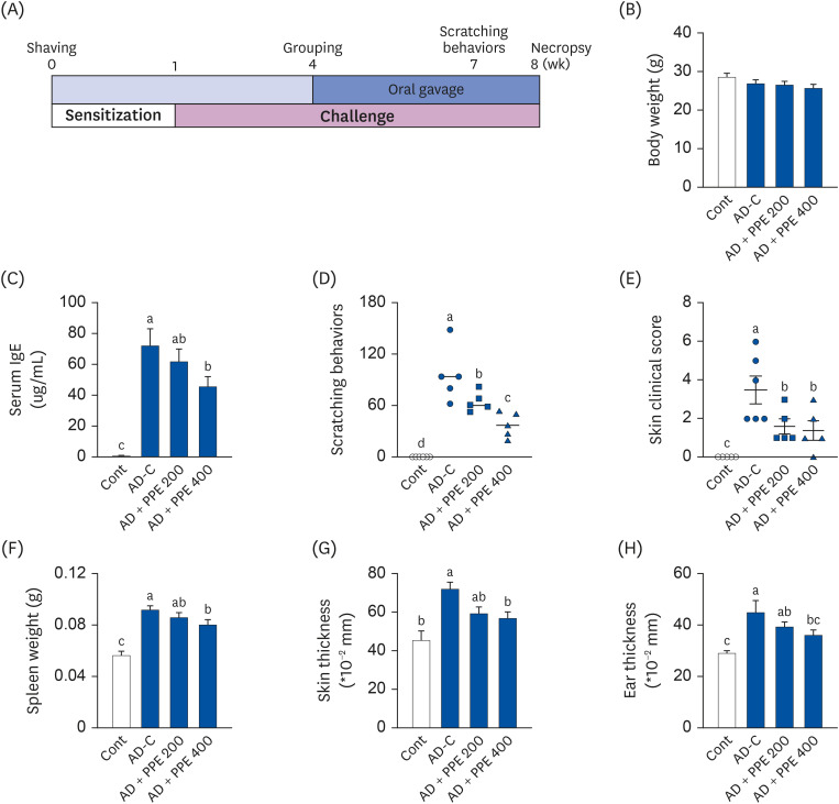
Fig. 1 Pear pomace ameliorated AD symptoms in DNCB-induced AD skin lesion in NC/Nga mice. (A) Experiment design. To induce AD-like lesions, DNCB was applied to NC/Nga mice (n = 7). The mice were sensitized with 1% DNCB for 1 week and then challenged with 0.4% DNCB solution for 7 weeks. The mice were administered PBS or PPE daily for 4 weeks. (B) Final body weight. (C) Serum levels of IgE. (D) Total scratching behavior scores 7 weeks after sensitization. (E) Clinical severity score for dermatitis on the necropsy day. (F) Spleen weight. (G) Skin thickness. (H) Ear thickness.AD, atopic dermatitis; DNCB, 2, 4-dinitrochlorobenzene; PBS, phosphate-buffed saline; PPE, pear pomace ethanol extract; Cont, no sensitization; AD-C, DNCB treatment with oral administration of PBS (AD control); AD + PPE 200, DNCB treatment with oral administration of PPE 200 mg/kg body weight; AD + PPE 400, DNCB treatment with oral administration of PPE 400 mg/kg body weight.Different letters are significantly different by Duncan’s multiple range test (P < 0.05).
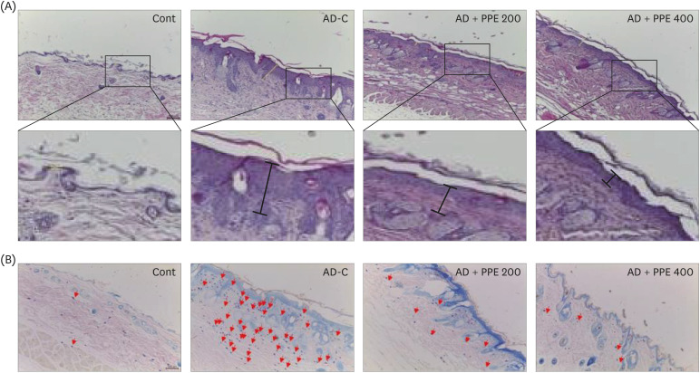
Fig. 2 Histological changes in the dorsal skin in pear pomace-treated NC/Nga mice. (A) H&E staining in skin tissue. (B) Toluidine blue staining. The arrows indicate mast cells in the skin tissue.AD, atopic dermatitis; DNCB, 2, 4-dinitrochlorobenzene; H&E, hematoxylin and eosin; PPE, pear pomace ethanol extract; Cont, no sensitization; AD-C, DNCB treatment with the oral administration of PBS (AD control); AD + PPE 200, DNCB treatment with the oral administration of PPE 200 mg/kg body weight; AD + PPE 400, DNCB treatment with oral administration of PPE 400 mg/kg body weight.
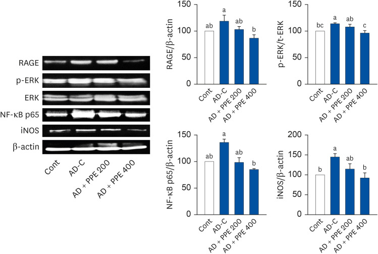
Fig. 3 Applications of pear pomace inhibited inflammation-related proteins in the skin tissue of NC/Nga mice. β-actin was used as the control for quantification.Cont, no sensitization; AD-C, DNCB treatment with oral administration of PBS (AD control); AD + PPE 200, DNCB treatment with the oral administration of PPE 200 mg/kg body weight; AD + PPE 400, DNCB treatment with the oral administration of PPE 400 mg/kg body weight; RAGE, receptor for advanced glycation end products; ERK, extracellular signal-regulated kinase, NF-κB, nuclear factor kappa B; iNOS, inducible nitrite oxide synthase; PPE, pear pomace ethanol extract.Different letters are significantly different by Duncan’s multiple range test (P < 0.05).
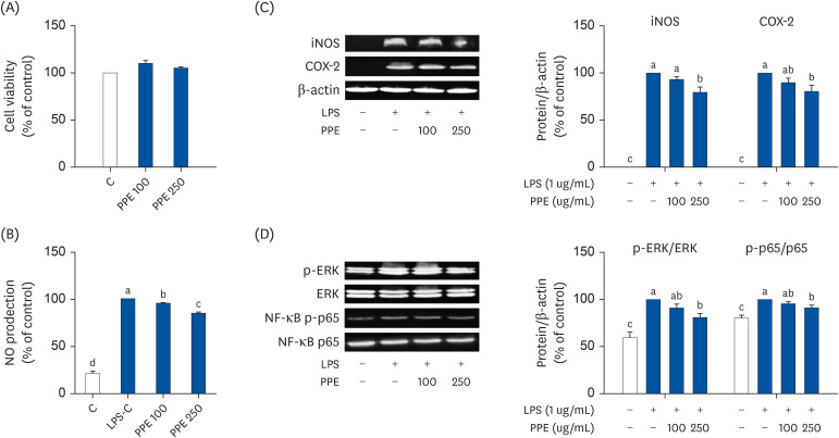
Fig. 4 Inhibitory effects of pear pomace on the inflammatory response in LPS-induced inflammation in RAW 264.7 macrophages. RAW 264.7 cells were pretreated with either 0, 100, or 250 ug/mL of PPE for 1 h before the addition of 1 ug/mL of LPS treatment for 24 h. (A) Cell viability. (B) NO production. (C) Protein expression of iNOS and COX-2. (D) Protein expression of ERK and NF-κB. β-actin was used as the control for quantification.LPS, lipopolysaccharide; NO, nitrite oxide; iNOS, inducible nitrite oxide synthase; COX-2, cyclooxygenase-2; ERK, extracellular signal-regulated kinase; NF-κB, nuclear factor kappa B; PPE, pear pomace ethanol extract.Different letters are significantly different by Duncan’s multiple range test (P < 0.05).
Reference
-
1. Leung DY, Boguniewicz M, Howell MD, Nomura I, Hamid QA. New insights into atopic dermatitis. J Clin Invest. 2004; 113:651–657. PMID: 14991059.
Article2. Hon KL, Leung AK. Use of moisturizers in patients with atopic dermatitis. Lodén M, Maibach HI, editors. Treatment of Dry Skin Syndrome. Berlin: Springer;2012. p. 59–73.3. Bylund S, Kobyletzki LB, Svalstedt M, Svensson Å. Prevalence and incidence of atopic dermatitis: a systematic review. Acta Derm Venereol. 2020; 100:adv00160. PMID: 32412646.
Article4. Brar K, Leung DY. Recent considerations in the use of recombinant interferon gamma for biological therapy of atopic dermatitis. Expert Opin Biol Ther. 2016; 16:507–514. PMID: 26694988.
Article5. Wahn U, Bos JD, Goodfield M, Caputo R, Papp K, Manjra A, Dobozy A, Paul C, Molloy S, Hultsch T, et al. Efficacy and safety of pimecrolimus cream in the long-term management of atopic dermatitis in children. Pediatrics. 2002; 110:e2. PMID: 12093983.
Article6. Tomi NS, Luger TA. The treatment of atopic dermatitis with topical immunomodulators. Clin Dermatol. 2003; 21:215–224. PMID: 12781439.
Article7. Gupta AK, Chow M. Pimecrolimus: a review. J Eur Acad Dermatol Venereol. 2003; 17:493–503. PMID: 12941081.
Article8. Reitamo S. Tacrolimus: a new topical immunomodulatory therapy for atopic dermatitis. J Allergy Clin Immunol. 2001; 107:445–448. PMID: 11240943.
Article9. Reitamo S, Wollenberg A, Schöpf E, Perrot JL, Marks R, Ruzicka T, Christophers E, Kapp A, Lahfa M, Rubins A, et al. Safety and efficacy of 1 year of tacrolimus ointment monotherapy in adults with atopic dermatitis. Arch Dermatol. 2000; 136:999–1006. PMID: 10926735.
Article10. Kondo K, Nagami T, Teramoto S. Differences in hematopoietic death among inbred strains of mice. Bond PV, Sugahara T, editors. Comparative Cellular and Species Radiosensitivity. Tokyo: Igakushoin;1969. p. 20–29.11. Vestergaard C, Yoneyama H, Matsushima K. The NC/Nga mouse: a model for atopic dermatitis. Mol Med Today. 2000; 6:209–210. PMID: 10782068.
Article12. Matsuda H, Watanabe N, Geba GP, Sperl J, Tsudzuki M, Hiroi J, Matsumoto M, Ushio H, Saito S, Askenase PW, et al. Development of atopic dermatitis-like skin lesion with IgE hyperproduction in NC/Nga mice. Int Immunol. 1997; 9:461–466. PMID: 9088984.
Article13. Inoue J, Aramaki Y. Suppression of skin lesions by transdermal application of CpG-oligodeoxynucleotides in NC/Nga mice, a model of human atopic dermatitis. J Immunol. 2007; 178:584–591. PMID: 17182599.
Article14. Park SJ, Lee HA, Kim JW, Lee BS, Kim EJ. Platycodon grandiflorus alleviates DNCB-induced atopy-like dermatitis in NC/Nga mice. Indian J Pharmacol. 2012; 44:469–474. PMID: 23087507.
Article15. Novak N, Bieber T, Leung DY. Immune mechanisms leading to atopic dermatitis. J Allergy Clin Immunol. 2003; 112:S128–S139. PMID: 14657843.
Article16. Nagai H, Teramachi H, Tuchiya T. Recent advances in the development of anti-allergic drugs. Allergol Int. 2006; 55:35–42. PMID: 17075284.
Article17. James-Martin G, Williams G, Stonehouse W, O'Callaghan N, Noakes M. Health and Nutritional Properties of Pears (Pyrus): A Literature Review. Adelaide: CSIRO;2015.18. Nawirska A, Kwaśniewska M. Dietary fibre fractions from fruit and vegetable processing waste. Food Chem. 2005; 91:221–225.
Article19. Martin-Cabrejas M, Esteban R, Lopez-Andreu F, Waldron K, Selvendran R. Dietary fiber content of pear and kiwi pomaces. J Agric Food Chem. 1995; 43:662–666.
Article20. Chang S, Cui X, Guo M, Tian Y, Xu W, Huang K, Zhang Y. Insoluble dietary fiber from pear pomace can prevent high-fat diet-induced obesity in rats mainly by improving the structure of the gut microbiota. J Microbiol Biotechnol. 2017; 27:856–867. PMID: 28173692.
Article21. Sharma K, Kang S, Gong D, Oh SH, Park EY, Oak MH, Yi E. Combination of Garcinia cambogia extract and pear pomace extract additively suppresses adipogenesis and enhances lipolysis in 3T3-L1 cells. Pharmacogn Mag. 2018; 14:220–226. PMID: 29720835.22. Rhyu J, Kim MS, You MK, Bang MA, Kim HA. Pear pomace water extract inhibits adipogenesis and induces apoptosis in 3T3-L1 adipocytes. Nutr Res Pract. 2014; 8:33–39. PMID: 24611103.
Article23. You MK, Rhuy J, Kim HA. Pear pomace water extract suppresses hepatic lipid peroxidation and protects against liver damage in rats fed a high fat/cholesterol diet. Food Sci Biotechnol. 2017; 26:801–806. PMID: 30263606.
Article24. You MK, Kim HJ, Rhyu J, Kim HA. Pear pomace ethanol extract improves insulin resistance through enhancement of insulin signaling pathway without lipid accumulation. Nutr Res Pract. 2017; 11:198–205. PMID: 28584576.
Article25. Yang G, Lee K, Lee MH, Kim SH, Ham IH, Choi HY. Inhibitory effects of Chelidonium majus extract on atopic dermatitis-like skin lesions in NC/Nga mice. J Ethnopharmacol. 2011; 138:398–403. PMID: 21963561.
Article26. Choi JH, Kim HG, Jin SW, Han EH, Khanal T, Do MT, Hwang YP, Choi JM, Chun SS, Chung YC, et al. Topical application of Pleurotus eryngii extracts inhibits 2,4-dinitrochlorobenzene-induced atopic dermatitis in NC/Nga mice by the regulation of Th1/Th2 balance. Food Chem Toxicol. 2013; 53:38–45. PMID: 23200891.
Article27. Park HS, Hwang YH, Kim MK, Hong GE, Lee HJ, Nagappan A, Yumnam S, Kim EH, Heo JD, Lee SJ, et al. Functional polysaccharides from Grifola frondosa aqueous extract inhibit atopic dermatitis-like skin lesions in NC/Nga mice. Biosci Biotechnol Biochem. 2015; 79:147–154. PMID: 25248662.
Article28. Ha H, Lee H, Seo CS, Lim HS, Lee JK, Lee MY, Shin H. Artemisia capillaris inhibits atopic dermatitis-like skin lesions in Dermatophagoides farinae-sensitized Nc/Nga mice. BMC Complement Altern Med. 2014; 14:100. PMID: 24624888.29. Ko E, Park S, Lee JH, Cui CH, Hou J, Kim MH, Kim SC. Ginsenoside Rh2 ameliorates atopic dermatitis in NC/Nga mice by suppressing NF-kappaB-mediated thymic stromal lymphopoietin expression and T helper type 2 differentiation. Int J Mol Sci. 2019; 20:6111.
Article30. Shin JH, Chung MJ, Seo JG. A multistrain probiotic formulation attenuates skin symptoms of atopic dermatitis in a mouse model through the generation of CD4+Foxp3+ T cells. Food Nutr Res. 2016; 60:32550. PMID: 27802847.
Article31. Galli SJ, Maurer M, Lantz CS. Mast cells as sentinels of innate immunity. Curr Opin Immunol. 1999; 11:53–59. PMID: 10047539.
Article32. Kim J, Lee I, Park S, Choue R. Effects of Scutellariae radix and Aloe vera gel extracts on immunoglobulin E and cytokine levels in atopic dermatitis NC/Nga mice. J Ethnopharmacol. 2010; 132:529–532. PMID: 20817082.
Article33. Emrick JJ, Mathur A, Wei J, Gracheva EO, Gronert K, Rosenblum MD, Julius D. Tissue-specific contributions of Tmem79 to atopic dermatitis and mast cell-mediated histaminergic itch. Proc Natl Acad Sci U S A. 2018; 115:E12091–E12100. PMID: 30463955.34. Saunders SP, Goh CS, Brown SJ, Palmer CN, Porter RM, Cole C, Campbell LE, Gierlinski M, Barton GJ, Schneider G, et al. Tmem79/Matt is the matted mouse gene and is a predisposing gene for atopic dermatitis in human subjects. J Allergy Clin Immunol. 2013; 132:1121–1129. PMID: 24084074.
Article35. Sasaki T, Shiohama A, Kubo A, Kawasaki H, Ishida-Yamamoto A, Yamada T, Hachiya T, Shimizu A, Okano H, Kudoh J, et al. A homozygous nonsense mutation in the gene for Tmem79, a component for the lamellar granule secretory system, produces spontaneous eczema in an experimental model of atopic dermatitis. J Allergy Clin Immunol. 2013; 132:1111–1120.e4. PMID: 24060273.
Article36. Ramasamy R, Vannucci SJ, Yan SS, Herold K, Yan SF, Schmidt AM. Advanced glycation end products and RAGE: a common thread in aging, diabetes, neurodegeneration, and inflammation. Glycobiology. 2005; 15:16R–28R.
Article37. Hofmann MA, Drury S, Fu C, Qu W, Taguchi A, Lu Y, Avila C, Kambham N, Bierhaus A, Nawroth P, et al. RAGE mediates a novel proinflammatory axis: a central cell surface receptor for S100/calgranulin polypeptides. Cell. 1999; 97:889–901. PMID: 10399917.38. O’Neill LA, Kaltschmidt C. NF-kappa B: a crucial transcription factor for glial and neuronal cell function. Trends Neurosci. 1997; 20:252–258. PMID: 9185306.39. Serasanambati M, Chilakapati SR. Function of nuclear factor kappa B (NF-kB) in human diseases-a review. South Indian J Biol Sci. 2016; 2:368–387.
Article40. MacMicking J, Xie QW, Nathan C. Nitric oxide and macrophage function. Annu Rev Immunol. 1997; 15:323–350. PMID: 9143691.
Article41. Reiland H, Slavin J. Systematic review of pears and health. Nutr Today. 2015; 50:301–305. PMID: 26663955.
Article42. Karuppagounder V, Arumugam S, Thandavarayan RA, Sreedhar R, Giridharan VV, Watanabe K. Molecular targets of quercetin with anti-inflammatory properties in atopic dermatitis. Drug Discov Today. 2016; 21:632–639. PMID: 26905599.
Article43. Weng Z, Zhang B, Asadi S, Sismanopoulos N, Butcher A, Fu X, Katsarou-Katsari A, Antoniou C, Theoharides TC. Quercetin is more effective than cromolyn in blocking human mast cell cytokine release and inhibits contact dermatitis and photosensitivity in humans. PLoS One. 2012; 7:e33805. PMID: 22470478.
Article44. Liang N, Kitts DD. Role of chlorogenic acids in controlling oxidative and inflammatory stress conditions. Nutrients. 2015; 8:16.
Article45. Hwang SJ, Kim YW, Park Y, Lee HJ, Kim KW. Anti-inflammatory effects of chlorogenic acid in lipopolysaccharide-stimulated RAW 264.7 cells. Inflamm Res. 2014; 63:81–90. PMID: 24127072.
Article46. Bisht A, Dickens M, Rutherfurd-Markwick K, Thota R, Mutukumira AN, Singh H. Chlorogenic acid potentiates the anti-inflammatory activity of curcumin in LPS-stimulated THP-1 cells. Nutrients. 2020; 12:2706.
Article
- Full Text Links
-
- Actions
-
Cited
- CITED
-
- Close
- Share
-
- Similar articles
-
- Effect of Anti-inflammation and Skin Hydration of AF-343 on Macrophage Raw 264.7 Cells and NC/Nga Mice with Atopic Dermatitis
- Anti-inflammatory and Anti-pruritic Effects of Portulaca oleracea L. Extract Using In Vitro and In Vivo Inflammation Model: LPS-treated Raw264.7 Cells, Keratinocytes, NC/Nga Mice and Hairless SKH-1 Mice
- Screening of Anti-Atopic Dermatitis Material by Using NC/Nga Mouse Whole Blood System
- Rifampicin Alleviates Atopic Dermatitis-Like Response in vivo and in vitro
- A Novel Model for Human Atopic Dermatitis: Application of Repeated DNCB Patch in BALB/c Mice, in Comparison with NC/Nga Mice
