Pattern Analysis of Laser Fiber Degradation According to the Laser Setting: In Vitro Study of the DoubleFiring Phenomenon
- Affiliations
-
- 1Department of Urology, Seoul National University Bundang Hospital, Seongnam, Korea
- 2College of Medicine, Seoul National University, Seoul, Korea
- 3Interdisciplinary Postgraduate Program in Biomedical Engineering, Jeju National University, Jeju, Korea
- 4Department of Electronic Engineering, Sogang University, Seoul, Korea
- 5Department of Medicine, School of Medicine, Jeju National University, Jeju, Korea
- 6Department of Urology, Seoul National University Hospital, Seoul, Korea
- KMID: 2533561
- DOI: http://doi.org/10.3346/jkms.2022.37.e280
Abstract
- Background
It is essential to understand the mechanism of the various causes of laser fiber damage and an ideal method of reducing endoscope damage induced by laser emission in multiple sites. This study classified the different patterns of laser fiber degradation according to laser settings and analyzed the role of cavitation bubbles to find a desirable way of minimizing endoscope damage.
Methods
A total of 118 laser fibers were analyzed after 1-,3-, and 5-min laser emission to artificial stones under the settings of 1 J-10 Hz, 1 J-20 Hz, 1 J-30 Hz, and 2 J-10 Hz. Every 3 cm from the fiber tip was marked and examined with a digital microscope and a high-speed camera. The images of the fibers and the movement of cavitation bubbles were taken with a distance of 1 to 5 mm from the gel.
Results
Seven types of fiber damage (charring, limited and extensive peeled-off, bumpy, whitish plaque, crack, and break-off ) coincided during laser emission. Damages rapidly increased with emission time > 3 minutes regardless of the laser settings. The damaged lengths covered 5 mm on average, and the fibers at 5-min emission were significantly shorter than others. The fiber durability of 1J-10Hz setting was better than other settings after 3-min laser emission. Backward movement of the cavitation bubbles was found at the 1-mm distance from the gel, and the damaged lengths were longer than the diameters of the cavitation bubbles because of their proximal movement.
Conclusion
The damage patterns of the laser fiber tips were classified into seven types. The heat damage around the surface of the laser fiber can be increased according to the highenergy or high-frequency laser setting, a short distance to the stone, a short distance from the tips of flexible ureteroscopes, no cutting laser fiber procedures, and the inappropriate use of irrigation fluid or laser fiber jacket.
Keyword
Figure
-
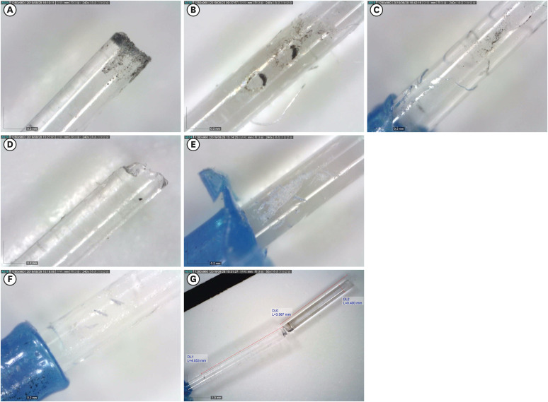
Fig. 1 Patterns of laser fiber degradation. Types of laser fiber damages. (A) Charring, (B) Limited peeled off, (C) Extensive peeled off, (D) Bumpy, (E) Bumpy, (F) Crack, (G) Break off.
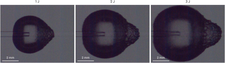
Fig. 2 Cavitation around the tip of the laser fiber. Single shot of laser emission with the energy of 1 J, 40,000 frames per second, and 1 microsec minimum exposure time.
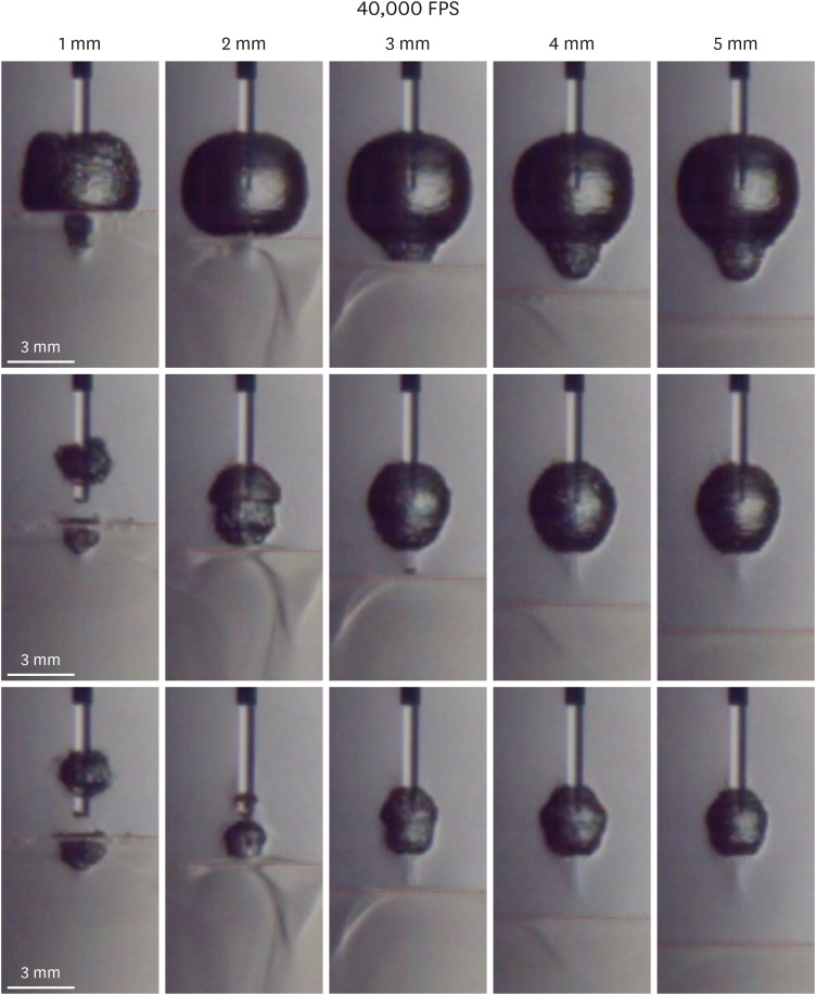
Fig. 3 Size differences of cavitation bubbles according to the laser power (J). Migration of the bubbles at the distance of 1 mm.
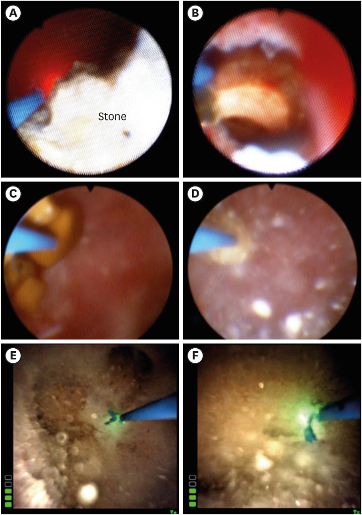
Fig. 4 Laser tip during flexible ureteroscopic surgery. The laser fiber cannot be advanced > 3 mm from the tip of the flexible ureteroscope to reach the stone when the laser fiber almost escapes from the stones because of the acute deflection angle in a lower pole (A) or in a diverticular space (B). Change in the lengths of the intact part of the laser fibers are shown before and after pop-dusting techniques for 5 minutes. The laser setting of 1 J-20 Hz (C, D) showed longer remnant laser fibers than 1 J-30 Hz (E, F).
Reference
-
1. Mues AC, Teichman JM, Knudsen BE. Quantification of holmium:yttrium aluminum garnet optical tip degradation. J Endourol. 2009; 23(9):1425–1428. PMID: 19698052.
Article2. Talso M, Emiliani E, Haddad M, Berthe L, Baghdadi M, Montanari E, et al. Laser fiber and flexible ureterorenoscopy: the safety distance concept. J Endourol. 2016; 30(12):1269–1274. PMID: 27733053.
Article3. Ryang SH, Ly TH, Yoon HS, Park DH, Cho SY. How to reduce ‘double-firing’-induced scope damage by investigating the relationship between laser fiber core degradation and fiber jacket burn? PLoS One. 2020; 15(5):e0233135. PMID: 32442200.
Article4. Lee JW, Park MG, Cho SY. How to perform the dusting technique for calcium oxalate stone phantoms during Ho:YAG laser lithotripsy. BMC Urol. 2018; 18(1):103. PMID: 30424765.
Article5. Denstedt JD, Razvi HA, Sales JL, Eberwein PM. Preliminary experience with holmium: YAG laser lithotripsy. J Endourol. 1995; 9(3):255–258. PMID: 7550269.6. Sooriakumaran P, Kaba R, Andrews HO, Buchholz NP. Evaluation of the mechanisms of damage to flexible ureteroscopes and suggestions for ureteroscope preservation. Asian J Androl. 2005; 7(4):433–438. PMID: 16281093.
Article7. Landman J, Lee DI, Lee C, Monga M. Evaluation of overall costs of currently available small flexible ureteroscopes. Urology. 2003; 62(2):218–222. PMID: 12893322.
Article8. Cecchetti W, Zattoni F, Nigro F, Tasca A. Plasma bubble formation induced by holmium laser: an in vitro study. Urology. 2004; 63(3):586–590. PMID: 15028473.
Article9. Sinibaldi G, Occhicone A, Pereira FA, Caprini D, Marino L, Michelotti F, et al. Laser induced cavitation: plasma generation and breakdown shockwave. Phys Fluids. 2019; 31(10):103302.
