Comparative histological study on the effect of tramadol abuse on the testis of juvenile and adult male albino mice
- Affiliations
-
- 1Department of Histology and Cell Biology, Faculty of Medicine, Assiut University, Assiut, Egypt
- 2Department of Histology and Cell Biology, Sphinx University, New Assiut City, Egypt
- 3Department of Anatomy and Embriology, Faculty of Medicine, Assiut University, Assiut, Egypt
- 4Department of Histology and Cell Biology, Faculty of Medicine, Aswan University, Aswan, Egypt
- 5Department of Histology and Cell Biology, Faculty of Veterinary Medicine, Assiut University, Assiut, Egypt
- KMID: 2533360
- DOI: http://doi.org/10.5115/acb.22.013
Abstract
- As a synthetic analog of codeine, tramadol is often prescribed to treat mild to moderate pains. This study was designed to estimate and compare the histological effect of tramadol on testes of both juvenile and adult male albino mice. A total number of 40 healthy male albino mice were classified into two main groups as follows: group I (juvenile group, includes 20 mice aged three weeks) subdivided equally into group Ia (control group received isotonic saline) and group Ib (tramadoltreated group received 40 mg/kg/d tramadol orally for 30 days); group II (adult group, includes 20 mice aged two months) subdivided equally into group IIa (control group received isotonic saline) and group IIb (tramadol-treated group). Juvenile and adult tramadol-treated groups showed numerous testicular changes, including blood vessels congestion, widening of intercellular spaces, vacuolization in interstitial tissues, luminal germ cells exfoliation, and increased expression of caspase-3 that indicated cellular apoptosis. In the ultrastructural examination, spermatogenic cells degenerated with the frequent appearance of apoptotic cells. Sertoli cells showed vacuolations, large lipid droplets, and disrupted intercellular cell junctions. These observed testicular changes were markedly observed in the juvenile group. Testicular abnormalities and apoptotic changes can be caused by tramadol administration. These abnormalities are more common in juvenile mice.
Keyword
Figure
-

Fig. 1 Photomicrographs of a sections of the testis from mice stained by H&E. Group Ia (A–C): (A) Normal closely packed seminiferous tubules (st) with normal contour (×100). (B) The interstitial cell of Leydig (L) are present surrounding blood vessels (Bv) (×400). (C) A magnified photomicrograph showing spermatogonia (Spg), primary spermatocytes (Pr), and rounded early spermatids (Spd). Inset shows a group of Leydig cells with rounded nuclei and acidophilic cytoplasm (×1,000). Group Ib (D–I): (D, E) Group of seminiferous tubules (st) surrounded by a connective tissue capsule (arrows), some tubules show depletion of spermatogenic cells (arrowheads). Other tubules appeared shrunken with obliteration of the lumen and increase acidophilia (tailed arrows). The blood vessels appear congested (Bv) (×100). (F, G) Disorganized germinal epithelium (arrows), focal vacuoles between the destructed cells (V), and swelling in some cells (double arrow) can be observed. Group of interstitial cells of Leydig (L) with dark irregular nuclei and amalgamated highly acidophilic cytoplasm are also seen. Notice fusion between some tubules (arrowhead) (×400). (H, I) Magnified photomicrographs showing a thick irregular basement membrane (Bm), disorientation of late spermatids (arrow), and multiple focal vacuoles (V) between cells. The interstitial cells of Leydig (L) appear with irregular dense nuclei and highly acidophilic cytoplasm (×1,000).

Fig. 2 Photomicrographs of a sections of the testis from mice from the stained by H&E. Group IIa (A, B): (A) Normal regular appearance of seminiferous tubules (st) surrounded by a regular capsule (Cs) (×400). (B) A magnified photomicrograph of the previous section showing spermatogenic cells formed of spermatogonia (Spg), primary spermatocytes (Pr), and early spermatids (Spd). Sertoli cell (S) and Leydig cells (L) can be seen (×1,000). Group IIb (C–F): (C) Irregular deformed highly acidophilic seminiferous tubules (st) some tubules have no lumen (arrow). They are surrounded by a thick irregular capsule (Cs) (×100). (D) Increased mitotic activity (arrow). Irregular basement membrane (arrowhead) can be noticed (×400). (E, F) Magnified photomicrographs showing vacuolation (V), disorganized germinal epithelium (double arrow), thick irregular basement membrane (arrowhead), and increased interstitial Leydig cell mass (L) (×1,000).
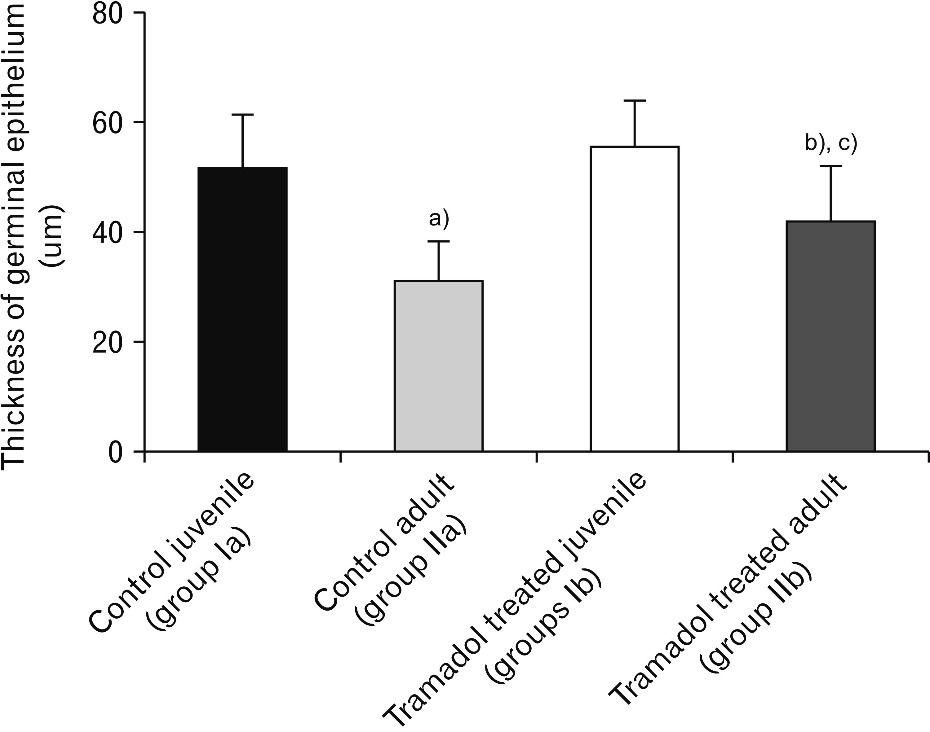
Fig. 3 The thickness of germinal epithelium was decreased significantly in Tramadol treated juvenile group when compared with the control juvenile group (a)P<0.001). Tramadol treated adult group showed a significantly decreased thickness of germinal epithelium versus the control adult group (b)P<0.001). The thickness of germinal epithelium was significantly reduced in Tramadol treated juvenile than Tramadol treated adult groups (c)P<0.001).

Fig. 4 The number of distorted tubules was increased significantly in Tramadol treated juvenile group in comparison with the control juvenile group (a)P<0.001). Tramadol treated adult group has a significantly increased number of distorted tubules than the control adult group (b)P<0.001). Tramadol treated juvenile group showed a significantly higher number of distorted tubules than the Tramadol treated adult group (c)P<0.001).

Fig. 5 (A–D) Photomicrographs of a sections of the testis from mice stained by Masson trichrome (×400). (A) In group Ia, normal distribution of collagen fibers in the testicular capsule (Cs) and basement membrane of seminiferous tubules (arrow). (B) In group Ib, excess collagen fibers in the testicular capsule (Cs) and around blood vessels (Bv). (C) In group IIa, normal distribution of collagen fibers in testicular capsule (Cs) and basement membrane of seminiferous tubules (arrow). (D) In group IIb, dense collagen fibers deposition in the capsule (Cs), and in the basement membrane of the seminiferous tubules (arrow). Note the severely congested blood vessel (Bv). (E) Graphical representation of the mean of the Seminiferous tubule capsule thickness. Data are expressed as mean±standard error of the mean. The capsule thickness was increased significantly in Tramadol treated juvenile group in comparison with the control juvenile group (a)P<0.001). Tramadol treated adult group has a significantly increased capsule thickness versus the control adult group (b)P<0.05). An insignificant difference was obtained between the Tramadol-treated juvenile and Tramadol-treated adult groups.

Fig. 6 (A–D) Photomicrographs of a sections of the testis from mice stained by caspase-3 (×400). (A, C) Few positive staining cells in control groups Ia and IIa respectively. (B, D) Increased positive cells in both tramadol-treated groups Ib and IIb. (E) Graphical representation of the mean of the number of positive cells. Data are expressed as mean±standard error of the mean (n=6). The tramadol-treated juvenile group showed significantly higher caspase-3 positive cells than the control juvenile group (a)P<0.001). Also, the Tramadol-treated adult group has a significantly increased number of caspase-3 positive cells than the control adult group (b)P<0.001). An insignificant difference was obtained between the Tramadol-treated juvenile and Tramadol-treated adult groups. The arrows refer to the positive immunostained cells

Fig. 7 Photomicrographs of a sections of the testis from mice stained by Toluidine blue (×1,000). (A) In group Ia, normal organization of spermatogenic cells. Spermatogonia (Spg), primary spermatocytes (Pr), and spermatids (Spd). Sertoli cell (S) appears with a regular vesicular nucleus and prominent nucleolus. Regular basement membrane (Bm) and myoid cell (my) are noticed. The interstitial cells of Leydig (L) are observed. (B, C) In group Ib, seminiferous tubules with hazard arranged germ cells with dense nuclei and multiple vacuolations (V). Some of Sertoli cells are detached from the basement membrane (head arrow). The basement membrane (Bm) is irregular. Increased cellularity of interstitial tissue (L) surrounding congested blood vessel (Bv). Sperm (Sp) could be seen. (D) Group IIa, seminiferous tubules are surrounded by a regular basement membrane (Bm) and have the normal organization of spermatogenic cells. Spermatogonia (Spg), primary spermatocytes (Pr), normal spermatids (Spd) with normal sperm (Sp) morphology and orientation. Sertoli cell (S) with a regular vesicular nucleus and prominent nucleolus. The interstitial cells of Leydig (L) can be observed. (E, F) In group IIb, spermatogonia (Spg) with dense nuclei, multiple vacuolations (V), darkly stained primary spermatocytes (Pr), degenerated irregular spermatids (Spd). Sertoli cells (S) show irregular nuclei. Myoid cells (my) with dense nuclei can be seen. Note the irregular basement membrane (Bm). The interstitial cells of Leydig (L) can be observed. Sperm (Sp) could be seen.

Fig. 8 Transmission electron microscopy of sections of the testis of mice (×3,600). A–D represent group Ia and E–G for group Ib. (A, B) The seminiferous tubules are surrounded by a thin regular basement membrane and the myoid cells (my) appear with their flattened nuclei. Sertoli cell (S) has a regular euchromatic nucleus (N), central nucleolus, smooth endoplasmic reticulum (sER), and mitochondria (m). Spermatogonia (Spg) is rested on the basement membrane. Primary spermatocyte (Pr) with a large rounded nucleus and finely dispersed chromatin and peripherally arranged mitochondria. An intact intercellular junction (double arrow) is observed. (C) Rounded spermatids (Spd) have spherical euchromatic nuclei and a cytoplasm showing peripherally arranged mitochondria (m). Acrosomal dense granule (arrow) and acrosomal cap (arrowhead) spread over the nucleus pole can be observed. (D) Group of Leydig cells with rounded euchromatic nuclei and cytoplasm rich in smooth endoplasmic reticulum (sER) surrounding blood vessel (Bv). Seminiferous tubules (st) and nucleus (N) could be seen. (E) Sertoli cell (S) with a large nucleus (N) and a prominent nucleolus, vacuolated cytoplasm (V), and small mitochondria (m). Large lipid droplet (arrow), irregular electron-dense spermatogonia (Spg) with vacuolated cytoplasm can be noticed (V). (F) Disorientation of elongated spermatids (Spd) and a group of degenerated cells with multiple empty spaces (V) are present. (G) Group of electron-dense Leydig cells with irregular dense nuclei (N) and dilated smooth endoplasmic reticulum (sER) can be seen around a blood vessel (Bv).
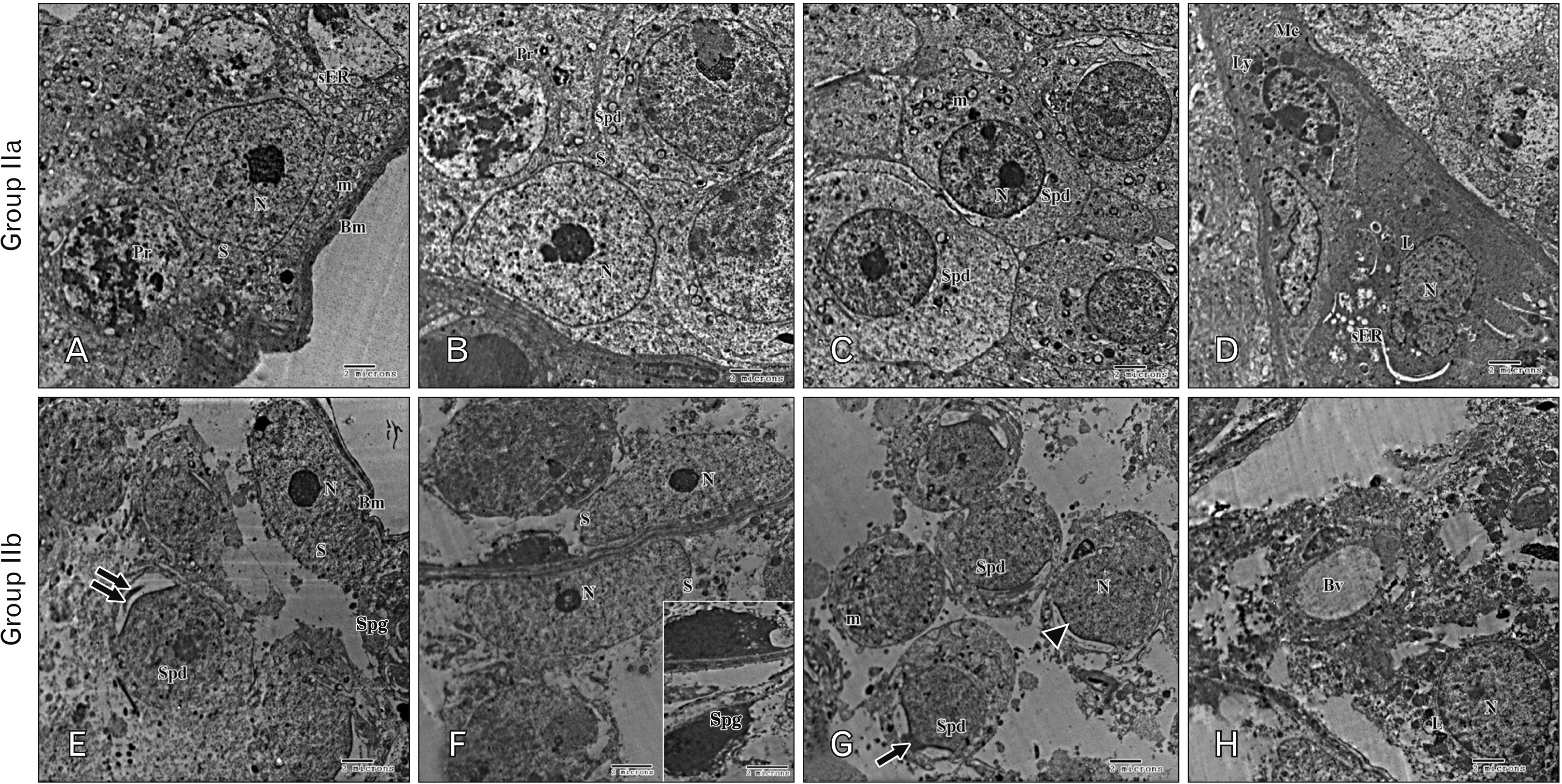
Fig. 9 Transmission electron microscopy of sections in the testis of mice (×3,600). A–D photos represent group IIa while E–H for group IIb. (A, B) Sertoli cells (S) with large rounded euchromatic nuclei (N), central nucleoli and finely dispersed chromatin, smooth endoplasmic reticulum (sER), and mitochondria (m). A primary spermatocyte (Pr) with a rounded nucleus is present. Round spermatid (Spd) with a rounded regular nucleus. The cells rest on a basement membrane (Bm). (C) Rounded spermatids (Spd) have spherical euchromatic nuclei (N) and cytoplasm rich in small-sized mitochondria (m). (D) The interstitial tissue containing Leydig cell (L) with oval to rounded euchromatic nuclei (N) and cytoplasm rich in smooth endoplasmic reticulum (sER). A macrophage (Mc) with an oval nucleus and cytoplasm engorged with lysosomes (Ly) can be noticed. (E, F) Sertoli cell (S) with large vesicular nuclei (N) and prominent nucleoli resting on an irregular basement membrane (Bm). The cytoplasm appears degenerated and rarified. Dark spermatogonia (Spg) and irregular spermatid (Spd) with capping deformity (double arrow) are observed. The inset shows spermatogonia (Spg) with electron-dense cytoplasm and an ill-defined nucleus. (G) Spermatids (Spd) appear with degenerated cytoplasm, irregular nuclei (N), peripherally arranged mitochondria (m), with capping deformity (arrowhead) and acrosomal granule (arrow). (H) Leydig cell (L) with rounded nucleus (N) and irregular degenerated cytoplasm. The interstitial blood vessel can be noticed (Bv).
Reference
-
References
1. Candeletti S, Lopetuso G, Cannarsa R, Cavina C, Romualdi P. 2006; Effects of prolonged treatment with the opiate tramadol on prodynorphin gene expression in rat CNS. J Mol Neurosci. 30:341–7. DOI: 10.1385/JMN:30:3:341. PMID: 17401159.
Article2. Hawton K, Bergen H, Simkin S, Wells C, Kapur N, Gunnell D. 2012; Six-year follow-up of impact of co-proxamol withdrawal in England and Wales on prescribing and deaths: time-series study. PLoS Med. 9:e1001213. DOI: 10.1371/journal.pmed.1001213. PMID: 22589703. PMCID: PMC3348153. PMID: 77833b219ee74089a2b3311167cc468e.
Article3. Miotto K, Cho AK, Khalil MA, Blanco K, Sasaki JD, Rawson R. 2017; Trends in tramadol: pharmacology, metabolism, and misuse. Anesth Analg. 124:44–51. DOI: 10.1213/ANE.0000000000001683. PMID: 27861439.4. Leppert W, Łuczak J. 2005; The role of tramadol in cancer pain treatment--a review. Support Care Cancer. 13:5–17. DOI: 10.1007/s00520-004-0720-4. PMID: 15668743.
Article5. Grond S, Sablotzki A. 2004; Clinical pharmacology of tramadol. Clin Pharmacokinet. 43:879–923. DOI: 10.2165/00003088-200443130-00004. PMID: 15509185.
Article6. Babalonis S, Lofwall MR, Nuzzo PA, Siegel AJ, Walsh SL. 2013; Abuse liability and reinforcing efficacy of oral tramadol in humans. Drug Alcohol Depend. 129:116–24. DOI: 10.1016/j.drugalcdep.2012.09.018. PMID: 23098678. PMCID: PMC3594406.
Article7. Scott LJ, Perry CM. 2000; Tramadol: a review of its use in perioperative pain. Drugs. 60:139–76. DOI: 10.2165/00003495-200060010-00008. PMID: 10929933.8. Aloisi AM, Ceccarelli I, Fiorenzani P, Maddalena M, Rossi A, Tomei V, Sorda G, Danielli B, Rovini M, Cappelli A, Anzini M, Giordano A. 2010; Aromatase and 5-alpha reductase gene expression: modulation by pain and morphine treatment in male rats. Mol Pain. 6:69. DOI: 10.1186/1744-8069-6-69. PMID: 20977699. PMCID: PMC2978140.
Article9. Ghoneim FM, Khalaf HA, Elsamanoudy AZ, Helaly AN. 2014; Effect of chronic usage of tramadol on motor cerebral cortex and testicular tissues of adult male albino rats and the effect of its withdrawal: histological, immunohistochemical and biochemical study. Int J Clin Exp Pathol. 7:7323–41. PMID: 25550769. PMCID: PMC4270590.10. Bar-Or D, Salottolo KM, Orlando A, Winkler JV. Tramadol ODT Study Group. 2012; A randomized double-blind, placebo-controlled multicenter study to evaluate the efficacy and safety of two doses of the tramadol orally disintegrating tablet for the treatment of premature ejaculation within less than 2 minutes. Eur Urol. 61:736–43. DOI: 10.1016/j.eururo.2011.08.039. PMID: 21889833.
Article11. Creasy DM. 2002; Histopathology of the male reproductive system I: techniques. Curr Protoc Toxicol. Chapter 16:Unit16.3. DOI: 10.1002/0471140856.tx1603s12. PMID: 20963755.
Article12. Bancroft JD, Layton C. Suvarna SK, Layton C, Bancroft JD, editors. 2013. The hematoxylins and eosin. Bancroft's Theory and Practice of Histological Techniques. 7th ed. Churchill Livingstone of Elsevier;Philadelphia: p. 173–86. DOI: 10.1016/B978-0-7020-4226-3.00010-X.
Article13. Glauert AM, Lewis PR. 2014. Biological specimen preparation for transmission electron microscopy. Princeton University Press;Princeton: DOI: 10.1002/0471140856.tx1603s12.14. O'Callaghan JP, Jensen KF. 1992; Enhanced expression of glial fibrillary acidic protein and the cupric silver degeneration reaction can be used as sensitive and early indicators of neurotoxicity. Neurotoxicology. 13:113–22. PMID: 1508411.15. Martyn-St James M, Cooper K, Kaltenthaler E, Dickinson K, Cantrell A, Wylie K, Frodsham L, Hood C. 2015; Tramadol for premature ejaculation: a systematic review and meta-analysis. BMC Urol. 15:6. DOI: 10.1186/1471-2490-15-6. PMID: 25636495. PMCID: PMC4417346.
Article16. Ahmed AI, El-Dawy K, Fawzy MM, Abdallah HA, Elsaid HN, Elmesslamy WO. 2018; Retrospective review of tramadol abuse. Slov Vet Res. 55(Suppl 20):471–83.17. Abdellatief RB, Elgamal DA, Mohamed EE. 2015; Effects of chronic tramadol administration on testicular tissue in rats: an experimental study. Andrologia. 47:674–9. DOI: 10.1111/and.12316. PMID: 25228095.
Article18. Cerilli LA, Kuang W, Rogers D. 2010; A practical approach to testicular biopsy interpretation for male infertility. Arch Pathol Lab Med. 134:1197–204. DOI: 10.5858/2009-0379-RA.1. PMID: 20670143.
Article19. Salama N, Bergh A, Damber JE. 2003; The changes in testicular vascular permeability during progression of the experimental varicocele. Eur Urol. 43:84–91. DOI: 10.1016/S0302-2838(02)00501-8. PMID: 12507549.
Article20. Awadalla EA, Salah-Eldin A. 2005; Histopathological and molecular studies on tramadol mediated hepato-renal toxicity in rats. J Pharm Biol Sci. 10:90–102.21. Essam Hafez M, Sahar Issa Y, Safaa Abdel Rahman M. 2015; Parenchymatous toxicity of tramadol: histopathological and biochemical study. J Alcohol Drug Depend. 3:225. DOI: 10.4172/2329-6488.1000225.
Article22. Elkhateeb A, El Khishin I, Megahed O, Mazen F. 2015; Effect of Nigella sativa Linn oil on tramadol-induced hepato- and nephrotoxicity in adult male albino rats. Toxicol Rep. 2:512–9. DOI: 10.1016/j.toxrep.2015.03.002. PMID: 28962386. PMCID: PMC5598165.
Article23. Mohamed D, Saber A, Omar A, Soliman A. 2014; Effect of cadmium on the testes of adult albino rats and the ameliorating effect of zinc and vitamin E. Br J Sci. 11:72–95.24. Nolte T, Harleman JH, Jahn W. 1995; Histopathology of chemically induced testicular atrophy in rats. Exp Toxicol Pathol. 47:267–86. DOI: 10.1016/S0940-2993(11)80260-5. PMID: 8855122.
Article25. Caju FM, Queiroz GCD, Torres SM, Tenório BM, Júnior VAS. 2011; Opioid system manipulation during testicular development: results on sperm production and sertoli cells population. Acta Sci Biol Sci. 33:219–25. DOI: 10.4025/actascibiolsci.v33i2.5940. PMID: 9379367d9bf24995aa270c4eff76ad63.26. Ahmed MA, Kurkar A. 2014; Effects of opioid (tramadol) treatment on testicular functions in adult male rats: the role of nitric oxide and oxidative stress. Clin Exp Pharmacol Physiol. 41:317–23. DOI: 10.1111/1440-1681.12213. PMID: 24472030.
Article27. Zhang YT, Zheng QS, Pan J, Zheng RL. 2004; Oxidative damage of biomolecules in mouse liver induced by morphine and protected by antioxidants. Basic Clin Pharmacol Toxicol. 95:53–8. DOI: 10.1111/j.1742-7843.2004.950202.x. PMID: 15379780.
Article28. Berruti G. 2006; Signaling events during male germ cell differentiation: update, 2006. Front Biosci. 11:2144–56. DOI: 10.2741/1957. PMID: 16720301.
Article29. Lombardo F, Sgrò P, Salacone P, Gilio B, Gandini L, Dondero F, Jannini EA, Lenzi A. 2005; Androgens and fertility. J Endocrinol Invest. 28(3 Suppl):51–5. DOI: 10.1111/j.1365-2605.2005.00585.x. PMID: 16236065.30. Soliman HM, Wagih HM, Attia GM, Algaidi SA. 2014; Light and electron microscopic study on the effect of antischizophrenic drugs on the structure of seminiferous tubules of adult male albino rats. Folia Histochem Cytobiol. 52:335–49. DOI: 10.5603/FHC.a2014.0038. PMID: 25535927.
Article31. Heidari Z, Mahmoudzadeh-Sagheb H, Kohan F. 2012; A quantitative and qualitative study of rat testis following administration of methadone and buprenorphine. Int J High Risk Behav Addict. 1:14–7. DOI: 10.5812/ijhrba.4119.
Article32. Sorge RE, Stewart J. 2006; The effects of long-term chronic buprenorphine treatment on the locomotor and nucleus accumbens dopamine response to acute heroin and cocaine in rats. Pharmacol Biochem Behav. 84:300–5. DOI: 10.1016/j.pbb.2006.05.013. PMID: 16806444.
Article33. Rashed RMA, Mohamed IK, El-Alfy SH. 2010; Effects of two different doses of melatonin on the spermatogenic cells of rat testes: a light and electron microscopic study. Egypt J Histol. 33:819–35.34. Manivannan B, Mittal R, Goyal S, Ansari AS, Lohiya NK. 2009; Sperm characteristics and ultrastructure of testes of rats after long-term treatment with the methanol subfraction of Carica papaya seeds. Asian J Androl. 11:583–99. DOI: 10.1038/aja.2009.25. PMID: 19648937. PMCID: PMC3735006.
Article35. Jezek D, Simunić-Banek L, Pezerović-Panijan R. 1993; Effects of high doses of testosterone propionate and testosterone enanthate on rat seminiferous tubules--a stereological and cytological study. Arch Toxicol. 67:131–40. DOI: 10.1007/BF01973684. PMID: 8481101.36. Sharpe RM, McKinnell C, Kivlin C, Fisher JS. 2003; Proliferation and functional maturation of Sertoli cells, and their relevance to disorders of testis function in adulthood. Reproduction. 125:769–84. DOI: 10.1530/rep.0.1250769. PMID: 12773099.
Article37. Mohamed HM, Mahmoud AM. 2019; Chronic exposure to the opioid tramadol induces oxidative damage, inflammation and apoptosis, and alters cerebral monoamine neurotransmitters in rats. Biomed Pharmacother. 110:239–47. DOI: 10.1016/j.biopha.2018.11.141. PMID: 30508735.
Article38. Drobnis EZ, Nangia AK. 2017; Pain medications and male reproduction. Adv Exp Med Biol. 1034:39–57. DOI: 10.1007/978-3-319-69535-8_6. PMID: 29256126.
Article39. Erkkilä K, Aito H, Aalto K, Pentikäinen V, Dunkel L. 2002; Lactate inhibits germ cell apoptosis in the human testis. Mol Hum Reprod. 8:109–17. DOI: 10.1093/molehr/8.2.109. PMID: 11818513.
Article40. Eddy EM. 1999; Role of heat shock protein HSP70-2 in spermatogenesis. Rev Reprod. 4:23–30. DOI: 10.1530/ror.0.0040023. PMID: 10051099.
Article41. Newton AP, Cadena SM, Rocha ME, Carnieri EG, Martinelli de Oliveira MB. 2005; Effect of triclosan (TRN) on energy-linked functions of rat liver mitochondria. Toxicol Lett. 160:49–59. DOI: 10.1016/j.toxlet.2005.06.004. PMID: 16023799.
Article42. Tamura I, Kanbara Y, Saito M, Horimoto K, Satoh M, Yamamoto H, Oyama Y. 2012; Triclosan, an antibacterial agent, increases intracellular Zn(2+) concentration in rat thymocytes: its relation to oxidative stress. Chemosphere. 86:70–5. DOI: 10.1016/j.chemosphere.2011.09.009. PMID: 22000841.
Article43. Abdel-Zaher AO, Abdel-Rahman MS, Elwasei FM. 2011; Protective effect of Nigella sativa oil against tramadol-induced tolerance and dependence in mice: role of nitric oxide and oxidative stress. Neurotoxicology. 32:725–33. DOI: 10.1016/j.neuro.2011.08.001. PMID: 21855572.
Article44. Halawa AM. 2010; Effect of sildenafil administration on ischemic reperfusion of the testis in adult albino rat light and electron microscopic study. Egypt J Histol. 2:380–95.45. Gouda ZA, Selim AO. 2013; A possible correlation between the testicular structure and short photoperiod exposure in young albino rats: light and electron microscopic study. Egypt J Histol. 36:28–38. DOI: 10.1097/01.EHX.0000423980.95382.0c.
Article46. Bassiony MM, Youssef UM, El-Gohari H. 2020; Free testosterone and prolactin levels and sperm morphology and function among male patients with tramadol abuse: a case-control study. J Clin Psychopharmacol. 40:405–8. DOI: 10.1097/JCP.0000000000001223. PMID: 32639294.
Article47. Popovic M, Janicijevic-Hudomal S, Kaurinovic B, Rasic J, Trivic S, Vojnović M. 2009; Antioxidant effects of some drugs on immobilization stress combined with cold restraint stress. Molecules. 14:4505–16. DOI: 10.3390/molecules14114505. PMID: 19924083. PMCID: PMC6254731.
Article48. El-Gaafarawi II. 2006; Biochemical toxicity induced by tramadol administration in male rats. Egypt J Hosp Med. 23:353–62. DOI: 10.21608/ejhm.2006.17946.
Article
