Myiasis with Larvae of Sarcophaga Species in a Diabetic Foot with Gangrene in Korea: A Case Report
- Affiliations
-
- 1Departments of Orthopedic Surgery, Yonsei University College of Medicine, Seoul, Korea
- 2Environmental Medical Biology, Yonsei University College of Medicine, Seoul, Korea
- KMID: 2533198
- DOI: http://doi.org/10.14193/jkfas.2022.26.3.148
Abstract
- Myiasis is the parasitic infestation of the body of a live animal by fly larvae that grow inside the host while feeding on its tissue. Necrotic tissue is a favorable environment for larvae to thrive, which can be seen easily in patients with a diabetic foot. Myiasis in a diabetic foot is rare but is constantly being reported. The common larvae genera causing myiasis are Calliphoridae, Sarcophagidae, and Muscidae. This paper reports a rare case of sarcophaga myiasis in a diabetic foot. To the best of the author’s knowledge, this is the first case report in Korea regarding human myiasis with the sarcophaga genus.
Keyword
Figure
-
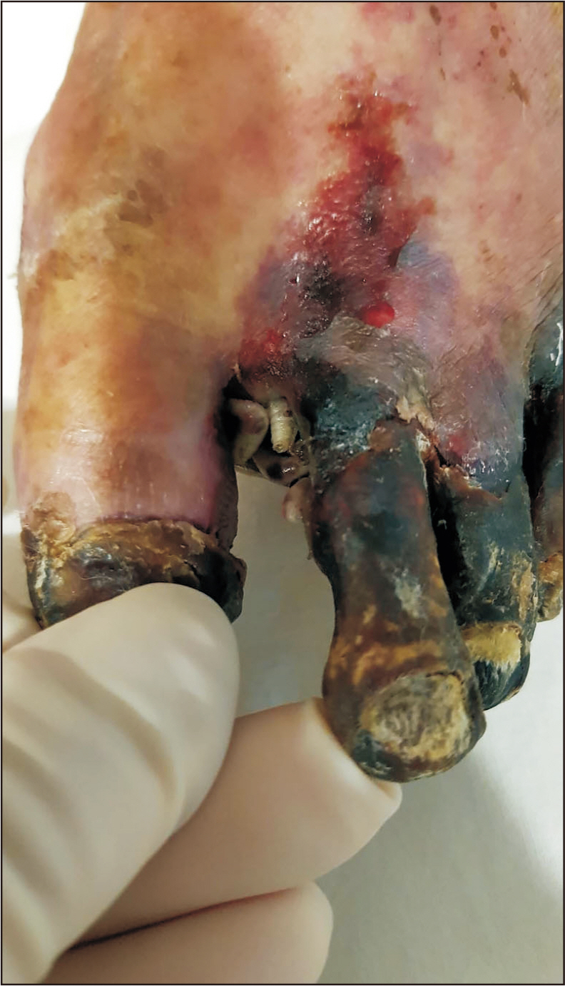
Figure 1 Initial clinical photograph of left foot with gangrenous change and maggots in the first webspace.

Figure 2 Preoperative image studies. (A) Dorsoplantar radiograph of left foot shows osteolysis of fourth proximal phalanx (arrow). (B) Internal oblique radiograph of left foot shows osteolysis of fifth proximal phalanx (arrow). (C) Computed tomography of left foot shows gas formation on fourth webspace (arrow).

Figure 3 Macroscopic view of extracted maggot. (A) The length of larvae was 11 mm. (B) Anterior view of larvae ×16 magnification. (C) Posterior view of larvae ×16 magnification.

Figure 4 Optic microscope view of extract maggot. (A) Three pairs of posterior spiracle and spiracular slits not pointing toward opening in peritreme can be seen in ×890 magnification. (B) Small pores listed in a row on anterior spiracle can be seen in ×1,400 magnification.
Reference
-
References
1. Singh A, Singh Z. 2015; Incidence of myiasis among humans-a review. Parasitol Res. 114:3183–99. doi: 10.1007/s00436-015-4620-y. DOI: 10.1007/s00436-015-4620-y. PMID: 26220558.
Article2. Francesconi F, Lupi O. 2012; Myiasis. Clin Microbiol Rev. 25:79–105. doi: 10.1128/CMR.00010-11. DOI: 10.1128/CMR.00010-11. PMID: 22232372. PMCID: PMC3255963.
Article3. Callaghan BC, Cheng HT, Stables CL, Smith AL, Feldman EL. 2012; Diabetic neuropathy: clinical manifestations and current treatments. Lancet Neurol. 11:521–34. doi: 10.1016/S1474-4422(12)70065-0. DOI: 10.1016/S1474-4422(12)70065-0.
Article4. Armstrong DG, Boulton AJM, Bus SA. 2017; Diabetic foot ulcers and their recurrence. N Engl J Med. 376:2367–75. doi: 10.1056/NEJMra1615439. DOI: 10.1056/NEJMra1615439. PMID: 28614678.
Article5. Rivers DB, Dahlem GA. The science of forensic entomology. 2014. p. 121–87. Wiley-Blackwell;Hoboken:6. Safdar N, Young DK, Andes D. 2003; Autochthonous furuncular myiasis in the United States: case report and literature review. Clin Infect Dis. 36:e73–80. doi: 10.1086/368183. DOI: 10.1086/368183. PMID: 12652404.
Article7. Uysal S, Ozturk AM, Tasbakan M, Simsir IY, Unver A, Turgay N, et al. 2018; Human myiasis in patients with diabetic foot: 18 cases. Ann Saudi Med. 38:208–13. doi: 10.5144/0256-4947.2018.208. DOI: 10.5144/0256-4947.2018.208. PMID: 29848939. PMCID: PMC6074300.
Article8. Demirel Kaya F, Orkun Ö, Çakmak A, İnkaya AÇ, Ergüven S. 2014; [Cutanous myiasis caused by Sarcophaga spp. larvae in a diabetic patient]. Mikrobiyol Bul. 48:356–61. Turkish. doi: 10.5578/mb.7107. DOI: 10.5578/mb.7107. PMID: 24819275.
Article9. Zaglool DA, Tayeb K, Khodari YA, Farooq MU. 2013; First case report of human myiasis with Sarcophaga species in Makkah city in the wound of a diabetic patient. J Nat Sci Biol Med. 4:225–8. doi: 10.4103/0976-9668.107301. DOI: 10.4103/0976-9668.107301. PMID: 23633868. PMCID: PMC3633283.
Article10. Sherman RA. 2002; Maggot therapy for foot and leg wounds. Int J Low Extrem Wounds. 1:135–42. doi: 10.1177/1534734602001002009. DOI: 10.1177/1534734602001002009. PMID: 15871964.
Article
- Full Text Links
-
- Actions
-
Cited
- CITED
-
- Close
- Share
-
- Similar articles
-
- Gastrointestinal Myiasis by Larvae of Sarcophaga sp. and Oestrus sp. in Egypt: Report of Cases, and Endoscopical and Morphological Studies
- Nosocomial Oral Myiasis by Sarcophaga sp. in Turkey
- A Case of Cutaneous Wound Myiasis Associated with Basal Cell Carcinoma by Sarcophaga africa
- Traumatic Myiasis Caused by an Association of Sarcophaga tibialis (Diptera: Sarcophagidae) and Lucilia sericata (Diptera: Calliphoridae) in a Domestic Cat in Italy
- Ignatzschineria larvae Bacteremia Following Lucilia sp. Myiasis in an Irregular Migrant: A Case Report
