Correlation between the Fibrillar Pattern in Ultrasonography and Clinical Factors after Achilles Tendon Repair
- Affiliations
-
- 1Department of Orthopedic Surgery, Inje University Sanggye Paik Hospital, Seoul, Korea
- 2Yonsei Hangang Hospital, Seoul, Korea
- KMID: 2533194
- DOI: http://doi.org/10.14193/jkfas.2022.26.3.123
Abstract
- Purpose
To evaluate the correlation between the fibrillar pattern of the Achilles tendon on ultrasonography (US) and functional outcomes in patients who underwent open tendon repair after Achilles tendon rupture.
Materials and Methods
Data of 44 patients who had been subjected to US at least 6 months after repair, during the period between July 2012 and July 2019 were reviewed. Those with bilateral tendon rupture, re-rupture, open injury, and chronic or insertional rupture, were excluded from the review. We divided them into two groups, the homogenous group (HoP) and the heterogenous group (HeP) based on the fibrillar pattern on US. We also divided the HoP into linear and wavy subgroups, and the HeP into no hypoechoic lesion and hypoechoic lesion subgroups. The rupture type of the Achilles tendon, radiographic factors including US and magnetic resonance images, patient-related, surgical factors, and clinical results at the last visit after repair were assessed retrospectively.
Results
The tendon thickness was 11.4±1.7 mm in the HoP, and 14.5±3.0 mm in the HeP (p<0.001). A shredded pattern was observed in 17 cases (65.4%) in the HoP, and in 17 cases (94.4%) in the HeP (p=0.031). The mean value of the number of sutures used was 8.9±3.05 in the HoP and 11.39±1.75 in the HeP (p=0.001). The mean value of the difference in calf circumference was 0.9±0.67 cm in the HoP and 1.36±0.71 cm in the HeP (p=0.037). There were no statistically significant differences in the fibrillar patterns and patient-related factors.
Conclusion
The fibrillar pattern observed after repair was correlated with the functional outcome and showed a significant relationship with the rupture pattern and the number of sutures used. Therefore, we suggest a careful individualized postoperative rehabilitation protocol to maximize functional outcomes by referring to the fibrillar pattern in US, especially in shredded ruptures.
Keyword
Figure
-

Figure 1 (A) Longitudinal scan of a homogenous fibrillary pattern showing parallel longitudinal architecture of the tendon. (B) Axial scan of a homogenous fibrillary pattern. (C) Longitudinal scan of a heterogenous fibrillary pattern showing not parallel longitudinal architecture of the tedndon. (D) Axial scan of a heterogenous fibrillary pattern showing multiple hypoechogenic area.

Figure 2 Flow charts of Achilles tendon rupture type and ultrasonographic findings.

Figure 3 Saggital magnetic resonance image of a Achilles tendon rupture. On T2-weighted image the Achilles tendon fiber is ruptured 5 cm above the calcaneal tubercle. Red arrow indicates the mixed high signal intensity of distal Achilles tendon stump meaning tendinopathy.
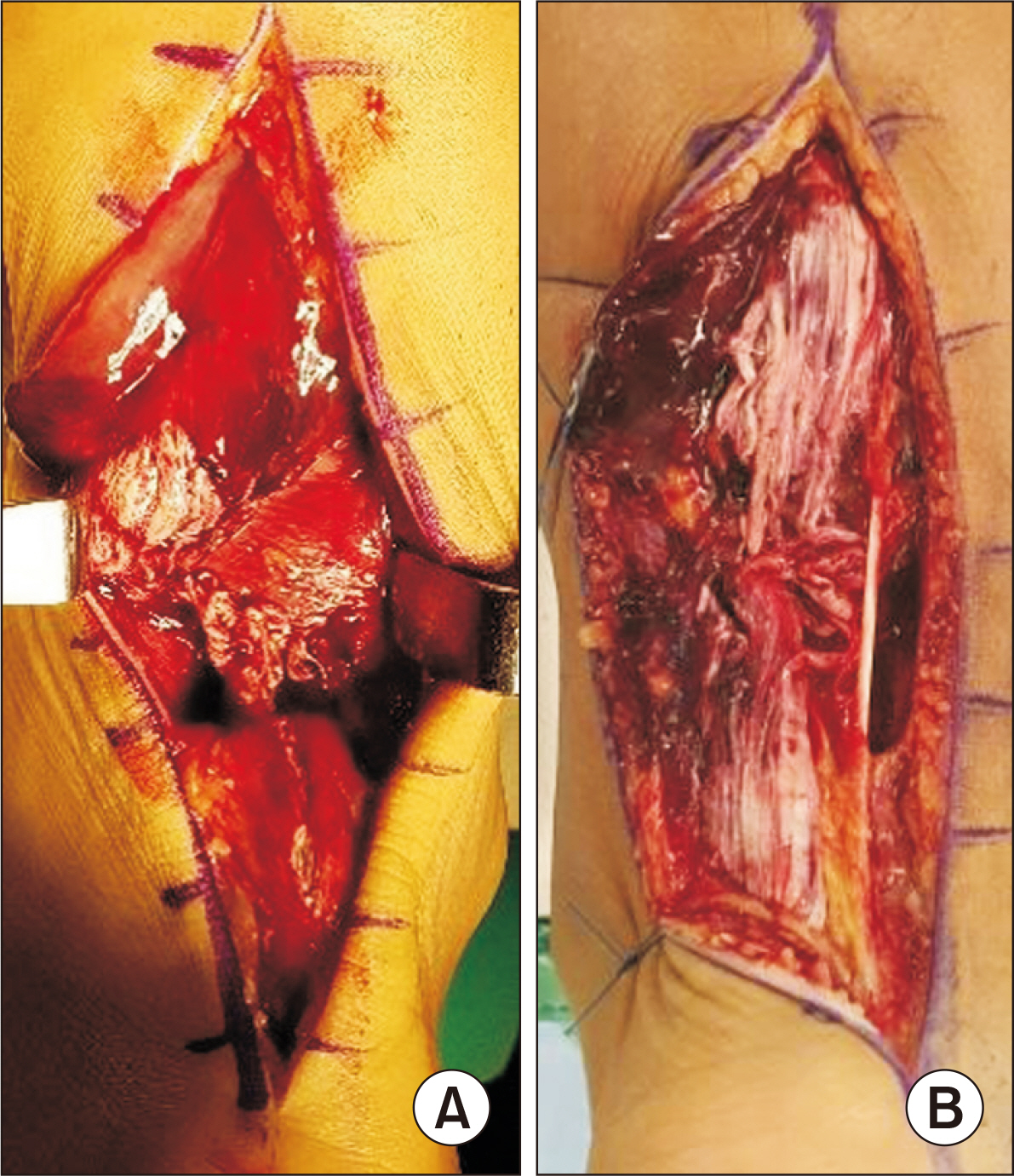
Figure 4 Intraoperative medical photo of Achilles tendon rupture fiber. (A) Simple rupture and (B) shredded rupture.
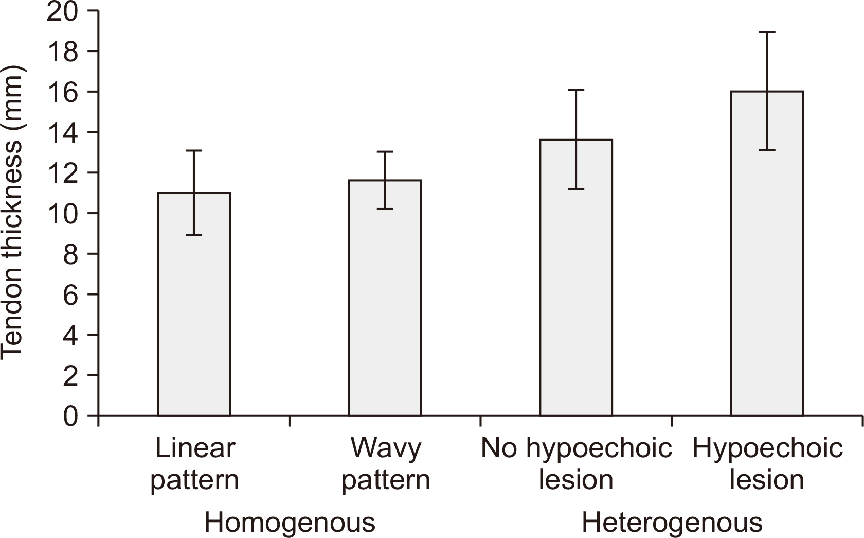
Figure 5 The mean value graph of tendon thickness of each group. The mean value showed an increase from homogenous group to heterogenous group. There is a significant difference between homogenous group and heterogenous group. There is no significant difference between linear pattern group and wavy pattern group, and no significant difference between no hypoechoic lesion group and hypoechoic lesion group.
Reference
-
References
1. Lantto I, Heikkinen J, Flinkkilä T, Ohtonen P, Leppilahti J. 2015; Epidemiology of Achilles tendon ruptures: increasing incidence over a 33-year period. Scand J Med Sci Sports. 25:e133–8. doi: 10.1111/sms.12253. DOI: 10.1111/sms.12253. PMID: 24862178.
Article2. Ciloglu O, Görgülü FF. 2020; Evaluation of a torn Achilles tendon after surgical repair: an ultrasound and elastographic study with 1-year follow-up. J Ultrasound Med. 39:1263–9. doi: 10.1002/jum.15214. DOI: 10.1002/jum.15214. PMID: 31943316.3. Nicholson G, Walker J, Dawson Z, Bissas A, Harris N. 2020; Morphological and functional outcomes of operatively treated Achilles tendon ruptures. Phys Sportsmed. 48:290–7. doi: 10.1080/00913847.2019.1685364. DOI: 10.1080/00913847.2019.1685364. PMID: 31662010.4. Braccagni M, Grange S, Arcade A, Klasan A, Boyer B, Farizon F, et al. 2021; What are the sonographic outcomes of acute Achilles tendon rupture? Nonoperative versus surgical repair? Surg Technol Int. 39:427–32. doi: 10.52198/21.STI.39.OS1505. DOI: 10.52198/21.STI.39.OS1505.5. Kim DH, Choi JH, Park CH, Park HJ, Yoon KJ, Lee YT. 2021; The diagnostic significance of ultrasonographic measurement of the Achilles tendon thickness for the insertional Achilles tendinopathy in patients with heel pain. J Clin Med. 10:2165. doi: 10.3390/jcm10102165. DOI: 10.3390/jcm10102165. PMID: 34067786. PMCID: PMC8157148.
Article6. Gitto S, Draghi AG, Bortolotto C, Draghi F. 2016; Sonography of the Achilles tendon after complete rupture repair: what the radiologist should know. J Ultrasound Med. 35:2529–36. doi: 10.7863/ultra.16.01092. DOI: 10.7863/ultra.16.01092. PMID: 27738294.7. Kauwe M. 2017; Acute Achilles tendon rupture: clinical evaluation, conservative management, and early active rehabilitation. Clin Podiatr Med Surg. 34:229–43. doi: 10.1016/j.cpm.2016.10.009. DOI: 10.1016/j.cpm.2016.10.009. PMID: 28257676.8. Rupp S, Tempelhof S, Fritsch E. 1995; Ultrasound of the Achilles tendon after surgical repair: morphology and function. Br J Radiol. 68:454–8. doi: 10.1259/0007-1285-68-809-454. DOI: 10.1259/0007-1285-68-809-454. PMID: 7788228.
Article9. Karjalainen PT, Ahovuo J, Pihlajamäki HK, Soila K, Aronen HJ. 1996; Postoperative MR imaging and ultrasonography of surgically repaired Achilles tendon ruptures. Acta Radiol. 37:639–46. doi: 10.1177/02841851960373P244. DOI: 10.1177/02841851960373P244. PMID: 8915267.
Article10. Rominger MB, Bachmann G, Schulte S, Zedler A. 1998; [Value of ultrasound and magnetic resonance imaging in the control of the postoperative progress after Achilles tendon rupture]. Rofo. 168:27–35. German. doi: 10.1055/s-2007-1015178. DOI: 10.1055/s-2007-1015178. PMID: 9501931.11. Bleakney RR, Tallon C, Wong JK, Lim KP, Maffulli N. 2002; Long-term ultrasonographic features of the Achilles tendon after rupture. Clin J Sport Med. 12:273–8. doi: 10.1097/00042752-200209000-00003. DOI: 10.1097/00042752-200209000-00003. PMID: 12394198.
Article12. Hollenberg GM, Adams MJ, Weinberg EP. 2000; Sonographic appearance of nonoperatively treated Achilles tendon ruptures. Skeletal Radiol. 29:259–64. doi: 10.1007/s002560050604. DOI: 10.1007/s002560050604. PMID: 10883444.
Article13. O’Reilly MA, Massouh H. 1993; Pictorial review: the sonographic diagnosis of pathology in the Achilles tendon. Clin Radiol. 48:202–6. doi: 10.1016/S0009-9260(05)80284-3. DOI: 10.1016/S0009-9260(05)80284-3.14. Gould HP, Bano JM, Akman JL, Fillar AL. 2021; Postoperative rehabilitation following Achilles tendon repair: a systematic review. Sports Med Arthrosc Rev. 29:130–45. doi: 10.1097/JSA.0000000000000309. DOI: 10.1097/JSA.0000000000000309. PMID: 33972490.
Article15. Krackow KA, Thomas SC, Jones LC. 1986; A new stitch for ligament-tendon fixation. Brief note. J Bone Joint Surg Am. 68:764–6. doi: 10.2106/00004623-198668050-00020. DOI: 10.2106/00004623-198668050-00020.
Article16. Chiodo CP, Glazebrook M, Bluman EM, Cohen BE, Femino JE, Giza E, et al. 2010; Diagnosis and treatment of acute Achilles tendon rupture. J Am Acad Orthop Surg. 18:503–10. doi: 10.5435/00124635-201008000-00007. DOI: 10.5435/00124635-201008000-00007. PMID: 20675643.
Article17. Margetić P, Miklić D, Rakić-Ersek V, Doko Z, Lubina ZI, Brkljacić B. 2007; Comparison of ultrasonographic and intraoperative findings in Achilles tendon rupture. Coll Antropol. 31:279–84.18. Maffulli N, Dymond NP, Regine R. 1990; Surgical repair of ruptured Achilles tendon in sportsmen and sedentary patients: a longitudinal ultrasound assessment. Int J Sports Med. 11:78–84. doi: 10.1055/s-2007-1024767. DOI: 10.1055/s-2007-1024767. PMID: 2180833.
Article19. Majewski M, Lehmann M, Dick W, Steinbrück K. 2003; [Value of sonography to monitor the course of Achilles tendon rupture after treatment--comparison of conservative therapy, percutaneous tendon adaptation, and open suture]. Unfallchirurg. 106:556–60. German. doi: 10.1007/s00113-003-0623-8. DOI: 10.1007/s00113-003-0623-8. PMID: 12883782.20. Deng S, Sun Z, Zhang C, Chen G, Li J. 2017; Surgical treatment versus conservative management for acute Achilles tendon rupture: a systematic review and meta-analysis of randomized controlled trials. J Foot Ankle Surg. 56:1236–43. doi: 10.1053/j.jfas.2017.05.036. DOI: 10.1053/j.jfas.2017.05.036. PMID: 29079238.21. Leppilahti J, Lähde S, Forsman K, Kangas J, Kauranen K, Orava S. 2000; Relationship between calf muscle size and strength after achilles rupture repair. Foot Ankle Int. 21:330–5. doi: 10.1177/107110070002100410. DOI: 10.1177/107110070002100410. PMID: 10808974.
Article22. Szaro P, Ghali Gataa K. 2021; The correlations between dimensions of the normal tendon and tendinopathy changed Achilles tendon in routine magnetic resonance imaging. Sci Rep. 11:6131. doi: 10.1038/s41598-021-85604-9. DOI: 10.1038/s41598-021-85604-9. PMID: 33731785. PMCID: PMC7969943.
Article23. Mahoney PG, James PD, Howell CJ, Swannell AJ. 1981; Spontaneous rupture of the Achilles tendon in a patient with gout. Ann Rheum Dis. 40:416–8. doi: 10.1136/ard.40.4.416. DOI: 10.1136/ard.40.4.416. PMID: 7259334. PMCID: PMC1000740.24. Hess GW. 2010; Achilles tendon rupture: a review of etiology, population, anatomy, risk factors, and injury prevention. Foot Ankle Spec. 3:29–32. doi: 10.1177/1938640009355191. DOI: 10.1177/1938640009355191. PMID: 20400437.
- Full Text Links
-
- Actions
-
Cited
- CITED
-
- Close
- Share
-
- Similar articles
-
- Long Term Result of Four Cases without a Staged Reconstruction of an Infected Achilles Tendon Following Repair
- Treatment of Massive Defect in Achilles Tendon with Tendon Allograft: A Case Report
- Heterotopic Ossification of a Partially Ruptured Achilles Tendon (A Case Report)
- Deep Vein Thrombosis after Achilles Tendon Repair: A Case Report
- Reapir of the Torn Achilles Tendon, Using the Plantaris Tendon
