Comprehensive investigations of key mitochondrial metabolic changes in senescent human fibroblasts
- Affiliations
-
- 1Chair of Medical and Molecular Genetics Research, Department of Clinical Laboratory Sciences, College of Applied Medical Sciences, King Saud University, Riyadh 12372, Saudi Arabia
- KMID: 2530946
- DOI: http://doi.org/10.4196/kjpp.2022.26.4.263
Abstract
- There is a paucity of detailed data related to the effect of senescence on the mitochondrial antioxidant capacity and redox state of senescent human cells. Activities of TCA cycle enzymes, respiratory chain complexes, hydrogen peroxide (H 2 O 2 ), superoxide anions (SA), lipid peroxides (LPO), protein carbonyl content (PCC), thioredoxin reductase 2 (TrxR2), superoxide dismutase 2 (SOD2), glutathione peroxidase 1 (GPx1), glutathione reductase (GR), reduced glutathione (GSH), and oxidized glutathione (GSSG), along with levels of nicotinamide cofactors and ATP content were measured in young and senescent human foreskin fibroblasts. Primary and senescent cultures were biochemically identified by monitoring the augmented cellular activities of key glycolytic enzymes including phosphofructokinase, lactate dehydrogenase, and glycogen phosphorylase, and accumulation of H2O2 , SA, LPO, PCC, and GSSG. Citrate synthase, aconitase, α-ketoglutarate dehydrogenase, succinate dehydrogenase, malate dehydrogenase, isocitrate dehydrogenase, and complex I-III, IIIII, and IV activities were significantly diminished in P25 and P35 cells compared to P5 cells. This was accompanied by significant accumulation of mitochondrial H2O2 , SA, LPO, and PCC, along with increased transcriptional and enzymatic activities of TrxR2, SOD2, GPx1, and GR. Notably, the GSH/GSSG ratio was significantly reduced whereas NAD+ /NADH and NADP+ /NADPH ratios were significantly elevated. Metabolic exhaustion was also evident in senescent cells underscored by the severely diminished ATP/ ADP ratio. Profound oxidative stress may contribute, at least in part, to senescence pointing at a potential protective role of antioxidants in aging-associated disease.
Keyword
Figure
-
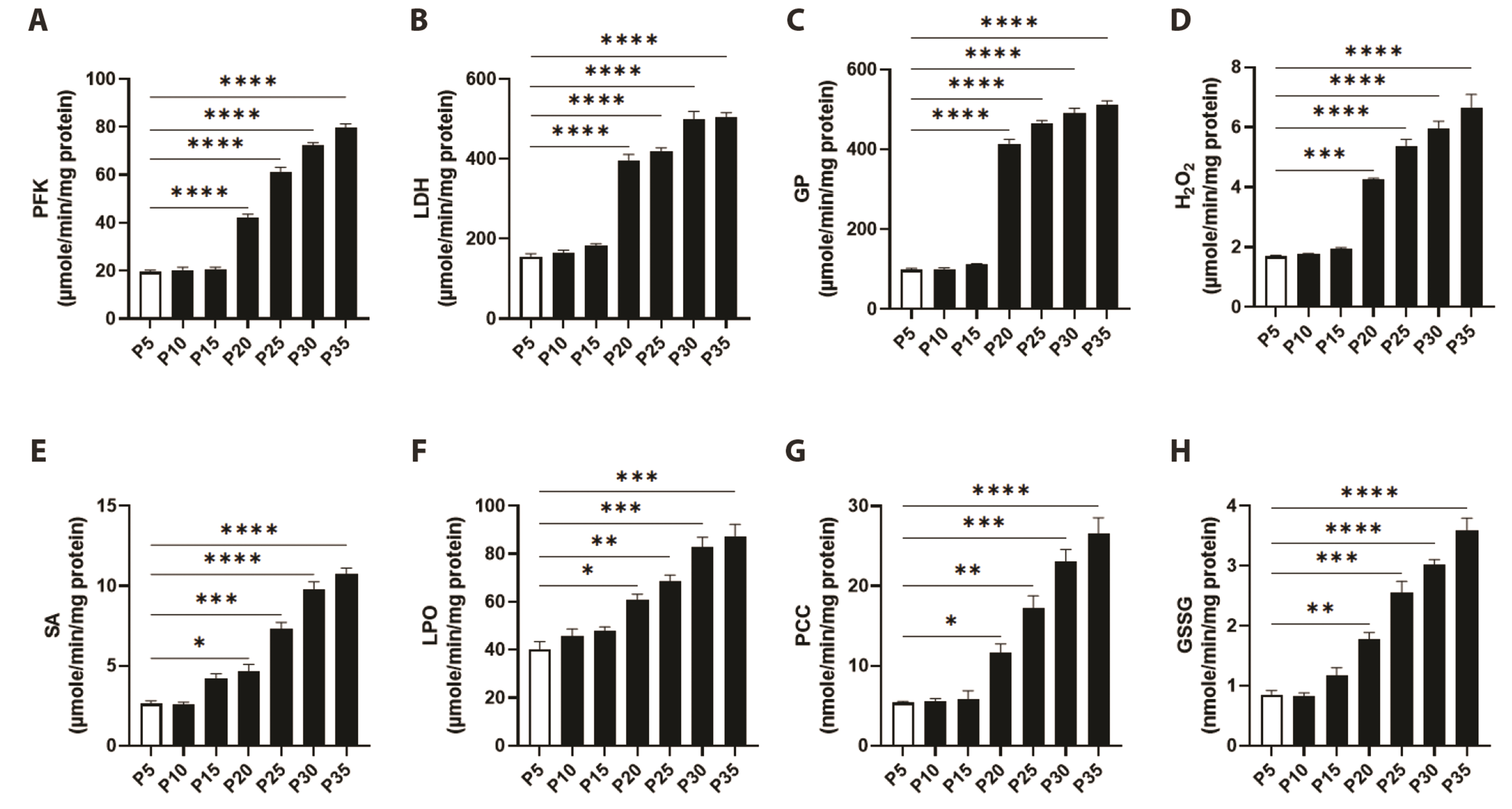
Fig. 1 Identification of senescent fibroblasts by monitoring total cellular glucose and glycogen degradative enzyme activities and oxidative stress markers in confluent serial subcultures. Enzyme activities of (A) phosphofructokinase (PFK), (B) lactate dehydrogenase (LDH), and (C) glycogen phosphorylase (GP), along with levels of (D) hydrogen peroxide (H2O2) and (E) superoxide anions (SA), (F) lipid peroxides (LPO), (G) protein carbonyl content (PCC), and (H) oxidized glutathione (GSSG) in P5-P35 confluent cells. *p < 0.05, **p < 0.01, ***p < 0.001, and ****p < 0.0001 indicate significant difference from control P5 values (n = 6).

Fig. 2 Mitochondrial specific activities of TCA cycle enzymes in primary P5 and senescent P25 and P35 fibroblasts. Enzyme activities of (A) aconitase (can), (B) α-ketoglutarate dehydrogenase (α-KGDH), (C) succinate dehydrogenase (SDH), (D) malate dehydrogenase (MDH), (E) NAD+- isocitrate dehydrogenase (ICD), and (F) NADP+-ICD in young (P5) and senescent (P25 and P35) confluent cells. ***p < 0.001 and ****p < 0.0001 indicate significant difference from control P5 values (n = 6).
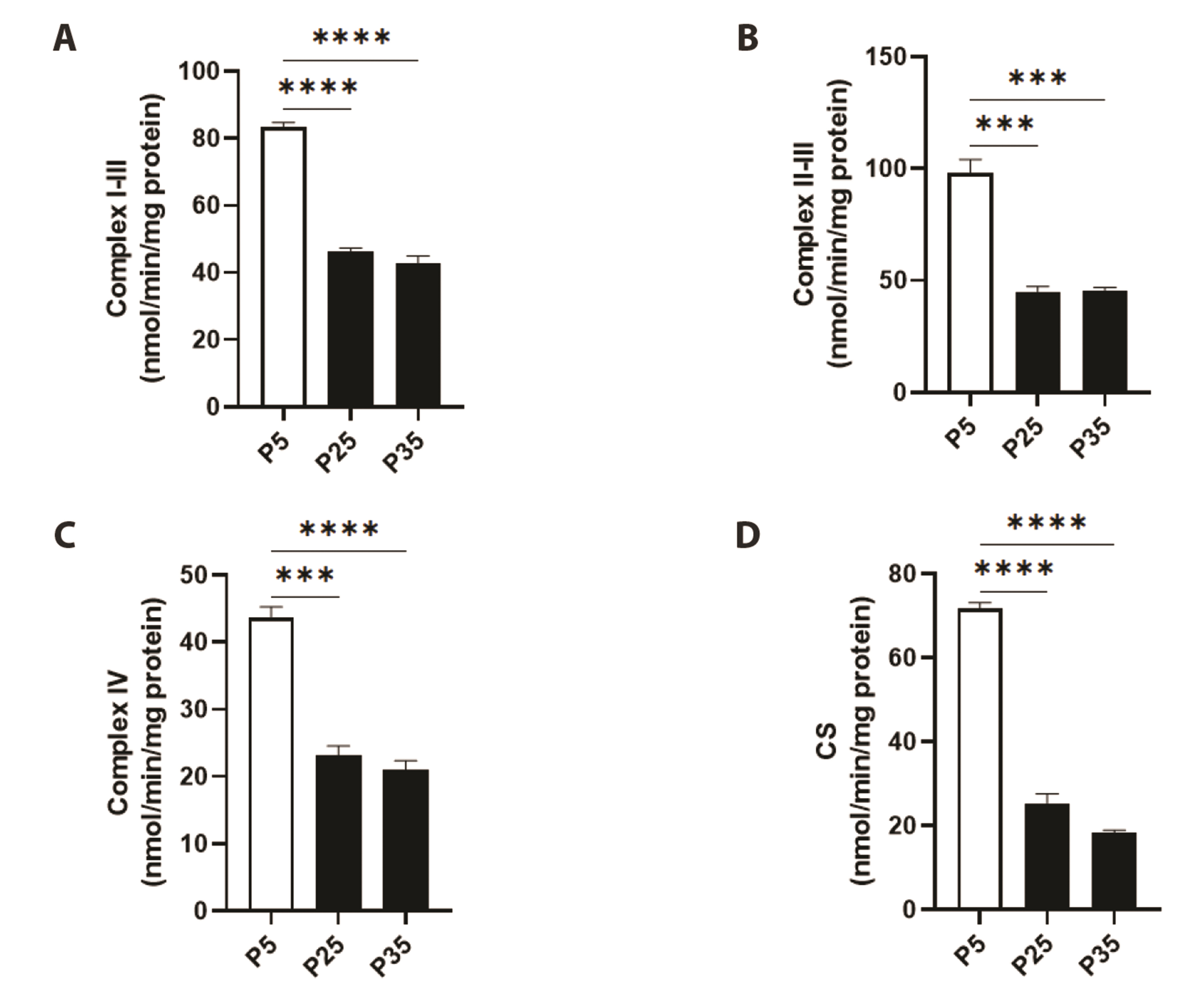
Fig. 3 Mitochondrial specific activities of electron transport chain (ETC) enzymes in primary P5 and senescent P25 and P35 fibroblasts. Enzyme activities of (A) complex I-III, (B) complex II-III, (C) complex IV, and (D) citrate synthase (CS) in young (P5) and senescent (P25 and P35) confluent cells. ***p < 0.001 and ****p < 0.0001 indicate significant difference from control P5 values (n = 6).
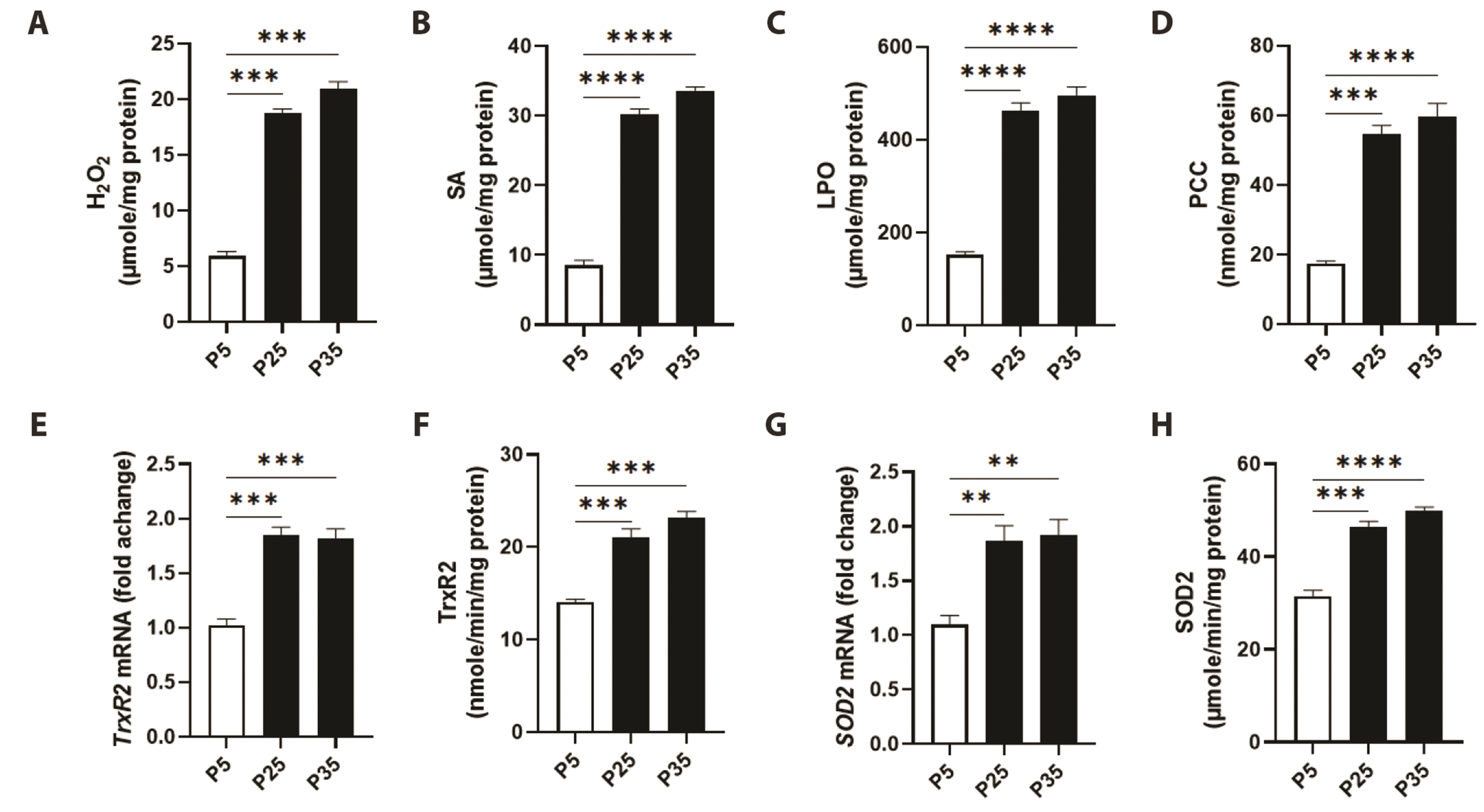
Fig. 4 Mitochondrial oxidative stress markers in primary P5 and senescent P25 and P35 fibroblasts. Generation levels of (A) hydrogen peroxide (H2O2), (B) superoxide anions (SA), (C) lipid peroxides (LPO), and (D) protein carbonyl content (PCC), and transcriptional and enzyme activities of thioredoxin reductase 2 (TrxR2) (E, F) and superoxide dismutase 2 (SOD2) (G, H) in young (P5) and senescent (P25 and P35) confluent cells. **p < 0.01), ***p < 0.001, and ****p < 0.0001 indicate significant difference from control P5 values (n = 6).
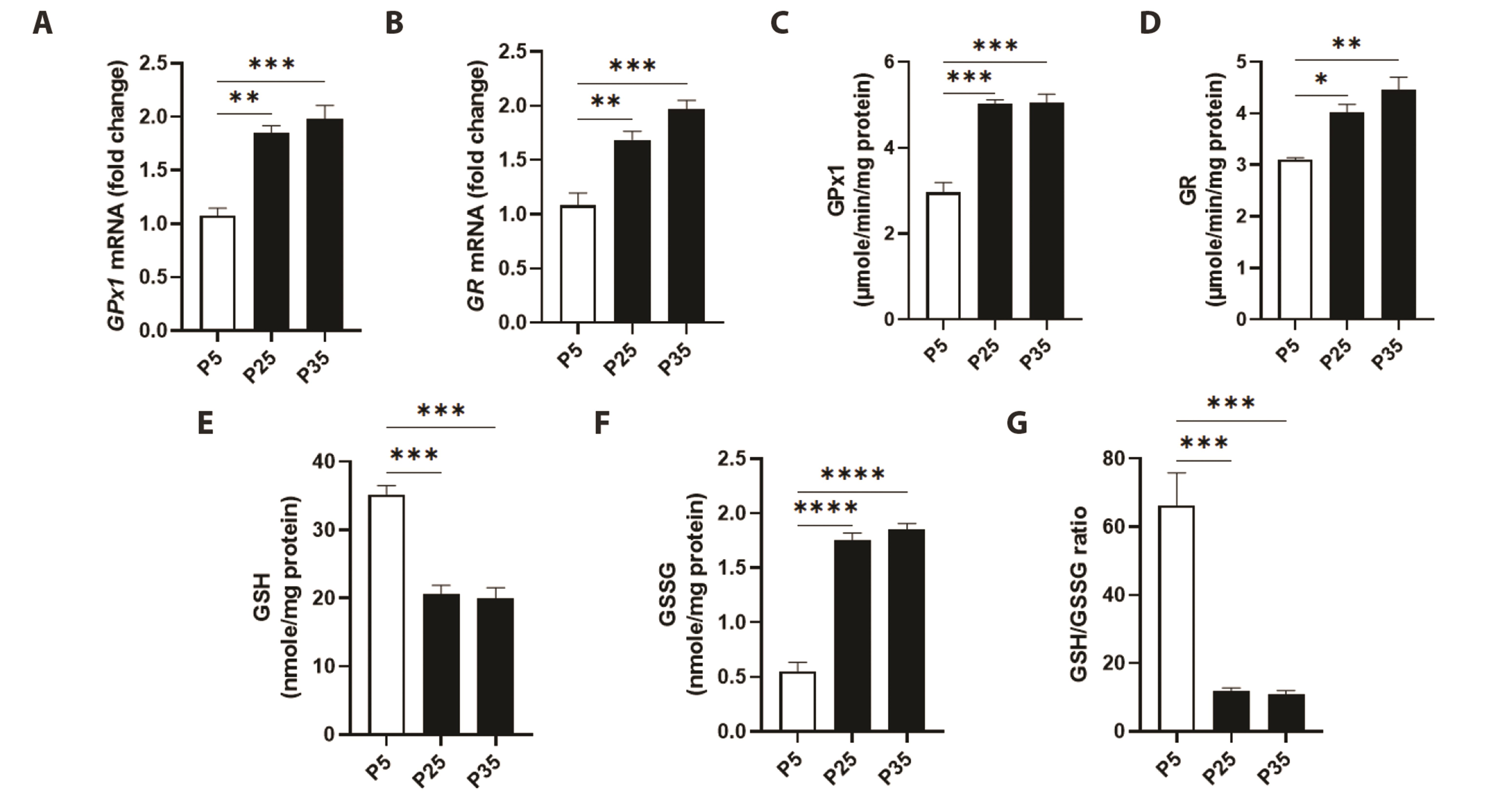
Fig. 5 Mitochondrial glutathione status in primary P5 and senescent P25 and P35 fibroblasts. Transcriptional activities of (A) glutathione peroxidase 1 (GPx1) and (B) glutathione reductase (GR), and enzyme activities of (C) GPx1 and (D) GR, along with levels of (E) reduced glutathione (GSH), (F) oxidized glutathione (GSSG), and (G) GSH/GSSG ratio in young (P5) and senescent (P25 and P35) confluent cells. *p < 0.05, **p < 0.01, ***p < 0.001, and ****p < 0.0001 indicate significant difference from control P5 values (n = 6).
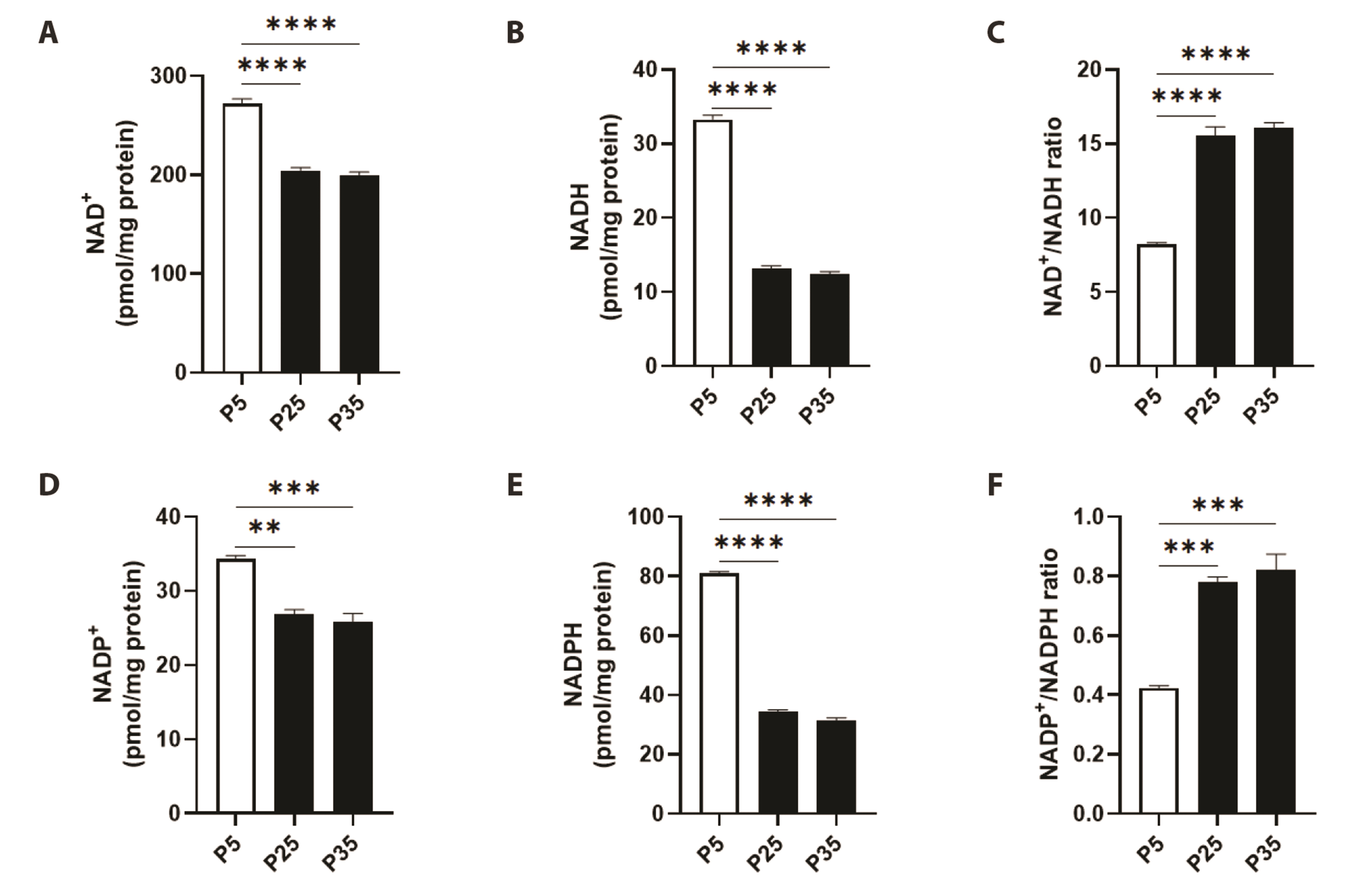
Fig. 6 Mitochondrial NAD+, NADH, NADP+, and NADPH levels in primary P5 and senescent P25 and P35 fibroblasts. Levels of (A) NAD+, (B) NADH, (C) NAD+/NADH ratio, (D) NADP+, (E) NADPH, and (F) NADP+/NADPH ratio in young (P5) and senescent (P25 and P35) confluent cells. **p < 0.01, ***p < 0.001, and ****p < 0.0001 indicate significant difference from control P5 values (n = 6).
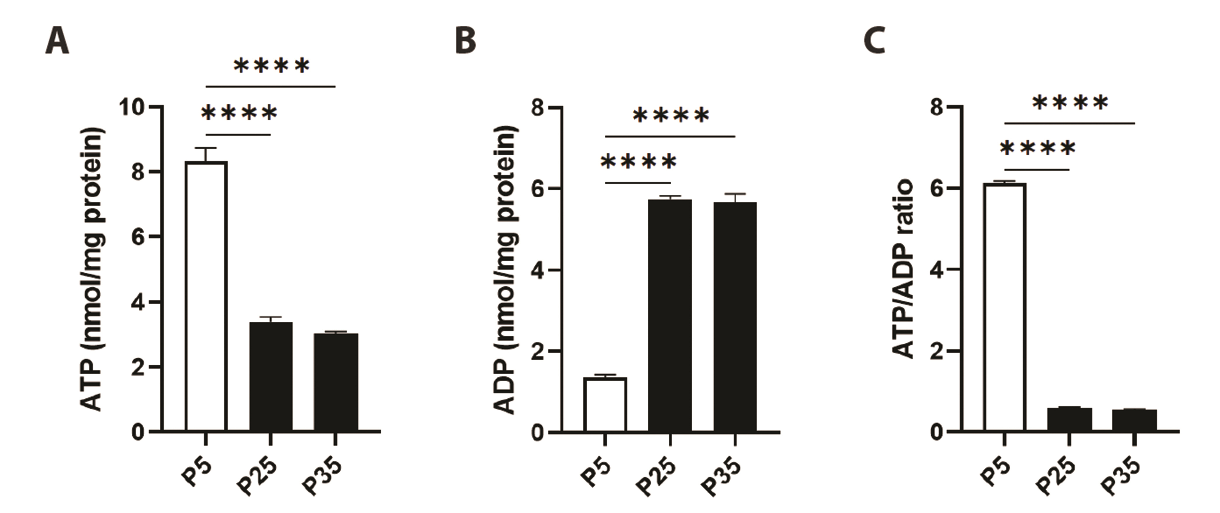
Fig. 7 Cellular ATP and ADP levels in primary P5 and senescent P25 and P35 fibroblasts. Levels of (A) ATP, (B) ADP, and (C) ATP/ADP ratio in young (P5) and senescent (P25 and P35) confluent cells. ****p < 0.0001 indicates significant difference from control P5 values (n = 6).

Fig. 8 A working model of mitochondrial senescence [65]. Replicative senescence induces metabolic changes in mitochondria evident as accumulation of ROS, shutdown of TCA and ETC pathways, glutathione depletion, and energy exhaustion. ROS, reactive oxygen species; ETC, electron transport chain; PFK, phosphofructokinase; LDH, lactate dehydrogenase; GP, glycogen phosphorylase; H2O2, hydrogen peroxide; SA, superoxide anions; LPO, lipid peroxides; PCC, protein carbonyl content; GSSG, oxidized glutathione; TrxR2, thioredoxin reductase 2; SOD2, superoxide dismutase 2; GPx1, glutathione peroxidase 1; GR, glutathione reductase; GSH, reduced glutathione.
Reference
-
1. Ziada AS, Smith MR, Côté HCF. 2020; Updating the free radical theory of aging. Front Cell Dev Biol. 8:575645. DOI: 10.3389/fcell.2020.575645. PMID: 33043009. PMCID: PMC7525146. PMID: e862f00f92eb43ca9149b66c33740037. PMID: https://www.scopus.com/inward/record.uri?partnerID=HzOxMe3b&scp=85091911747&origin=inward.
Article2. Sanz A, Stefanatos RK. 2008; The mitochondrial free radical theory of aging: a critical view. Curr Aging Sci. 1:10–21. DOI: 10.2174/1874609810801010010. PMID: 20021368. PMID: https://www.scopus.com/inward/record.uri?partnerID=HzOxMe3b&scp=77449156533&origin=inward.
Article3. Shinmura K. 2013; Effects of caloric restriction on cardiac oxidative stress and mitochondrial bioenergetics: potential role of cardiac sirtuins. Oxid Med Cell Longev. 2013:528935. DOI: 10.1155/2013/528935. PMID: 23577224. PMCID: PMC3614061. PMID: https://www.scopus.com/inward/record.uri?partnerID=HzOxMe3b&scp=84876522971&origin=inward.
Article4. Lu J, Holmgren A. 2012; Thioredoxin system in cell death progression. Antioxid Redox Signal. 17:1738–1747. DOI: 10.1089/ars.2012.4650. PMID: 22530689. PMID: https://www.scopus.com/inward/record.uri?partnerID=HzOxMe3b&scp=84867711090&origin=inward.
Article5. Kong Y, Trabucco SE, Zhang H. 2014; Oxidative stress, mitochondrial dysfunction and the mitochondria theory of aging. Interdiscip Top Gerontol. 39:86–107. DOI: 10.1159/000358901. PMID: 24862016. PMID: https://www.scopus.com/inward/record.uri?partnerID=HzOxMe3b&scp=84925873316&origin=inward.
Article6. Kwon SM, Hong SM, Lee YK, Min S, Yoon G. 2019; Metabolic features and regulation in cell senescence. BMB Rep. 52:5–12. DOI: 10.5483/BMBRep.2019.52.1.291. PMID: 30526768. PMCID: PMC6386228. PMID: https://www.scopus.com/inward/record.uri?partnerID=HzOxMe3b&scp=85060935100&origin=inward.
Article7. Myrianthopoulos V, Evangelou K, Vasileiou PVS, Cooks T, Vassilakopoulos TP, Pangalis GA, Kouloukoussa M, Kittas C, Georgakilas AG, Gorgoulis VG. 2019; Senescence and senotherapeutics: a new field in cancer therapy. Pharmacol Ther. 193:31–49. DOI: 10.1016/j.pharmthera.2018.08.006. PMID: 30121319. PMID: https://www.scopus.com/inward/record.uri?partnerID=HzOxMe3b&scp=85052745957&origin=inward.
Article8. Hernandez-Segura A, Nehme J, Demaria M. 2018; Hallmarks of cellular senescence. Trends Cell Biol. 28:436–453. DOI: 10.1016/j.tcb.2018.02.001. PMID: 29477613. PMID: https://www.scopus.com/inward/record.uri?partnerID=HzOxMe3b&scp=85042223621&origin=inward.
Article9. Wiley CD, Velarde MC, Lecot P, Liu S, Sarnoski EA, Freund A, Shirakawa K, Lim HW, Davis SS, Ramanathan A, Gerencser AA, Verdin E, Campisi J. 2016; Mitochondrial dysfunction induces senescence with a distinct secretory phenotype. Cell Metab. 23:303–314. DOI: 10.1016/j.cmet.2015.11.011. PMID: 26686024. PMCID: PMC4749409. PMID: https://www.scopus.com/inward/record.uri?partnerID=HzOxMe3b&scp=84957952429&origin=inward.
Article10. Baker DJ, Childs BG, Durik M, Wijers ME, Sieben CJ, Zhong J, Saltness RA, Jeganathan KB, Verzosa GC, Pezeshki A, Khazaie K, Miller JD, van Deursen JM. 2016; Naturally occurring p16(Ink4a)-positive cells shorten healthy lifespan. Nature. 530:184–189. DOI: 10.1038/nature16932. PMID: 26840489. PMCID: PMC4845101. PMID: https://www.scopus.com/inward/record.uri?partnerID=HzOxMe3b&scp=84958093401&origin=inward.
Article11. Schafer MJ, White TA, Iijima K, Haak AJ, Ligresti G, Atkinson EJ, Oberg AL, Birch J, Salmonowicz H, Zhu Y, Mazula DL, Brooks RW, Fuhrmann-Stroissnigg H, Pirtskhalava T, Prakash YS, Tchkonia T, Robbins PD, Aubry MC, Passos JF, Kirkland JL, et al. 2017; Cellular senescence mediates fibrotic pulmonary disease. Nat Commun. 8:14532. DOI: 10.1038/ncomms14532. PMID: 28230051. PMCID: PMC5331226. PMID: 24b84bad3904485caad4533a2d6fd946. PMID: https://www.scopus.com/inward/record.uri?partnerID=HzOxMe3b&scp=85013807598&origin=inward.
Article12. Ogrodnik M, Miwa S, Tchkonia T, Tiniakos D, Wilson CL, Lahat A, Day CP, Burt A, Palmer A, Anstee QM, Grellscheid SN, Hoeijmakers JHJ, Barnhoorn S, Mann DA, Bird TG, Vermeij WP, Kirkland JL, Passos JF, von Zglinicki T, Jurk D. 2017; Cellular senescence drives age-dependent hepatic steatosis. Nat Commun. 8:15691. DOI: 10.1038/ncomms15691. PMID: 28608850. PMCID: PMC5474745. PMID: 59da7366bd92469288a45b90a4b396f9. PMID: https://www.scopus.com/inward/record.uri?partnerID=HzOxMe3b&scp=85020726721&origin=inward.
Article13. Byun HO, Lee YK, Kim JM, Yoon G. 2015; From cell senescence to age-related diseases: differential mechanisms of action of senescence-associated secretory phenotypes. BMB Rep. 48:549–558. Erratum in: BMB Rep. 2016;49:641-650. DOI: 10.5483/BMBRep.2016.49.11.122. PMID: 27881217. PMCID: PMC4911181. PMID: https://www.scopus.com/inward/record.uri?partnerID=HzOxMe3b&scp=85009192963&origin=inward.
Article14. Dong D, Cai GY, Ning YC, Wang JC, Lv Y, Hong Q, Cui SY, Fu B, Guo YN, Chen XM. 2017; Alleviation of senescence and epithelial-mesenchymal transition in aging kidney by short-term caloric restriction and caloric restriction mimetics via modulation of AMPK/mTOR signaling. Oncotarget. 8:16109–16121. DOI: 10.18632/oncotarget.14884. PMID: 28147330. PMCID: PMC5369951. PMID: https://www.scopus.com/inward/record.uri?partnerID=HzOxMe3b&scp=85014671397&origin=inward.
Article15. Davalli P, Mitic T, Caporali A, Lauriola A, D'Arca D. 2016; ROS, cell senescence, and novel molecular mechanisms in aging and age-related diseases. Oxid Med Cell Longev. 2016:3565127. DOI: 10.1155/2016/3565127. PMID: 27247702. PMCID: PMC4877482. PMID: https://www.scopus.com/inward/record.uri?partnerID=HzOxMe3b&scp=84973300708&origin=inward.
Article16. Nacarelli T, Sell C. 2017; Targeting metabolism in cellular senescence, a role for intervention. Mol Cell Endocrinol. 455:83–92. DOI: 10.1016/j.mce.2016.08.049. PMID: 27591812. PMID: https://www.scopus.com/inward/record.uri?partnerID=HzOxMe3b&scp=84992327128&origin=inward.
Article17. Birch J, Passos JF. 2017; Targeting the SASP to combat ageing: mitochondria as possible intracellular allies? Bioessays. 39:1600235. DOI: 10.1002/bies.201600235. PMID: 28217839. PMID: https://www.scopus.com/inward/record.uri?partnerID=HzOxMe3b&scp=85013411055&origin=inward.
Article18. Byun HO, Jung HJ, Seo YH, Lee YK, Hwang SC, Hwang ES, Yoon G. 2012; GSK3 inactivation is involved in mitochondrial complex IV defect in transforming growth factor (TGF) β1-induced senescence. Exp Cell Res. 318:1808–1819. DOI: 10.1016/j.yexcr.2012.04.012. PMID: 22652454. PMID: https://www.scopus.com/inward/record.uri?partnerID=HzOxMe3b&scp=84863785173&origin=inward.
Article19. Byun HO, Jung HJ, Kim MJ, Yoon G. 2014; PKCδ phosphorylation is an upstream event of GSK3 inactivation-mediated ROS generation in TGF-β1-induced senescence. Free Radic Res. 48:1100–1108. DOI: 10.3109/10715762.2014.929120. PMID: 24917460. PMID: https://www.scopus.com/inward/record.uri?partnerID=HzOxMe3b&scp=84906050719&origin=inward.
Article20. Victorelli S, Passos JF. 2019; Reactive oxygen species detection in senescent cells. Methods Mol Biol. 1896:21–29. DOI: 10.1007/978-1-4939-8931-7_3. PMID: 30474836. PMID: https://www.scopus.com/inward/record.uri?partnerID=HzOxMe3b&scp=85057129774&origin=inward.
Article21. Passos JF, Nelson G, Wang C, Richter T, Simillion C, Proctor CJ, Miwa S, Olijslagers S, Hallinan J, Wipat A, Saretzki G, Rudolph KL, Kirkwood TB, von Zglinicki T. 2010; Feedback between p21 and reactive oxygen production is necessary for cell senescence. Mol Syst Biol. 6:347. DOI: 10.1038/msb.2010.5. PMID: 20160708. PMCID: PMC2835567. PMID: https://www.scopus.com/inward/record.uri?partnerID=HzOxMe3b&scp=77149164811&origin=inward.
Article22. Correia-Melo C, Passos JF. 2015; Mitochondria: are they causal players in cellular senescence? Biochim Biophys Acta. 1847:1373–1379. DOI: 10.1016/j.bbabio.2015.05.017. PMID: 26028303. PMID: https://www.scopus.com/inward/record.uri?partnerID=HzOxMe3b&scp=84941730889&origin=inward.
Article23. Pernas L, Scorrano L. 2016; Mito-morphosis: mitochondrial fusion, fission, and cristae remodeling as key mediators of cellular function. Annu Rev Physiol. 78:505–531. DOI: 10.1146/annurev-physiol-021115-105011. PMID: 26667075. PMID: https://www.scopus.com/inward/record.uri?partnerID=HzOxMe3b&scp=84958673479&origin=inward.
Article24. Tai H, Wang Z, Gong H, Han X, Zhou J, Wang X, Wei X, Ding Y, Huang N, Qin J, Zhang J, Wang S, Gao F, Chrzanowska-Lightowlers ZM, Xiang R, Xiao H. 2017; Autophagy impairment with lysosomal and mitochondrial dysfunction is an important characteristic of oxidative stress-induced senescence. Autophagy. 13:99–113. DOI: 10.1080/15548627.2016.1247143. PMID: 27791464. PMCID: PMC5240829. PMID: https://www.scopus.com/inward/record.uri?partnerID=HzOxMe3b&scp=84996598519&origin=inward.
Article25. Habiballa L, Salmonowicz H, Passos JF. 2019; Mitochondria and cellular senescence: Implications for musculoskeletal ageing. Free Radic Biol Med. 132:3–10. DOI: 10.1016/j.freeradbiomed.2018.10.417. PMID: 30336251. PMID: https://www.scopus.com/inward/record.uri?partnerID=HzOxMe3b&scp=85055134454&origin=inward.
Article26. Studencka M, Schaber J. 2017; Senoptosis: non-lethal DNA cleavage as a route to deep senescence. Oncotarget. 8:30656–30671. DOI: 10.18632/oncotarget.15693. PMID: 28427150. PMCID: PMC5458157. PMID: https://www.scopus.com/inward/record.uri?partnerID=HzOxMe3b&scp=85019150025&origin=inward.
Article27. Ghneim HK, Al-Sheikh YA. 2010; The effect of aging and increasing ascorbate concentrations on respiratory chain activity in cultured human fibroblasts. Cell Biochem Funct. 28:283–292. DOI: 10.1002/cbf.1653. PMID: 20517892. PMID: https://www.scopus.com/inward/record.uri?partnerID=HzOxMe3b&scp=77953490083&origin=inward.
Article28. Ghneim HK. 2017; Selenium concentrations for maximisation of thioredoxin reductase 2 activity and upregulation of its gene transcripts in senescent human fibroblasts. Antioxidants (Basel). 6:83. DOI: 10.3390/antiox6040083. PMID: 29084149. PMCID: PMC5745493. PMID: https://www.scopus.com/inward/record.uri?partnerID=HzOxMe3b&scp=85033567597&origin=inward.
Article29. Ghneim HK. 2017; The effect of Echis coloratus venom on biochemical and molecular markers of the antioxidant capacity in human fibroblasts. Libyan J Med. 12:1304515. DOI: 10.1080/19932820.2017.1304515. PMID: 28347204. PMCID: PMC5418940. PMID: https://www.scopus.com/inward/record.uri?partnerID=HzOxMe3b&scp=85025614807&origin=inward.30. Reznick AZ, Packer L. 1994; Oxidative damage to proteins: spectrophotometric method for carbonyl assay. Methods Enzymol. 233:357–363. DOI: 10.1016/S0076-6879(94)33041-7. PMID: 8015470. PMID: https://www.scopus.com/inward/record.uri?partnerID=HzOxMe3b&scp=0028239163&origin=inward.31. Al-Sheikh YA, Ghneim HK. 2011; 'The effect of micronutrients on superoxide dismutase in senescent fibroblasts'. Cell Biochem Funct. 29:384–393. DOI: 10.1002/cbf.1761. PMID: 21538411. PMID: https://www.scopus.com/inward/record.uri?partnerID=HzOxMe3b&scp=79960077327&origin=inward.
Article32. Ghneim HK, Alshebly MM. 2016; Biochemical markers of oxidative stress in Saudi women with recurrent miscarriage. J Korean Med Sci. 31:98–105. DOI: 10.3346/jkms.2016.31.1.98. PMID: 26770044. PMCID: PMC4712587. PMID: https://www.scopus.com/inward/record.uri?partnerID=HzOxMe3b&scp=84954150765&origin=inward.
Article33. Ghneim HK. 2016; The kinetics of the effect of manganese supplementation on SOD2 activity in senescent human fibroblasts. Eur Rev Med Pharmacol Sci. 20:1866–1880. PMID: 27212182. PMID: https://www.scopus.com/inward/record.uri?partnerID=HzOxMe3b&scp=85026302951&origin=inward.34. Ghneim HK, Al-Sheikh YA. 2011; Effect of selenium supplementation on glutathione peroxidase and catalase activities in senescent cultured human fibroblasts. Ann Nutr Metab. 59:127–138. DOI: 10.1159/000334069. PMID: 22142804. PMID: https://www.scopus.com/inward/record.uri?partnerID=HzOxMe3b&scp=82655188068&origin=inward.
Article35. Carlberg I, Mannervik B. 1985; Glutathione reductase. Methods Enzymol. 113:484–490. DOI: 10.1016/S0076-6879(85)13062-4. PMID: 3003504. PMID: https://www.scopus.com/inward/record.uri?partnerID=HzOxMe3b&scp=0022272480&origin=inward.36. Hausladen A, Fridovich I. 1996; Measuring nitric oxide and superoxide: rate constants for aconitase reactivity. Methods Enzymol. 269:37–41. DOI: 10.1016/S0076-6879(96)69007-7. PMID: 8791635. PMID: https://www.scopus.com/inward/record.uri?partnerID=HzOxMe3b&scp=0029819752&origin=inward.37. Goncalves S, Paupe V, Dassa EP, Brière JJ, Favier J, Gimenez-Roqueplo AP, Bénit P, Rustin P. 2010; Rapid determination of tricarboxylic acid cycle enzyme activities in biological samples. BMC Biochem. 11:5. DOI: 10.1186/1471-2091-11-5. PMID: 20109171. PMCID: PMC2823639. PMID: https://www.scopus.com/inward/record.uri?partnerID=HzOxMe3b&scp=77249172928&origin=inward.
Article38. Lai JC, Cooper AJ. 1986; Brain alpha-ketoglutarate dehydrogenase complex: kinetic properties, regional distribution, and effects of inhibitors. J Neurochem. 47:1376–1386. DOI: 10.1111/j.1471-4159.1986.tb00768.x. PMID: 3760866. PMID: https://www.scopus.com/inward/record.uri?partnerID=HzOxMe3b&scp=0022972426&origin=inward.
Article39. Tan AK, Ramsay RR, Singer TP, Miyoshi H. 1993; Comparison of the structures of the quinone-binding sites in beef heart mitochondria. J Biol Chem. 268:19328–19333. DOI: 10.1016/S0021-9258(19)36517-2. PMID: 8396133.
Article40. Ghneim HK, Al-Sheikh YA, Aboul-Soud MA. 2015; The effect of Walterinnesia aegyptia venom proteins on TCA cycle activity and mitochondrial NAD+-redox state in cultured human fibroblasts. Biomed Res Int. 2015:738147. DOI: 10.1155/2015/738147. PMID: 25705684. PMCID: PMC4331154. PMID: https://www.scopus.com/inward/record.uri?partnerID=HzOxMe3b&scp=84924105584&origin=inward.41. Akiel M, Alsughayyir J, Basudan AM, Alamri HS, Dera A, Barhoumi T, Al Subayyil AM, Basmaeil YS, Aldakheel FM, Alakeel R, Ghneim HK, Al-Sheikh YA, Alraey Y, Asiri S, Alfhili MA. 2021; Physcion induces hemolysis and premature phosphatidylserine externalization in human erythrocytes. Biol Pharm Bull. 44:372–378. DOI: 10.1248/bpb.b20-00744. PMID: 33431739. PMID: https://www.scopus.com/inward/record.uri?partnerID=HzOxMe3b&scp=85101768149&origin=inward.
Article42. Chao CH, Wu WC, Lai YC, Tsai PJ, Perng GC, Lin YS, Yeh TM. 2019; Dengue virus nonstructural protein 1 activates platelets via Toll-like receptor 4, leading to thrombocytopenia and hemorrhage. PLoS Pathog. 15:e1007625. DOI: 10.1371/journal.ppat.1007625. PMID: 31009511. PMCID: PMC6497319. PMID: 2f2802fe11204f1aa0489ae9a3fdc718. PMID: https://www.scopus.com/inward/record.uri?partnerID=HzOxMe3b&scp=85065555312&origin=inward.
Article43. Alfhili MA, Hussein HAM, Park Y, Lee MH, Akula SM. 2021; Triclosan induces apoptosis in Burkitt lymphoma-derived BJAB cells through caspase and JNK/MAPK pathways. Apoptosis. 26:96–110. DOI: 10.1007/s10495-020-01650-0. PMID: 33387145. PMID: https://www.scopus.com/inward/record.uri?partnerID=HzOxMe3b&scp=85098520815&origin=inward.
Article44. Ghneim HK, Al-Sheikh YA, Alshebly MM, Aboul-Soud MA. 2016; Superoxide dismutase activity and gene expression levels in Saudi women with recurrent miscarriage. Mol Med Rep. 13:2606–2612. DOI: 10.3892/mmr.2016.4807. PMID: 26821085. PMCID: PMC4768979. PMID: https://www.scopus.com/inward/record.uri?partnerID=HzOxMe3b&scp=84958721787&origin=inward.
Article45. Aljaser FS, Ghneim HK, ALshubaily MM, Abudawood M, Almajed F, Fatima S, ALsheikh YA. 2021; Glutathione and oxidized nicotinamide adenine dinucleotide NAD+ redox status in plasma and placental tissue of Saudi patients with intrauterine growth restriction. Saudi Med J. 42:491–498. DOI: 10.15537/smj.2021.42.5.20200685. PMID: 33896778. PMCID: PMC9149688. PMID: https://www.scopus.com/inward/record.uri?partnerID=HzOxMe3b&scp=85105904119&origin=inward.
Article46. Zeng H, Uthus EO, Combs GF Jr. 2005; Mechanistic aspects of the interaction between selenium and arsenic. J Inorg Biochem. 99:1269–1274. DOI: 10.1016/j.jinorgbio.2005.03.006. PMID: 15917080. PMID: https://www.scopus.com/inward/record.uri?partnerID=HzOxMe3b&scp=19444383615&origin=inward.
Article47. Tang MJ, Tannen RL. 1990; Relationship between proliferation and glucose metabolism in primary cultures of rabbit proximal tubules. Am J Physiol. 259(3 Pt 1):C455–C461. DOI: 10.1152/ajpcell.1990.259.3.C455. PMID: 2399968. PMID: https://www.scopus.com/inward/record.uri?partnerID=HzOxMe3b&scp=0025080227&origin=inward.
Article48. Matsuo K, Galson DL, Zhao C, Peng L, Laplace C, Wang KZ, Bachler MA, Amano H, Aburatani H, Ishikawa H, Wagner EF. 2004; Nuclear factor of activated T-cells (NFAT) rescues osteoclastogenesis in precursors lacking c-Fos. J Biol Chem. 279:26475–26480. DOI: 10.1074/jbc.M313973200. PMID: 15073183. PMID: https://www.scopus.com/inward/record.uri?partnerID=HzOxMe3b&scp=2942731511&origin=inward.
Article49. Legrain V, Iannetti GD, Plaghki L, Mouraux A. 2011; The pain matrix reloaded: a salience detection system for the body. Prog Neurobiol. 93:111–124. DOI: 10.1016/j.pneurobio.2010.10.005. PMID: 21040755. PMID: https://www.scopus.com/inward/record.uri?partnerID=HzOxMe3b&scp=78649782961&origin=inward.50. Campisi J, d'Adda di Fagagna F. 2007; Cellular senescence: when bad things happen to good cells. Nat Rev Mol Cell Biol. 8:729–740. DOI: 10.1038/nrm2233. PMID: 17667954. PMID: https://www.scopus.com/inward/record.uri?partnerID=HzOxMe3b&scp=34548186667&origin=inward.
Article51. Rodier F, Campisi J. 2011; Four faces of cellular senescence. J Cell Biol. 192:547–556. DOI: 10.1083/jcb.201009094. PMID: 21321098. PMCID: PMC3044123. PMID: https://www.scopus.com/inward/record.uri?partnerID=HzOxMe3b&scp=79951912532&origin=inward.
Article52. Finkel T, Holbrook NJ. 2000; Oxidants, oxidative stress and the biology of ageing. Nature. 408:239–247. DOI: 10.1038/35041687. PMID: 11089981. PMID: https://www.scopus.com/inward/record.uri?partnerID=HzOxMe3b&scp=0034626735&origin=inward.
Article53. Dimri GP, Lee X, Basile G, Acosta M, Scott G, Roskelley C, Medrano EE, Linskens M, Rubelj I, Pereira-Smith O, Peacocke M, Campisi J. 1995; A biomarker that identifies senescent human cells in culture and in aging skin in vivo. Proc Natl Acad Sci U S A. 92:9363–9367. DOI: 10.1073/pnas.92.20.9363. PMID: 7568133. PMCID: PMC40985. PMID: https://www.scopus.com/inward/record.uri?partnerID=HzOxMe3b&scp=0029047362&origin=inward.
Article54. Sharma P, Mongan PD. 2001; Ascorbate reduces superoxide production and improves mitochondrial respiratory chain function in human fibroblasts with electron transport chain deficiencies. Mitochondrion. 1:191–198. Erratum in: Mitochondrion. 2001;1:293. DOI: 10.1016/S1567-7249(01)00032-0. PMID: 16120278.
Article55. Kramer-Zucker AG, Wiessner S, Jensen AM, Drummond IA. 2005; Organization of the pronephric filtration apparatus in zebrafish requires Nephrin, Podocin and the FERM domain protein Mosaic eyes. Dev Biol. 285:316–329. DOI: 10.1016/j.ydbio.2005.06.038. PMID: 16102746. PMCID: PMC2836015. PMID: https://www.scopus.com/inward/record.uri?partnerID=HzOxMe3b&scp=25844519579&origin=inward.
Article56. Boffoli D, Scacco SC, Vergari R, Solarino G, Santacroce G, Papa S. 1994; Decline with age of the respiratory chain activity in human skeletal muscle. Biochim Biophys Acta. 1226:73–82. DOI: 10.1016/0925-4439(94)90061-2. PMID: 8155742. PMID: https://www.scopus.com/inward/record.uri?partnerID=HzOxMe3b&scp=0028349236&origin=inward.
Article57. Gupta S, Cheng H, Mollah AK, Jamison E, Morris S, Chance MR, Khrapunov S, Brenowitz M. 2007; DNA and protein footprinting analysis of the modulation of DNA binding by the N-terminal domain of the Saccharomyces cerevisiae TATA binding protein. Biochemistry. 46:9886–9898. DOI: 10.1021/bi7003608. PMID: 17683121. PMID: https://www.scopus.com/inward/record.uri?partnerID=HzOxMe3b&scp=34548499082&origin=inward.
Article58. Wendelaar Bonga SE. 1997; The stress response in fish. Physiol Rev. 77:591–625. DOI: 10.1152/physrev.1997.77.3.591. PMID: 9234959. PMID: https://www.scopus.com/inward/record.uri?partnerID=HzOxMe3b&scp=0037752925&origin=inward.
Article59. Conrad M, Schneider M, Seiler A, Bornkamm GW. 2007; Physiological role of phospholipid hydroperoxide glutathione peroxidase in mammals. Biol Chem. 388:1019–1025. DOI: 10.1515/BC.2007.130. PMID: 17937615. PMID: https://www.scopus.com/inward/record.uri?partnerID=HzOxMe3b&scp=35348840292&origin=inward.
Article60. Dröge W. 2002; Free radicals in the physiological control of cell function. Physiol Rev. 82:47–95. DOI: 10.1152/physrev.00018.2001. PMID: 11773609. PMID: https://www.scopus.com/inward/record.uri?partnerID=HzOxMe3b&scp=0036086130&origin=inward.
Article61. Ruder EH, Hartman TJ, Blumberg J, Goldman MB. 2008; Oxidative stress and antioxidants: exposure and impact on female fertility. Hum Reprod Update. 14:345–357. DOI: 10.1093/humupd/dmn011. PMID: 18535004. PMCID: PMC2772106. PMID: https://www.scopus.com/inward/record.uri?partnerID=HzOxMe3b&scp=45549098881&origin=inward.
Article62. Calcinotto A, Kohli J, Zagato E, Pellegrini L, Demaria M, Alimonti A. 2019; Cellular senescence: aging, cancer, and injury. Physiol Rev. 99:1047–1078. DOI: 10.1152/physrev.00020.2018. PMID: 30648461. PMID: https://www.scopus.com/inward/record.uri?partnerID=HzOxMe3b&scp=85060035237&origin=inward.
Article63. Sacitharan PK. 2019; Ageing and osteoarthritis. Subcell Biochem. 91:123–159. DOI: 10.1007/978-981-13-3681-2_6. PMID: 30888652. PMID: https://www.scopus.com/inward/record.uri?partnerID=HzOxMe3b&scp=85063261885&origin=inward.
Article64. Belikov AV. 2019; Age-related diseases as vicious cycles. Ageing Res Rev. 49:11–26. DOI: 10.1016/j.arr.2018.11.002. PMID: 30458244. PMID: https://www.scopus.com/inward/record.uri?partnerID=HzOxMe3b&scp=85056905200&origin=inward.
Article65. Servier Medical Art. Intracellular components [Internet]. Servier Medical Art;Available from: https://smart.servier.com. cited 2022 Mar 15.
- Full Text Links
-
- Actions
-
Cited
- CITED
-
- Close
- Share
-
- Similar articles
-
- Down-regulation of Type I Collagen Gene Expression in Human Skin Fibroblasts by G1 Cell Cycle Arrest
- The Senolytic Drug JQ1 Removes Senescent Cells via Ferroptosis
- Effects of Replicative Senescence on the Cell Cycle Regulation in Human Gingival Fibroblasts
- Enhanced Viral Replication by Cellular Replicative Senescence
- Expression of Senescence-Associated Secretory Phenotype in Senescent Gingival Fibroblasts
