Neonatologist-Performed Cranial Ultrasonography in the Neonatal Intensive Care Unit
- Affiliations
-
- 1Department of Pediatrics, Gachon University Gil Medical Center, Gachon University College of Medicine, Incheon, Korea
- KMID: 2530608
- DOI: http://doi.org/10.5385/nm.2022.29.2.57
Abstract
- Cranial ultrasound (CUS) is an initial screening imaging tool used to evaluate the neonatal brain. It is an accessible, inexpensive, and harmless technique that can be used at bedside as frequently as required. Timely focused CUS in the neonatal care unit can play a major role in the diagnosis, follow-up, and management of brain damage. Despite the increasing use of point-of-care ultrasonography by intensive care physicians, neonatologist-performed CUS remains unusual. This review aims to provide an overview of neonatal CUS to neonatologists, focusing on the optimal settings, standard planes of the brain, and main pathologies in preterm infants. Adding Doppler studies allows evaluation of the patency of intracranial arteries and veins, flow velocities, and indices. This may provide an opportunity for earlier targeted circulatory support to prevent brain injury and improve long-term neurodevelopmental outcomes.
Keyword
Figure
-
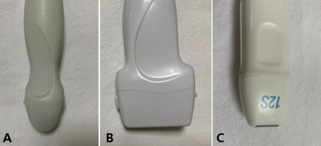
Figure 1. Three different types of probes. (A) Convex (curved) probe. (B) Linear probe. (C) Sector (phased array) probe.
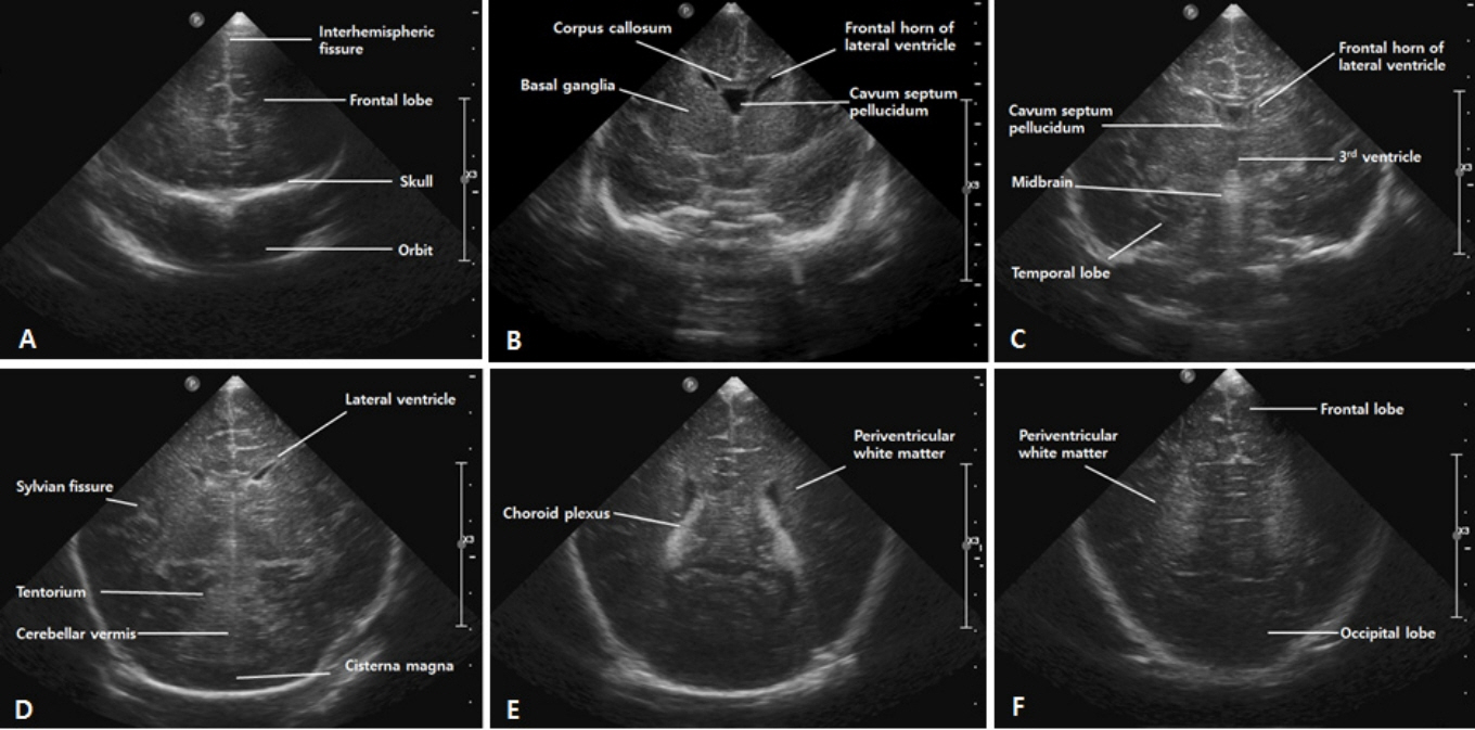
Figure 2. Standard coronal images of the brain through the anterior fontanelle. (A) First standard coronal image through the frontal lobes. (B) Second coronal image through the frontal horns of the lateral ventricles. (C) Third coronal image through the third ventricle. (D) Fourth coronal image at the level of the cerebellum. (E) Fifth coronal image through the trigone of the lateral ventricle. (F) Sixth coronal image through the occipital lobes.
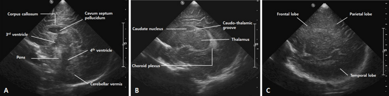
Figure 3. Standard sagittal images of the brain through the anterior fontanelle. (A) Midline sagittal image. (B) Parasagittal image. (C) Lateral parasagittal image.
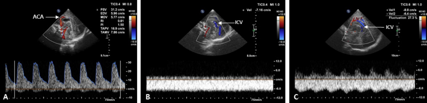
Figure 4. Color and pulsed Doppler evaluation in the midline sagittal plane. (A) Resistive index (RI), peak systolic velocity (PSV) and end diastolic velocity (EDV) in the anterior cerebral artery (ACA) are automatically evaluated using spectral Doppler tracings. (B) Perfusion waveform of the internal cerebral vein (ICV) shows a steady waveform with a constant perfusion speed (Grade 0 fluctuation). (C) Perfusion waveform of the ICV fluctuates; however, the minimum speed is never less than half the maximum speed (Grade 1 fluctuation).
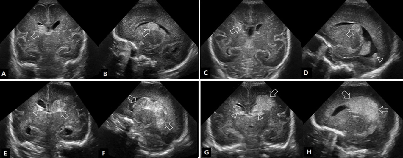
Figure 5. Ultrasound findings of germinal matrix-intraventricular hemorrhage (GMH-IVH). (A,B) Grade I GMH-IVH. The location of the echogenic clot (arrows) at the right caudothalamic notch is typical of grade I GMH-IVH. (C,D) Grade II GMH-IVH. Coronal scan shows echogenic clot involving the caudate nucleus (arrows). Parasagittal scan shows the same clot centered at the caudothalamic notch (arrows) and the intraventricular hemorrhage within the occipital horn separate to choroid plexus (arrowhead). (E,F) Grade III GMH-IVH. Coronal scan shows a large left GMH-IVH (arrows) with intraventricular blood acutely distending its lateral ventricle. Note also the enlarged temporal ventricular horns. A small amount of blood in the right lateral ventricle (arrowhead) is present. Parasagittal view shows that the left grade III GMH-IVH fills >50% of the distended lateral ventricle. (G,H) Grade IV GMH-IVH. Coronal scan showing a left-sided grade III GMH-IVH (arrowhead) and large echodensity in the left frontoparietal white matter (arrow). Smaller right-sided grade II GMH-IVH (arrowhead). Left parasagittal scan showing the periventricular echodensity (arrows) extending from the posterior frontal white matter to the parietal white matter.
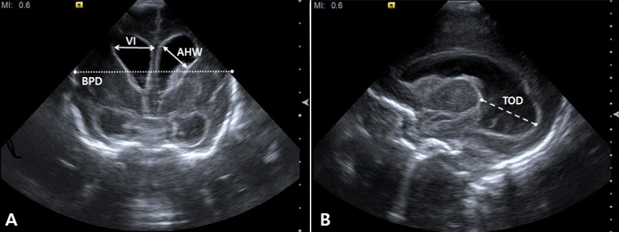
Figure 6. Measurements of the ventricle size in post-hemorrhagic ventricular dilatation. (A) Measurements of the ventricular index (VI), anterior horn width (AHW), and ventricle/brain ratio on coronal scan. (B) Measurements of the thalamo-occipital distance (TOD) on sagittal scan.
Figure 7. Ultrasound findings in a preterm infant (24 weeks of gestational age) with periventricular leukomalacia. (A) Coronal scan shows scattered hyperechoic lesions in the periventricular white matter. (B) Coronal scan 4 weeks later shows multiple cysts extending from the frontal lobe to the trigone area.
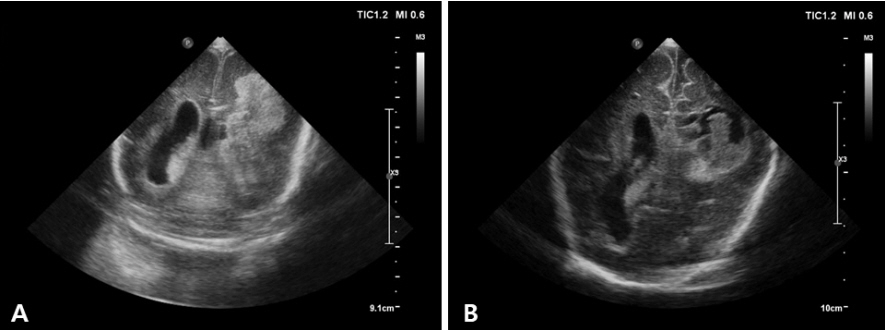
Figure 8. Ultrasound findings in a preterm infant (25 weeks of gestational age) with bilateral intraventricular hemorrhage and left periventricular hemorrhagic infarction (PVHI). (A) Coronal scan shows a large echodensity in the left frontoparietal white matter. (B) Coronal scan 2 weeks later shows cystic degeneration following the PVHI with echogenic clot debris.
Reference
-
1. Kurepa D, Boyar V, Zaghloul N, Beachy J, Zaytseva A, Teng D, et al. Sructured neonatal point-of-care ultrasound training program. Am J Perinatol. 2021; 38:e284–91.2. Ben Fadel N, Pulgar L, Khurshid F. Point of care ultrasound (POCUS) in Canadian neonatal intensive care units (NICUs): where are we? J Ultrasound. 2019; 22:201–6.3. Dudink J, Jeanne Steggerda S, Horsch S; eurUS.brain group. State-of-the-art neonatal cerebral ultrasound: technique and reporting. Pediatr Res. 2020; 87(Suppl 1):3–12.4. Riedesel EL. Neonatal cranial ultrasound: avanced techniques and image interpretation. J Pediatr Neurol. 2018; 16:106–24.5. Evans N, Gournay V, Cabanas F, Kluckow M, Leone T, Groves A, et al. Point-of-care ultrasound in the neonatal intensive care unit: international perspectives. Semin Fetal Neonatal Med. 2011; 16:61–8.6. Singh Y, Tissot C, Fraga MV, Yousef N, Cortes RG, Lopez J, et al. International evidence-based guidelines on Point of Care Ultrasound (POCUS) for critically ill neonates and children issued by the POCUS Working Group of the European Society of Paediatric and Neonatal Intensive Care (ESPNIC). Crit Care. 2020; 24:65.7. Miller LE, Stoller JZ, Fraga MV. Point-of-care ultrasound in the neonatal ICU. Curr Opin Pediatr. 2020; 32:216–27.8. AIUM practice parameter for the performance of neurosonography in neonates and infants. J Ultrasound Med. 2020; 39:E57–61.9. McLean G, Malhotra A, Lombardo P, Schneider M. Cranial ultrasound screening protocols for very preterm infants. Ultrasound Med Biol. 2021; 47:1645–56.10. Hand IL, Shellhaas RA, Milla SS; Committee on Fetus and Newborn, Section on Neurology, Section on Radiology. Routine neuroimaging of the preterm brain. Pediatrics. 2020; 146:e2020029082.11. Inder TE, de Vries LS, Ferriero DM, Grant PE, Ment LR, Miller SP, et al. Neuroimaging of the preterm brain: review and recommendations. J Pediatr. 2021; 237:276–87.12. Mohammad K, Scott JN, Leijser LM, Zein H, Afifi J, Piedboeuf B, et al. Consensus approach for standardizing the screening and classification of preterm brain injury diagnosed with cranial ultrasound: a Canadian perspective. Front Pediatr. 2021; 9:618236.13. The Korean Society of Neonatology. Manual of Neonatal Care. 4 ed. Seoul: The Korean Society of Neonatology;2021. p. 376–84.14. Beltempo M, Wintermark P, Lemyre B, Shalish W, Martel-Bucci A, Narvey M, et al. Predictors of severe neurologic injury on ultrasound scan of the head and risk factor-based screening for infants born preterm. J Pediatr. 2019; 214:27–33.15. Ecury-Goossen GM, Camfferman FA, Leijser LM, Govaert P, Dudink J. State of the art cranial ultrasound imaging in neonates. J Vis Exp. 2015; 96:e52238.16. O'Dell MC, Cassady C, Logsdon G, Varich L. Cinegraphic versus combined static and cinegraphic imaging for initial cranial ultrasound screening in premature infants. Pediatr Radiol. 2015; 45:1706–11.17. James AC. Practical guide to neonatal cranial ultrasound (crus): basics. Paediatr Child Health. 2018; 28:424–30.18. Caro-Dominguez P, Lecacheux C, Hernandez-Herrera C, Llorens-Salvador R. Cranial ultrasound for beginners. Transl Pediatr. 2021; 10:1117–37.19. Maller VV, Cohen HL. Neurosonography: assessing the premature infant. Pediatr Radiol. 2017; 47:1031–45.20. Couture A, Veyrac C, Baud C, Saguintaah M, Ferran JL. Advanced cranial ultrasound: transfontanellar Doppler imaging in neonates. Eur Radiol. 2001; 11:2399–410.21. Horgan JG, Rumack CM, Hay T, Manco-Johnson ML, Merenstein GB, Esola C. Absolute intracranial blood-flow velocities evaluated by duplex Doppler sonography in asymptomatic preterm and term neonates. AJR Am J Roentgenol. 1989; 152:1059–64.22. Romagnoli C, Giannantonio C, De Carolis MP, Gallini F, Zecca E, Papacci P. Neonatal color Doppler US study: normal values of cerebral blood flow velocities in preterm infants in the first month of life. Ultrasound Med Biol. 2006; 32:321–31.23. Ecury-Goossen GM, Raets MM, Camfferman FA, Vos RH, van Rosmalen J, Reiss IK, et al. Resistive indices of cerebral arteries in very preterm infants: values throughout stay in the neonatal intensive care unit and impact of patent ductus arteriosus. Pediatr Radiol. 2016; 46:1291–300.24. Lowe LH, Bailey Z. State-of-the-art cranial sonography: part 1, modern techniques and image interpretation. AJR Am J Roentgenol. 2011; 196:1028–33.25. Taylor GA. Intracranial venous system in the newborn: evaluation of normal anatomy and flow characteristics with color Doppler US. Radiology. 1992; 183:449–52.26. Camfferman FA, de Goederen R, Govaert P, Dudink J, van Bel F, Pellicer A, et al. Diagnostic and predictive value of Doppler ultrasound for evaluation of the brain circulation in preterm infants: a systematic review. Pediatr Res. 2020; 87(Suppl 1):50–8.27. Papile LA, Burstein J, Burstein R, Koffler H. Incidence and evolution of subependymal and intraventricular hemorrhage: a study of infants with birth weights less than 1,500 gm. J Pediatr. 1978; 92:529–34.28. Dorner RA, Burton VJ, Allen MC, Robinson S, Soares BP. Preterm neuroimaging and neurodevelopmental outcome: a focus on intraventricular hemorrhage, post-hemorrhagic hydrocephalus, and associated brain injury. J Perinatol. 2018; 38:1431–43.29. Taylor GA, Madsen JR. Neonatal hydrocephalus: hemodynamic response to fontanelle compression: correlation with intracranial pressure and need for shunt placement. Radiology. 1996; 201:685–9.30. Nakamura Y, Okudera T, Hashimoto T. Microvasculature in germinal matrix layer: its relationship to germinal matrix hemorrhage. Mod Pathol. 1991; 4:475–80.31. Ghazi-Birry HS, Brown WR, Moody DM, Challa VR, Block SM, Reboussin DM. Human germinal matrix: venous origin of hemorrhage and vascular characteristics. AJNR Am J Neuroradiol. 1997; 18:219–29.32. Van Bel F, Van de Bor M, Stijnen T, Baan J, Ruys JH. Aetiological rôle of cerebral blood-flow alterations in development and extension of peri-intraventricular haemorrhage. Dev Med Child Neurol. 1987; 29:601–14.33. Julkunen M, Parviainen T, Janas M, Tammela O. End-diastolic block in cerebral circulation may predict intraventricular hemorrhage in hypotensive extremely-low-birth-weight infants. Ultrasound Med Biol. 2008; 34:538–45.34. Ikeda T, Amizuka T, Ito Y, Mikami R, Matsuo K, Kawamura N, et al. Changes in the perfusion waveform of the internal cerebral vein and intraventricular hemorrhage in the acute management of extremely low-birth-weight infants. Eur J Pediatr. 2015; 174:331–8.35. Ikeda T, Ito Y, Mikami R, Matsuo K, Kawamura N, Yamoto A, et al. Fluctuations in internal cerebral vein and central side veins of preterm infants. Pediatr Int. 2021; 63:1319–26.36. Tanaka K, Sakamoto R, Imamura H, Naramura T, Matsumoto S, Iwai M, et al. Reversal of blood flow in deep cerebral vein in preterm intraventricular hemorrhage: two case reports. BMC Pediatr. 2020; 20:517.37. Dean LM, Taylor GA. The intracranial venous system in infants: normal and abnormal findings on duplex and color Doppler sonography. AJR Am J Roentgenol. 1995; 164:151–6.38. Pooh RK, Pooh KH, Nakagawa Y, Maeda K, Fukui R, Aono T. Transvaginal Doppler assessment of fetal intracranial venous flow. Obstet Gynecol. 1999; 93(5 Pt 1):697–701.39. Ikeda T, Ito Y, Mikami R, Matsuo K, Kawamura N, Yamoto A. Hemodynamics of infants with strong fluctuations of internal cerebral vein. Pediatr Int. 2019; 61:475–81.40. Yoon SA. Is it time to add point-of-care ultrasound education to pediatric residency curriculum? Clin Exp Pediatr. 2022; 65:33–4.
- Full Text Links
-
- Actions
-
Cited
- CITED
-
- Close
- Share
-
- Similar articles
-
- Current Status of Neonatologist Staffing and Workload in Korean Neonatal Intensive Care Units
- Bedside ultrasound-guided percutaneous cystostomy in an infant in the neonatal intensive care unit
- Organisation of Special and Intensive Care Facilities for Babies
- Quality Improvement in Neonatal Intensive Care Units
- Evaluating Nursing Needs in the Neonatal Intensive Care Unit with the Korean Patient Classification System for Neonatal Intensive Care Nurses
