Human Umbilical Cord Mesenchymal Stem Cells Improve the Necrosis and Osteocyte Apoptosis in Glucocorticoid-Induced Osteonecrosis of the Femoral Head Model through Reducing the Macrophage Polarization
- Affiliations
-
- 1Department of Orthopedics, Weihai Central Hospital Affiliated to Qingdao University & Qingdao University, Weihai, China
- 2Xinxiang Medical University, Xinxiang, China
- 3Weihai Key Laboratory of Autoimmunity & Central Laboratory of Weihai Central Hospital, Weihai, China
- 4Department of Sports Medicine, Weihai Central Hospital Affiliated to Qingdao University, Weihai, China
- 5Department of Trauma Surgery, Weihai Central Hospital Affiliated to Qingdao University, Weihai, China
- 6Department of Orthopedics, Weihai Central Hospital Affiliated to Qingdao University & Weihai Key Laboratory of Autoimmunity, Weihai, China
- KMID: 2529834
- DOI: http://doi.org/10.15283/ijsc21120
Abstract
- Background and Objectives
Apoptosis is an outstanding determinant of glucocorticoid (GC)-induced osteonecrosis of the femoral head (ONFH). Human umbilical cord mesenchymal stem cells (hUC-MSCs) have been demonstrated to be associated with apoptosis in diseases models. However, the role of hUC-MSCs in GC-induced ONFH via regulating apoptosis still needs further study.
Methods and Results
In the present study, a GC-induced ONFH model was built in vivo through a consecutive injection with lipopolysaccharide (LPS) and methylprednisolone. The necrosis and apoptosis of the femoral head was evaluated by histological and Terminal-deoxynucleoitidyl Transferase Mediated Nick End Labeling (TUNEL) assay. The level of collagen and TRAP positive cells were determined by Masson and TRAP staining, respectively. M1 macrophage polarization was assessed using immunofluorescence assay. The level of proinflammatory cytokines including tumor necrosis factor (TNF)‐α, Interleukin (IL)‐1β and IL-6 of femoral head was determined by enzyme-linked immunosorbent assay (ELISA) kits. The protein expression of AKT, mTOR, p-AKT and p-mTOR was detected using western blot assay. The results showed that hUC-MSCs treatment prominently promoted the GC-induced the decrease of the collagen level and the increase of TRAP positive cells. Besides, hUC-MSCs treatment decreased necrosis and apoptosis, macrophage polarization, the level of TNF‐α, IL‐1β and IL-6, the protein expression of p-AKT and p-mTOR, and the radio of p-AKT to AKT and p-mTOR to mTOR of femoral head in vivo.
Conclusions
Therefore, the present study revealed that hUC-MSCs improved the necrosis and osteocyte apoptosis in GC-induced ONFH model through reducing the macrophage polarization, which was associated with the inhibition of AKT/mTOR signaling pathway.
Keyword
Figure
-

Fig. 1 hUC-MSCs mitigated osteonecrosis and apoptosis in a GC-induced ONFH rat model. (A) Histological analysis was determined by H&E staining (magnification, ×100 or ×400). Scale bar: 50 μm. (B) The apoptosis rate of femoral head tissues was determined by TUNEL (magnification, ×400). Scale bar: 20 μm. (C) The collagen level of femoral head was determined by Masson staining (magnification, ×400). Scale bar: 20 μm. (D) The Number of osteoclasts was detected by TRAP staining (magnification, ×400). Scale bar: 20 μm. The means±SD of six independent samples were shown. *p<0.05 and **p<0.01, compared with control group; #p<0.05 and ##p<0.01, compared with model group.

Fig. 2 hUC-MSCs reduced M1 polarization in femoral heads. (A) The CD86 and F4/80 of macrophages were stained using immunofluorescence assay (magnification, x400). Scale bar: 20 μm. (B) The CD206 and F4/80 of macrophages were stained using immunofluorescence assay (magnification, x400). Scale bar: 20 μm. The means±SD of six independent samples were shown. *p<0.05 and **p< 0.01, compared with control group; ##p<0.01, compared with model group.
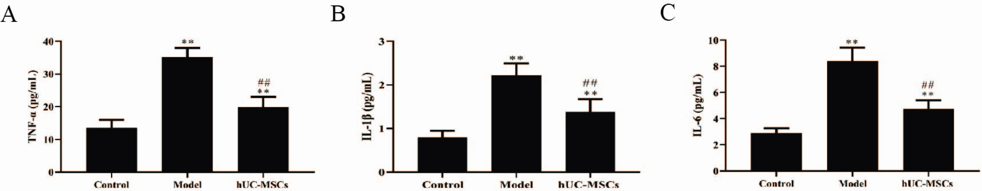
Fig. 3 hUC-MSCs reduced the level of proinflammatory cytokines in femoral heads. The serum level of TNF‐α (A), IL‐1β (B) and IL-6 (C) was determined by ELISA kits. The means±SD of six independent samples were shown. **p<0.01, compared with control group; ##p<0.01, compared with model group.

Fig. 4 hUC-MSCs repressed the activation of AKT/mTOR signaling pathway. The protein expression of AKT, mTOR, p-AKT and p-mTOR was detected using western blot. The data were expressed after being normalized to β-actin. The means±SD of six independent samples were shown. *p<0.05 and **p<0.01, compared with control group; ##p<0.01, compared with model group.
Reference
-
References
1. Zhao D, Zhang F, Wang B, Liu B, Li L, Kim SY, Goodman SB, Hernigou P, Cui Q, Lineaweaver WC, Xu J, Drescher WR, Qin L. 2020; Guidelines for clinical diagnosis and treatment of osteonecrosis of the femoral head in adults (2019 version). J Orthop Translat. 21:100–110. DOI: 10.1016/j.jot.2019.12.004. PMID: 32309135. PMCID: PMC7152793.
Article2. Tan G, Kang PD, Pei FX. 2012; Glucocorticoids affect the metabolism of bone marrow stromal cells and lead to osteonecrosis of the femoral head: a review. Chin Med J (Engl). 125:134–139. DOI: 10.3901/JME.2012.14.134. PMID: 22340480.3. Seguro LP, Rosario C, Shoenfeld Y. 2013; Long-term complications of past glucocorticoid use. Autoimmun Rev. 12:629–632. DOI: 10.1016/j.autrev.2012.12.002. PMID: 23261815.
Article4. Pan TT, Pan F, Gao W, Hu SS, Wang D. 2021; Involvement of macrophages and spinal microglia in osteoarthritis pain. Curr Rheumatol Rep. 23:29. DOI: 10.1007/s11926-021-00997-w. PMID: 33893883.
Article5. Yunna C, Mengru H, Lei W, Weidong C. 2020; Macrophage M1/M2 polarization. Eur J Pharmacol. 877:173090. DOI: 10.1016/j.ejphar.2020.173090. PMID: 32234529.
Article6. Chen G, Ni Y, Nagata N, Zhuge F, Xu L, Nagashimada M, Yamamoto S, Ushida Y, Fuke N, Suganuma H, Kaneko S, Ota T. 2019; Lycopene alleviates obesity-induced inflammation and insulin resistance by regulating M1/M2 status of macrophages. Mol Nutr Food Res. 63:e1900602. DOI: 10.1002/mnfr.201900602. PMID: 31408586.
Article7. Zhou X, Li W, Wang S, Zhang P, Wang Q, Xiao J, Zhang C, Zheng X, Xu X, Xue S, Hui L, Ji H, Wei B, Wang H. 2019; YAP aggravates inflammatory bowel disease by regulating M1/M2 macrophage polarization and gut microbial homeostasis. Cell Rep. 27:1176–1189.e5. DOI: 10.1016/j.celrep.2019.03.028. PMID: 31018132.
Article8. Chen S, Lu Z, Wang F, Wang Y. 2018; Cathelicidin-WA polarizes E. coli K88-induced M1 macrophage to M2-like macrophage in RAW264.7 cells. Int Immunopharmacol. 54:52–59. DOI: 10.1016/j.intimp.2017.10.013. PMID: 29101873.
Article9. Mathis D. 2013; Immunological goings-on in visceral adipose tissue. Cell Metab. 17:851–859. DOI: 10.1016/j.cmet.2013.05.008. PMID: 23747244. PMCID: PMC4264591.
Article10. Weinstein RS, Nicholas RW, Manolagas SC. 2000; Apoptosis of osteocytes in glucocorticoid-induced osteonecrosis of the hip. J Clin Endocrinol Metab. 85:2907–2912. DOI: 10.1210/jc.85.8.2907. PMID: 10946902.
Article11. Calder JD, Buttery L, Revell PA, Pearse M, Polak JM. 2004; Apoptosis--a significant cause of bone cell death in osteonecrosis of the femoral head. J Bone Joint Surg Br. 86:1209–1213. DOI: 10.1302/0301-620X.86B8.14834. PMID: 15568539.12. Lim JY, Jeong CH, Jun JA, Kim SM, Ryu CH, Hou Y, Oh W, Chang JW, Jeun SS. 2011; Therapeutic effects of human umbilical cord blood-derived mesenchymal stem cells after intrathecal administration by lumbar puncture in a rat model of cerebral ischemia. Stem Cell Res Ther. 2:38. DOI: 10.1186/scrt79. PMID: 21939558. PMCID: PMC3308035.
Article13. Yang C, Wang G, Ma F, Yu B, Chen F, Yang J, Feng J, Wang Q. 2018; Repeated injections of human umbilical cord blood-derived mesenchymal stem cells significantly promotes functional recovery in rabbits with spinal cord injury of two noncontinuous segments. Stem Cell Res Ther. 9:136. DOI: 10.1186/s13287-018-0879-0. PMID: 29751769. PMCID: PMC5948759.
Article14. Ma J, Liu N, Yi B, Zhang X, Gao BB, Zhang Y, Xu R, Li X, Dai Y. 2015; Transplanted hUCB-MSCs migrated to the damaged area by SDF-1/CXCR4 signaling to promote functional recovery after traumatic brain injury in rats. Neurol Res. 37:50–56. DOI: 10.1179/1743132814Y.0000000399. PMID: 24919714.
Article15. Peng Y, Pan W, Ou Y, Xu W, Kaelber S, Borlongan CV, Sun M, Yu G. 2016; Extracardiac-lodged mesenchymal stromal cells propel an inflammatory response against myocardial infarction via paracrine effects. Cell Transplant. 25:929–935. DOI: 10.3727/096368915X689758. PMID: 26498018.
Article16. Bernardo ME, Fibbe WE. 2013; Mesenchymal stromal cells: sensors and switchers of inflammation. Cell Stem Cell. 13:392–402. DOI: 10.1016/j.stem.2013.09.006. PMID: 24094322.
Article17. Peng Y, Chen B, Zhao J, Peng Z, Xu W, Yu G. 2019; Effect of intravenous transplantation of hUCB-MSCs on M1/M2 subtype conversion in monocyte/macrophages of AMI mice. Biomed Pharmacother. 111:624–630. DOI: 10.1016/j.biopha.2018.12.095. PMID: 30611986.
Article18. Vergadi E, Ieronymaki E, Lyroni K, Vaporidi K, Tsatsanis C. 2017; Akt signaling pathway in macrophage activation and M1/M2 polarization. J Immunol. 198:1006–1014. DOI: 10.4049/jimmunol.1601515. PMID: 28115590.
Article19. Weinstein RS. 2010; Glucocorticoids, osteocytes, and skeletal fragility: the role of bone vascularity. Bone. 46:564–570. DOI: 10.1016/j.bone.2009.06.030. PMID: 19591965. PMCID: PMC2823999.
Article20. Mont MA, Cherian JJ, Sierra RJ, Jones LC, Lieberman JR. 2015; Nontraumatic osteonecrosis of the femoral head: where do we stand today? A ten-year update. J Bone Joint Surg Am. 97:1604–1627. DOI: 10.2106/JBJS.O.00071. PMID: 26446969.21. Mankin HJ. 1992; Nontraumatic necrosis of bone (osteonecrosis). N Engl J Med. 326:1473–1479. DOI: 10.1056/NEJM199205283262206. PMID: 1574093.
Article22. Nie Z, Chen S, Peng H. 2019; Glucocorticoid induces osteonecrosis of the femoral head in rats through GSK3β-mediated osteoblast apoptosis. Biochem Biophys Res Commun. 511:693–699. DOI: 10.1016/j.bbrc.2019.02.118. PMID: 30827503.
Article23. Peng P, Nie Z, Sun F, Peng H. 2021; Glucocorticoids induce femoral head necrosis in rats through the ROS/JNK/c-Jun pathway. FEBS Open Bio. 11:312–321. DOI: 10.1002/2211-5463.13037. PMID: 33190410. PMCID: PMC7780117.
Article24. Chen M, Xiang Z, Cai J. 2013; The anti-apoptotic and neuro-protective effects of human umbilical cord blood mesenchymal stem cells (hUCB-MSCs) on acute optic nerve injury is transient. Brain Res. 1532:63–75. DOI: 10.1016/j.brainres.2013.07.037. PMID: 23933426.
Article25. Busillo JM, Cidlowski JA. 2013; The five Rs of glucocorticoid action during inflammation: ready, reinforce, repress, resolve, and restore. Trends Endocrinol Metab. 24:109–119. DOI: 10.1016/j.tem.2012.11.005. PMID: 23312823. PMCID: PMC3667973.
Article26. Wu X, Feng X, He Y, Gao Y, Yang S, Shao Z, Yang C, Wang H, Ye Z. 2016; IL-4 administration exerts preventive effects via suppression of underlying inflammation and TNF-α-induced apoptosis in steroid-induced osteonecrosis. Osteoporos Int. 27:1827–1837. DOI: 10.1007/s00198-015-3474-6. PMID: 26753542.
Article27. Okazaki S, Nishitani Y, Nagoya S, Kaya M, Yamashita T, Matsumoto H. 2009; Femoral head osteonecrosis can be caused by disruption of the systemic immune response via the toll-like receptor 4 signalling pathway. Rheumatology (Oxford). 48:227–232. DOI: 10.1093/rheumatology/ken462. PMID: 19129349.
Article28. Austyn JM, Gordon S. 1981; F4/80, a monoclonal antibody directed specifically against the mouse macrophage. Eur J Immunol. 11:805–815. DOI: 10.1002/eji.1830111013. PMID: 7308288.
Article29. Yin Y, Hao H, Cheng Y, Zang L, Liu J, Gao J, Xue J, Xie Z, Zhang Q, Han W, Mu Y. 2018; Human umbilical cord-derived mesenchymal stem cells direct macrophage polarization to alleviate pancreatic islets dysfunction in type 2 diabetic mice. Cell Death Dis. 9:760. DOI: 10.1038/s41419-018-0801-9. PMID: 29988034. PMCID: PMC6037817.
Article30. Lu X, Li N, Zhao L, Guo D, Yi H, Yang L, Liu X, Sun D, Nian H, Wei R. 2020; Human umbilical cord mesenchymal stem cells alleviate ongoing autoimmune dacryoadenitis in rabbits via polarizing macrophages into an anti-inflammatory phenotype. Exp Eye Res. 191:107905. DOI: 10.1016/j.exer.2019.107905. PMID: 31891674. PMCID: PMC8612174.
Article31. Ma C, Zhu L, Wang J, He H, Chang X, Gao J, Shumin W, Yan T. 2015; Anti-inflammatory effects of water extract of Taraxacum mongolicum hand.-Mazz on lipopolysacchari-de-induced inflammation in acute lung injury by suppressing PI3K/Akt/mTOR signaling pathway. J Ethnopharmacol. 168:349–355. DOI: 10.1016/j.jep.2015.03.068. PMID: 25861954.
Article32. Banerjee N, Kim H, Talcott S, Mertens-Talcott S. 2013; Pomegranate polyphenolics suppressed azoxymethane-induced colorectal aberrant crypt foci and inflammation: possible role of miR-126/VCAM-1 and miR-126/PI3K/AKT/mTOR. Carci-nogenesis. 34:2814–2822. DOI: 10.1093/carcin/bgt295. PMID: 23996930.
Article33. Zhou J, Zhang A, Fan L. 2020; HSPA12B secreted by tumor-as-sociated endothelial cells might induce M2 polarization of macrophages via activating PI3K/Akt/mTOR signaling. Onco Targets Ther. 13:9103–9111. DOI: 10.2147/OTT.S254985. PMID: 32982299. PMCID: PMC7494226.34. Wei Y, Liang M, Xiong L, Su N, Gao X, Jiang Z. 2021; PD-L1 induces macrophage polarization toward the M2 phenotype via Erk/Akt/mTOR. Exp Cell Res. 402:112575. DOI: 10.1016/j.yexcr.2021.112575. PMID: 33771483.
Article35. Zhao F, Qin Y, Yang J, Liu P, He X, Zhou L, Zhou S, Gui L, Zhang H, Wang X, Jiang S, Zhong Q, Zhou Y, Shi Y. 2020; R-CHOP immunochemotherapy plus surgery is associated with a superior prognosis in Chinese primary intestinal diffuse large B-cell lymphoma. Asia Pac J Clin Oncol. 16:385–391. DOI: 10.1111/ajco.13396. PMID: 32779387.
Article36. Tang J, Yu H, Wang Y, Duan G, Wang B, Li W, Zhu Z. 2021; miR-27a promotes osteogenic differentiation in glucocorticoid-treated human bone marrow mesenchymal stem cells by targeting PI3K. J Mol Histol. 52:279–288. DOI: 10.1007/s10735-020-09947-9. PMID: 33532936.
Article
- Full Text Links
-
- Actions
-
Cited
- CITED
-
- Close
- Share
-
- Similar articles
-
- Role of Mesenchymal Stem Cells in Patients with Nontraumatic Osteonecrosis of the Femoral Head
- The Therapeutic Effects of Exosomes Derived from Human Umbilical Cord Mesenchymal Stem Cells on Scleroderma
- Exosomes Derived from Human Umbilical Cord Mesenchymal Stem Cells Regulate Macrophage Polarization to Attenuate Systemic Lupus Erythematosus-Associated Diffuse Alveolar Hemorrhage in Mice
- Differentiation of Osteoblast Progenitor Cells from Human Umbilical Cord Blood
- Clinical Use of Mesenchymal Stem Cells in Bone Regeneration
