Small Bowel Hemangioma Complicated with Obstruction
- Affiliations
-
- 1Division of Gastroenterology and Hepatology, Department of Internal Medicine, Korea University College of Medicine, Seoul, Korea
- KMID: 2529370
- DOI: http://doi.org/10.4166/kjg.2022.035
Figure
-
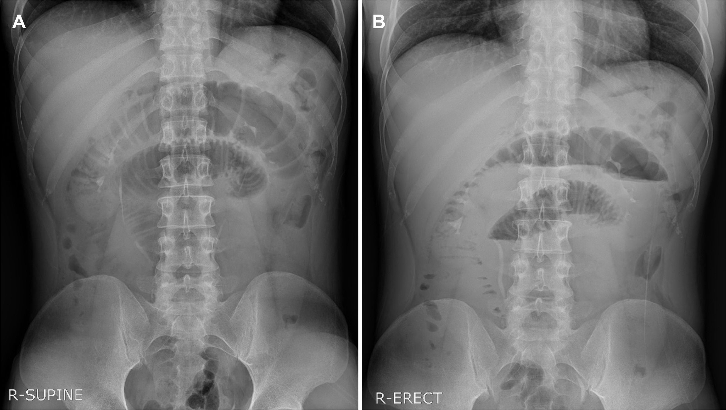
Fig. 1 Simple abdominal X-ray. (A) Supine simple X-ray image shows small bowel dilatation. (B) Erect simple X-ray image shows multiple air-fluid levels suggesting small bowel obstruction.

Fig. 2 Abdominal computed tomography on Admission date. (A) Coronal reconstruction images showed segmental enhancing wall thickening of small bowel loop with perilesional infiltration (arrow). (B) Axial reconstruction shows the segmental wall thickening of jejunum (arrow).
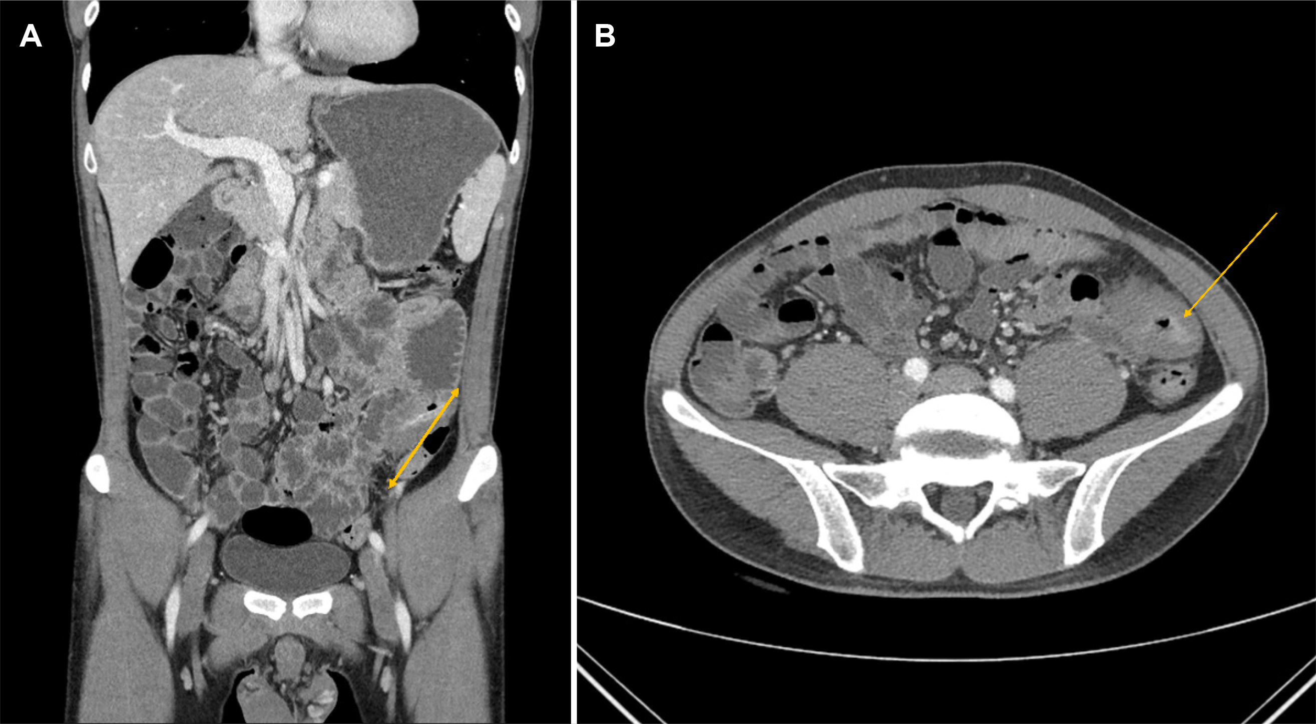
Fig. 3 Abdominal computed tomography on 3 days later. (A) Coronal reconstruction images showed slightly decompressed state of proximal small bowel dilatations with perilesional infiltration (arrow). (B) Axial reconstruction shows the no change of segmental wall thickening of jejunum (arrow).
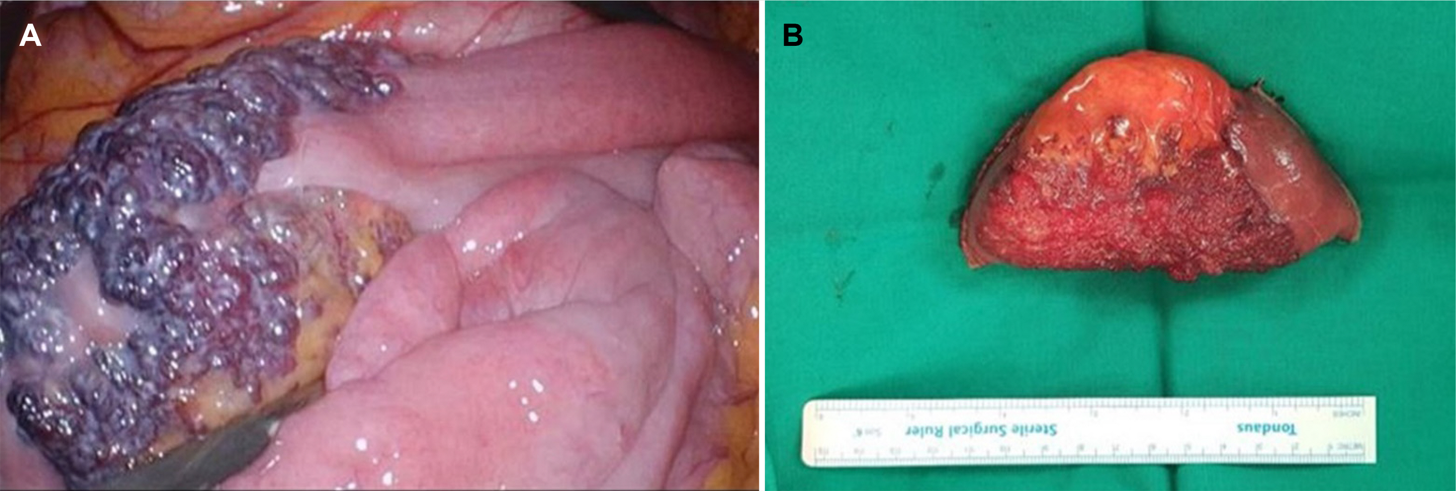
Fig. 4 (A, B) Intraoperative image showing that there was 6.2 cm lesion on the jejunum with bluish purple coloration and compressible varices on its surface.
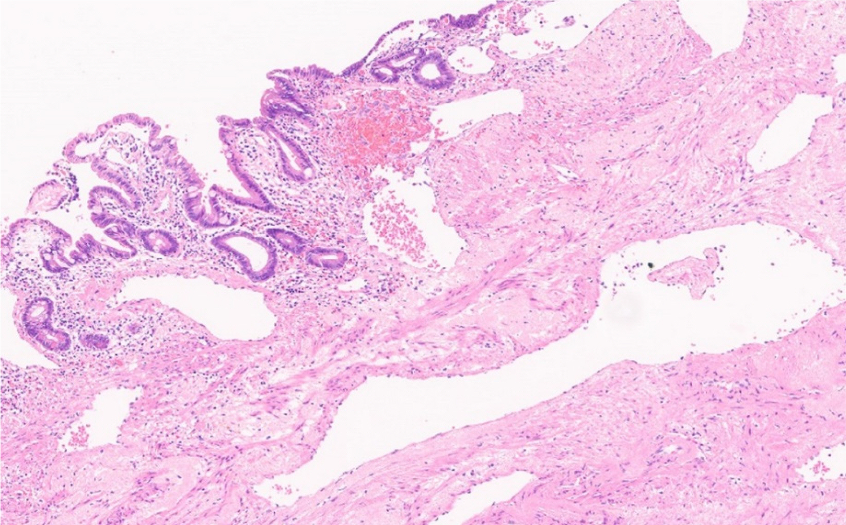
Fig. 5 Postoperative histopathological image shows the presence of arterial wall and venous wall in muscle layer (hematoxylin and eosin stain, ×40). It reveals a small bowel hemangioma, arteriovenous malformation.
Reference
-
1. Durer C, Durer S, Sharbatji M, Comba IY, Aharoni I, Majeed U. 2018; Cavernous hemangioma of the small bowel: a case report and literature review. Cureus. 10:e3113. DOI: 10.7759/cureus.3113.
Article2. Hu PF, Chen H, Wang XH, Wang WJ, Su N, Shi B. 2018; Small intestinal hemangioma: Endoscopic or surgical intervention? A case report and review of literature. World J Gastrointest Oncol. 10:516–521. DOI: 10.4251/wjgo.v10.i12.516. PMID: 30595805. PMCID: PMC6304305.
Article3. Ocampo Toro WA, Corral Ramos B, Concejo Iglesias P, Cubero Carralero J, Blanco García DF, Barón Ródiz P. 2018; Haemangiomas of the small intestine: poorly known cause of gastrointestinal bleeding of uncertain origin. Cureus. 10:e3155. DOI: 10.7759/cureus.3155. PMID: 30349762. PMCID: PMC6193570.
Article4. Levy AD, Abbott RM, Rohrmann CA Jr, Frazier AA, Kende A. 2001; Gastrointestinal hemangiomas: imaging findings with pathologic correlation in pediatric and adult patients. AJR Am J Roentgenol. 177:1073–1081. DOI: 10.2214/ajr.177.5.1771073. PMID: 11641173.5. Takase N, Fukui K, Tani T, et al. 2017; Preoperative detection and localization of small bowel hemangioma: two case reports. World J Gastroenterol. 23:3752–3757. DOI: 10.3748/wjg.v23.i20.3752. PMID: 28611528. PMCID: PMC5449432.
Article6. Igawa A, Oka S, Tanaka S, Kunihara S, Nakano M, Chayama K. 2016; Polidocanol injection therapy for small-bowel hemangioma by using double-balloon endoscopy. Gastrointest Endosc. 84:163–167. DOI: 10.1016/j.gie.2016.02.021. PMID: 26907744.
Article7. Chen HH, Tu CH, Lee PC, et al. 2015; Endoscopically diagnosed cavernous hemangioma in the deep small intestine: a case report. Advances in Digestive Medicine. 2:74–78. DOI: 10.1016/j.aidm.2014.03.009.
Article8. Fu JX, Zou YN, Han ZH, Yu H, Wang XJ. 2020; Small bowel racemose hemangioma complicated with obstruction and chronic anemia: a case report and review of literature. World J Gastroenterol. 26:1674–1682. DOI: 10.3748/wjg.v26.i14.1674. PMID: 32327915. PMCID: PMC7167414.
Article
- Full Text Links
-
- Actions
-
Cited
- CITED
-
- Close
- Share
-
- Similar articles
-
- A Case of a Cavernous Hemangioma in the Distal Jejunum Detected by Double-Balloon Enteroscopy in a Patient with Small Bowel Obstruction
- An Ileocolic Intussusception Caused by Small Bowel Hemangioma
- Small bowel intubation using guide wire: use in decompression of small bowel obstruction
- Laparoscopic Adhesiolysis for Small Bowel Obstruction: Effective Alternatives or Immoderate Challenge?
- Small Bowel Obstruction and Capsule Retention by a Small Bowel Ulcer That Was Not Found on Capsule Endoscopy
