Congenital web of the common bile duct combined with multiple intrahepatic duct stricture: a case report of successful radiological intervention
- Affiliations
-
- 1Department of Surgery, Asan Medical Center, University of Ulsan College of Medicine, Seoul, Korea
- 2Department of Radiology, Asan Medical Center, University of Ulsan College of Medicine, Seoul, Korea
- KMID: 2529275
- DOI: http://doi.org/10.12701/yujm.2021.01179
Abstract
- Congenital web formations are extremely rare anomalies of the extrahepatic biliary tree. We herein report a case of common bile duct septum combined with multiple intrahepatic bile duct strictures in a 74‐year‐old female patient who was successfully treated with radiological intervention. The patient initially visited the hospital because of upper abdominal pain. Imaging studies revealed multifocal strictures with dilatation in both intra- and extrahepatic ducts; the final clinical diagnosis was congenital common bile duct web combined with multiple intrahepatic duct strictures. Surgical treatment was not indicated because multiple biliary strictures were untreatable, and the disease was clinically diagnosed as benign. The multiple strictures were extensively dilated twice through bilateral percutaneous transhepatic biliary drainage (PTBD) for 2 months. After 1 month of observation, PTBD catheters were successfully removed. The patient is doing well at 6 months after completion of the radiological intervention, with the maintenance of normal liver function. Congenital web of the bile duct is very rare, and its treatment may vary depending on the patterns of biliary stenosis. In cases where surgical intervention is not indicated for congenital web and its associated disease, radiological intervention with balloon dilatation can be a viable therapeutic option.
Keyword
Figure
-
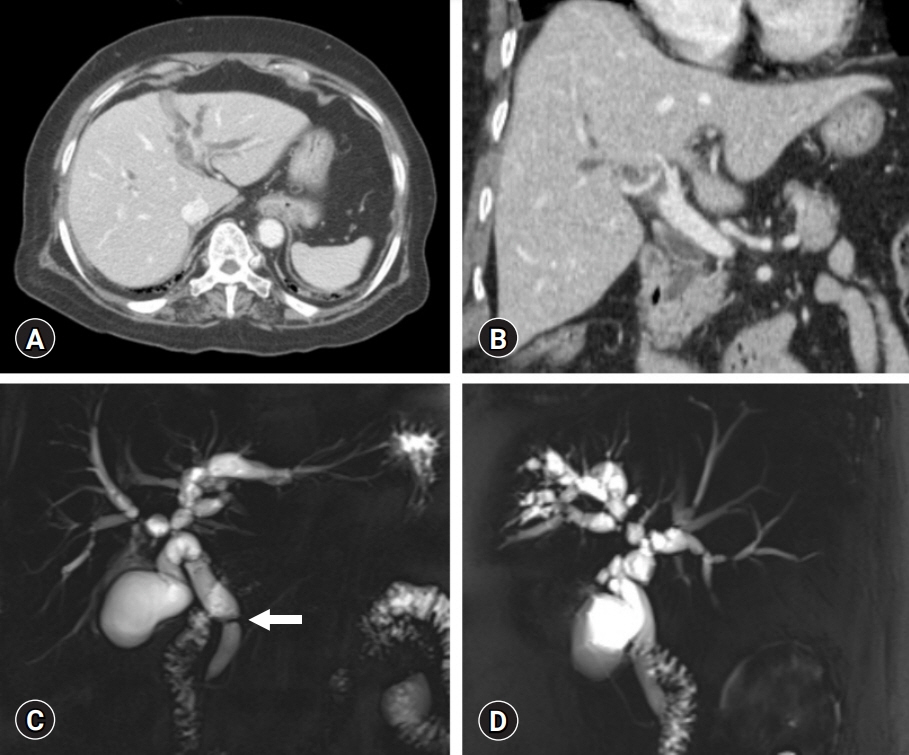
Fig. 1. Initial imaging studies. (A, B) Abdominal computed tomography shows diffuse dilatation of the intra- and extrahepatic bile ducts. (C, D) Magnetic resonance cholangiopancreatography shows multifocal biliary webs without anomalous pancreaticobiliary junction. Arrow indicates the location of a common bile duct web.
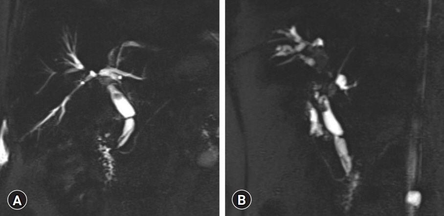
Fig. 2. The second magnetic resonance cholangiopancreatography shows multifocal stricture with dilatation in both intra- and extrahepatic ducts, suggesting that the stricture is more likely to be benign than malignant. The (A) location and (B) degree of the multifocal strictures are visualized.
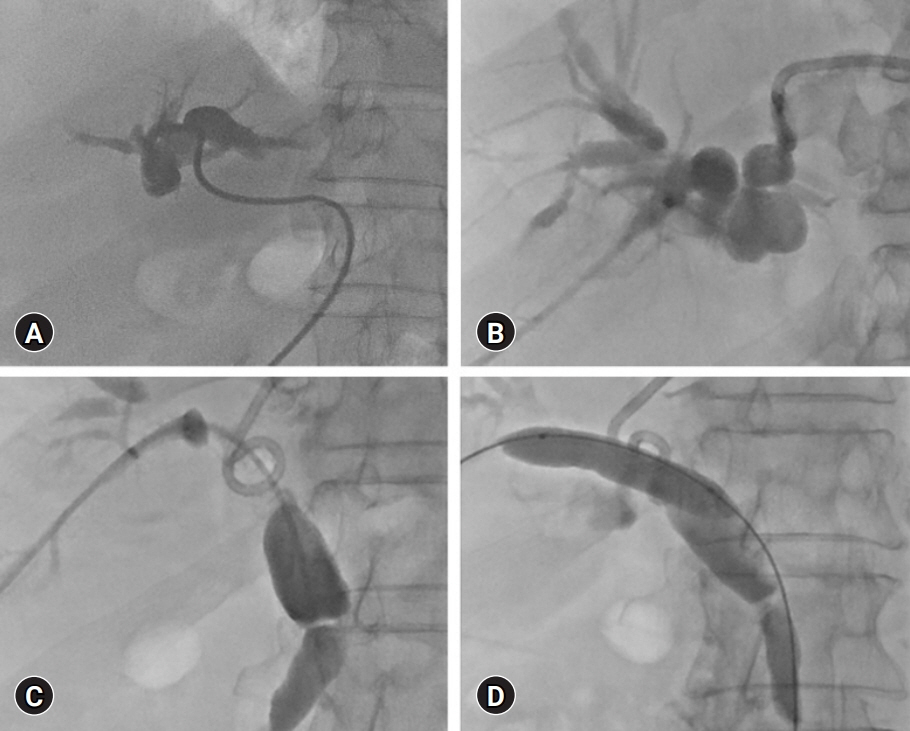
Fig. 3. Direct cholangiography findings. (A) The left-sided tubogram shows complete occlusion of the left hepatic duct. (B) The right-sided tubogram shows bile duct occlusion at the hepatic hilum. (C, D) The common bile duct web is finally cannulated after repeated trials, and then balloon dilatation of the intra- and extrahepatic stricture is performed.
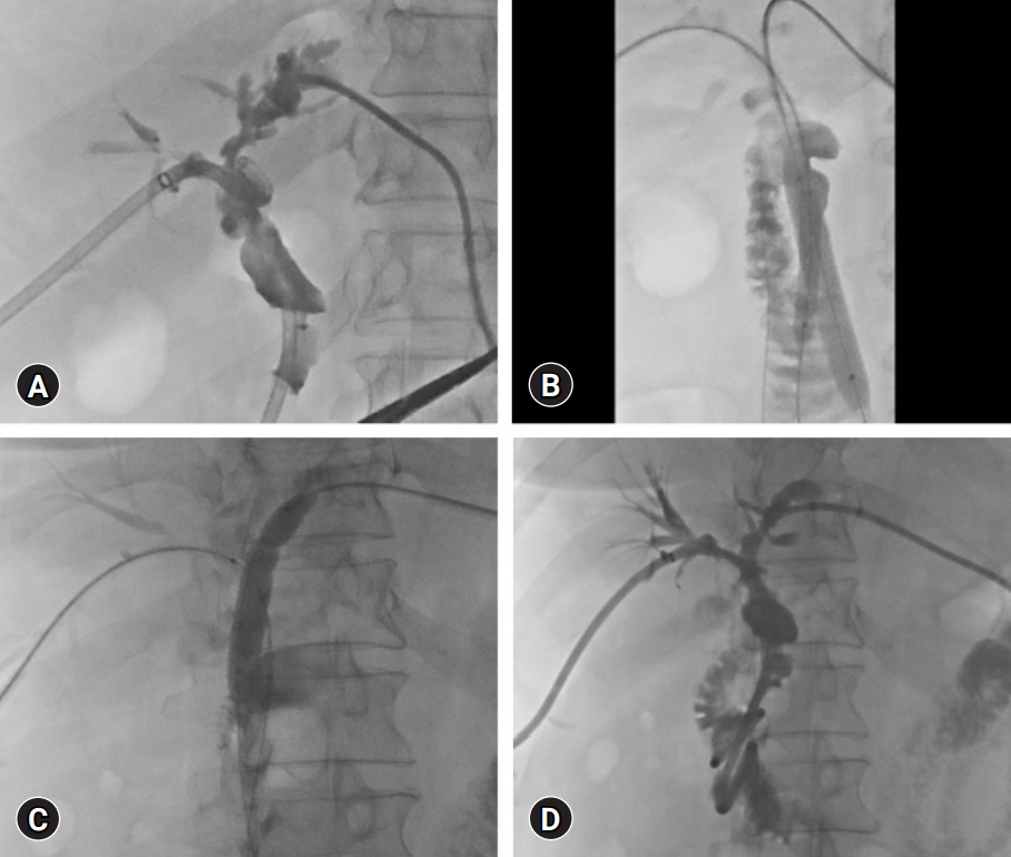
Fig. 4. The second balloon dilatation of the biliary stricture. (A) Tubogram shows the presence of residual strictures. (B, C) The second balloon dilatation of the intra- and extrahepatic stricture is extensively performed. (D) Tubogram shows improvement in multiple strictures.
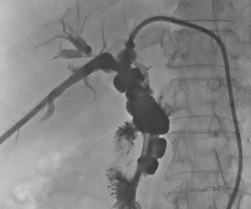
Fig. 5. Follow-up tubogram showing the good passage of the bile duct.
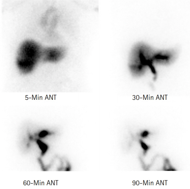
Fig. 6. Follow-up hepatobiliary scintigraphy taken at 6 months after the treatment showing a 90-minute excretion rate of 80% without significant obstruction. ANT, anterior view.

Fig. 7. Follow-up computed tomography taken at 6 months after the treatment showing a thin doughnut-shaped web at the distal bile duct. Dotted arrows indicate the proximal and distal levels of the bile duct to the web. Solid arrow indicates the level of a web at the distal bile duct.
Reference
-
References
1. Lamah M, Dickson GH. Congenital anatomical abnormalities of the extrahepatic biliary duct: a personal audit. Surg Radiol Anat. 1999; 21:325–7.2. Choi SH, Kim KW, Kwon HJ, Kim SY, Kwon JH, Song GW, et al. Clinical usefulness of gadoxetic acid-enhanced MRI for evaluating biliary anatomy in living donor liver transplantation. Eur Radiol. 2019; 29:6508–18.3. Takeishi K, Shirabe K, Yoshida Y, Tsutsui Y, Kurihara T, Kimura K, et al. Correlation between portal vein anatomy and bile duct variation in 407 living liver donors. Am J Transplant. 2015; 15:155–60.4. Furukawa H, Hara T, Taniguchi T. A case of septum formation of the common hepatic duct combined with an anomalous hepatic duct of the caudate lobe. Gastroenterol Jpn. 1992; 27:102–7.5. Kato S, Nakagawa T, Kobayashi H, Arai E, Isetani K. Septum formation of the common hepatic duct associated with an anomalous junction of the pancreaticobiliary ductal system and gallbladder cancer: report of a case. Surg Today. 1994; 24:534–7.6. Dolar ME, Ates KB, Dalay AR, Caner ME, Sasmaz N, Sahin B. Congenital stricture of the common hepatic duct due to a web: an unusual case without jaundice. Hepatogastroenterology. 1993; 40:194–5.7. Fisher MM, Chen SH, Dekker A. Congenital diaphragm of the common hepatic duct. Gastroenterology. 1968; 54:605–10.8. Kottoor R, Alvares JF, Devarbhavi H. Successful endoscopic therapy of an obstructing common bile duct web. Gastrointest Endosc. 2001; 53:126–8.9. Papaziogas B, Lazaridis C, Pavlidis T, Galanis I, Paraskevas G, Papaziogas T. Congenital web of the common bile duct in association with cholelithiasis. J Hepatobiliary Pancreat Surg. 2002; 9:271–3.10. Margolis M, Schein M. Biliary web: a rare cause of extrahepatic biliary obstruction. Dig Surg. 2001; 18:317–9.11. Gulliver DJ, Baker ME, Putnam W, Baillie J, Rice R, Cotton PB. Bile duct diverticula and webs: nonspecific cholangiographic features of primary sclerosing cholangitis. Am J Roentgenol. 1991; 157:281–5.12. Panés J, Bordas JM, Bruguera M, Cortés M, Rodés J. Localized sclerosing cholangitis? Endoscopy. 1985; 17:121–2.13. Yoon KH, Ha HK, Kim MH, Seo DW, Kim CG, Bang SW, et al. Biliary stricture caused by blunt abdominal trauma: clinical and radiologic features in five patients. Radiology. 1998; 207:737–41.14. Cherqui D, Palazzo L, Piedbois P, Charlotte F, Duvoux C, Duron JJ, et al. Common bile duct stricture as a late complication of upper abdominal radiotherapy. J Hepatol. 1994; 20:693–7.15. Shih SL, Lin JC, Lee HC, Blickman JG. Unusual causes of obstructive jaundice in children: diagnosis on CT. Pediatr Radiol. 1992; 22:512–4.16. Yamamoto R, Tazuma S, Kanno K, Igarashi Y, Inui K, Ohara H, et al. Ursodeoxycholic acid after bile duct stone removal and risk factors for recurrence: a randomized trial. J Hepatobiliary Pancreat Sci. 2016; 23:132–6.17. Wang SY, Tang HM, Chen GQ, Xu JM, Zhong L, Wang ZW, et al. Effect of ursodeoxycholic acid administration after liver transplantation on serum liver tests and biliary complications: a randomized clinical trial. Digestion. 2012; 86:208–17.
- Full Text Links
-
- Actions
-
Cited
- CITED
-
- Close
- Share
-
- Similar articles
-
- Type IV-A Choledochal Cyst with Intrahepatic Bile Duct Stricture
- Intrahepatic Bile Duct Dilatation Caused by Pancreatic Pseudocyst: A Case Report
- 4 Cases of Web of Common Bile Duct
- A Common Bile Duct Web Presenting with Obstructive Jaundice without Common Bile Duct Stone
- Solitary intrahepatic bile-duct cyst presenting with jaundice
