Cytological, histochemical, and ultrastructural study of human foetal liver of various gestation with future implications in segmental resection: an anatomical perspective
- Affiliations
-
- 1Department of Anatomy, All India Institute of Medical Sciences, Deoghar, India
- 2Department of Anatomy, All India Institute of Medical Sciences, Bhubaneswar, India
- 3Department of Anatomy, All India Institute of Medical Sciences, Bathinda, India
- 4Department of Anatomy, ESI-PGIMSR, Kolkata, India
- KMID: 2527673
- DOI: http://doi.org/10.5115/acb.21.152
Abstract
- The liver is the largest gland of the gastrointestinal tract having both exocrine and endocrine functions. Developmentally it arises as a ventral outgrowth from the gut endoderm during 3rd week of intrauterine life. The foetal liver is very important because of its synthetic and hemopoietic potential. The present work aimed to see the detailed histogenesis and development of the foetal liver by cytological, immunohistochemical and ultrastructural study. The liver tissue of nine aborted foetuses of various gestational age were studied. For cytology: special stains like Masson trichrome, periodic acid Schiff and reticulin were used, immunohistochemical staining was performed with triple antibodies (c-myc, Ki-67 and Ber-H2), and for ultrastructure: aluminium mounted specimens were coated with gold and argon gas and observed under scanning electron microscopy (EM). Cytology and immunohistochemistry showed the development of duct patterns and hemopoietic patterns in all stages of fetogenesis. The ductal plate was marked by the layer of dark brown staining cells at the edge of two portal tracts. The haemopoietic cells with sinusoids and aggregation of hepatocytes were observed in the early weeks of gestation. EM showed tree-like branching of a portal canal depicting hepatic segmentation of foetal liver. The organizational changes in lobular pattern, duct pattern, and microstructure of liver during fetogenesis are very crucial to achieve the adult morphology in feature. Histogenesis of the foetal liver follows a multistep process depending upon the gestational age, any deviation from normalcy may lead to structural and functional abnormality later in life.
Keyword
Figure
-
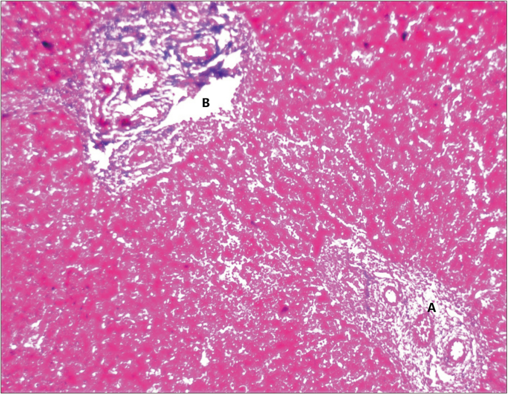
Fig. 1 Photomicrograph (Masson trichrome, ×10) of cytology showing the formation of the portal triad (A) and future central vein (B) in 1st-trimester foetal liver.
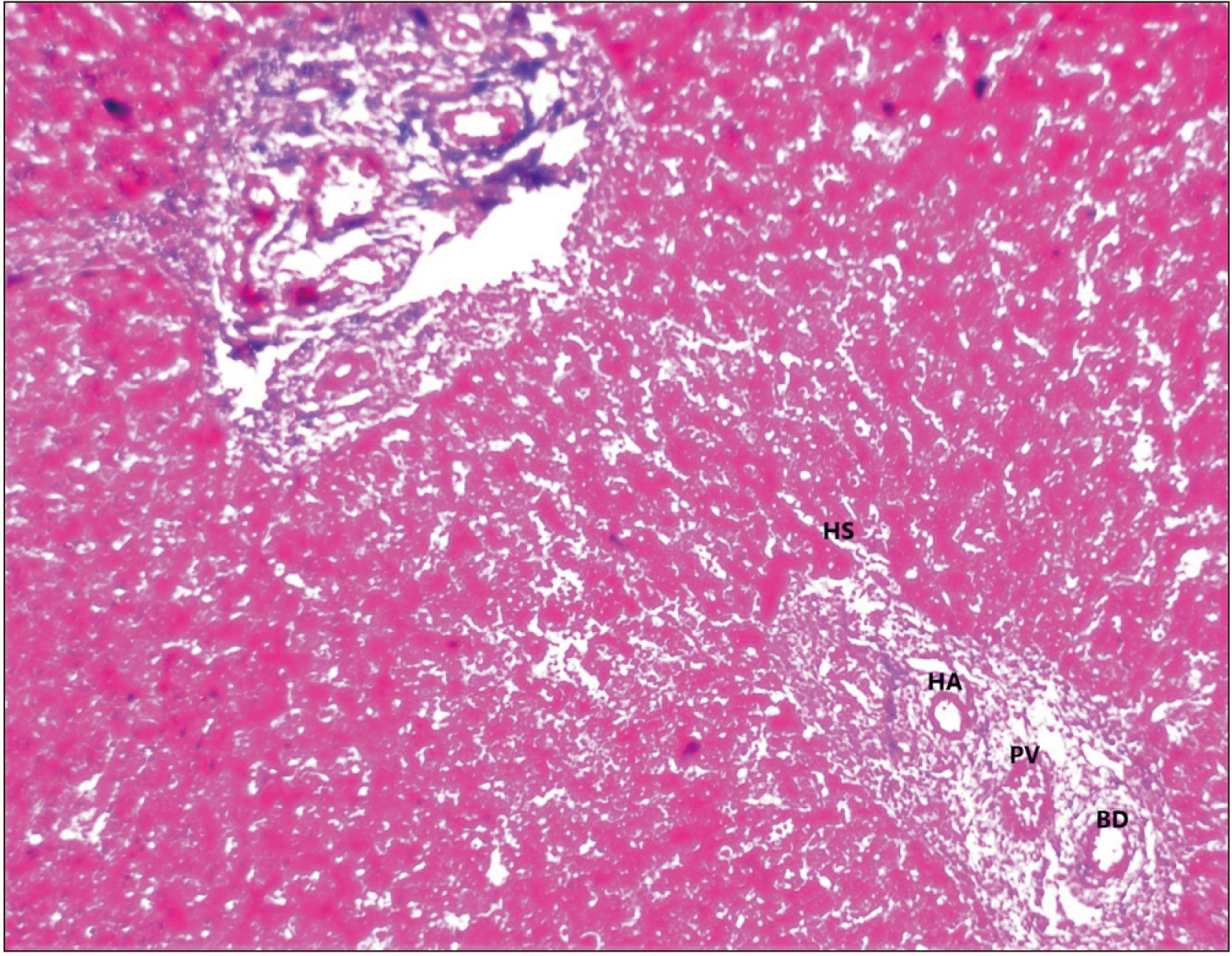
Fig. 2 Photomicrograph (Masson trichrome, ×10) of cytology showing the formation of the portal triad (the hepatic artery [HA], portal vein [PV] and bile duct [BD], hepatic sinusoids [HS] in 2nd-trimester foetal liver.
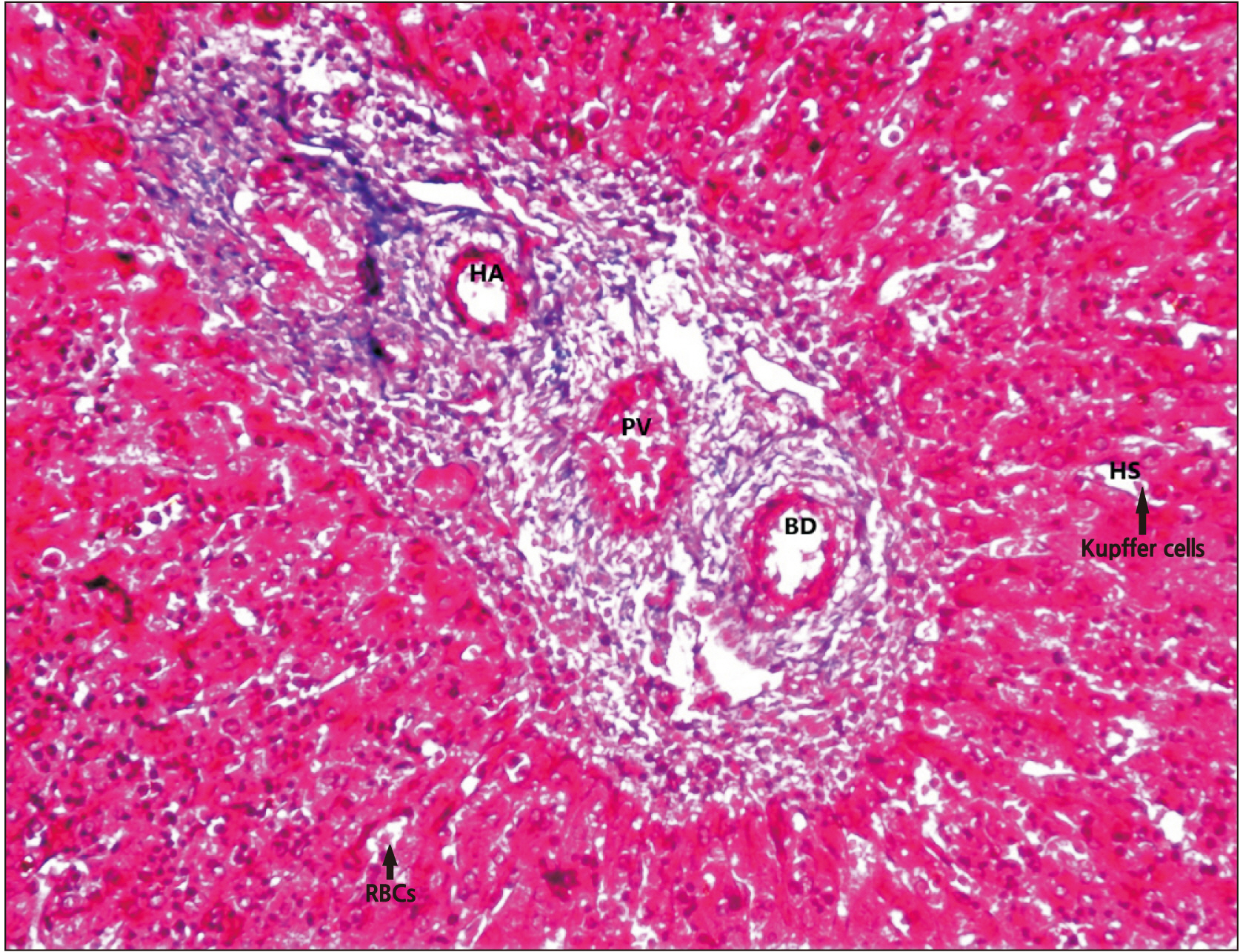
Fig. 3 Photomicrograph (Masson trichrome, ×20) of cytology showing Kupffer cells and red blood cells (RBCs) in hepatic sinusoids (HS) along with plates of hepatocytes in 3rd-trimester foetal liver. HA, hepatic artery; PV, portal vein; BD, bile duct.

Fig. 4 Photomicrograph (×10) of immunohistochemistry with CD30 (Ber-H2): brown discolouration of hepatocytes (arrow) labelled with Ber-H2, indicate immunopositivity. These dark brown staining cells at the edge of two portal tracts shows the formation of normal ductal plate. CV, central vein.
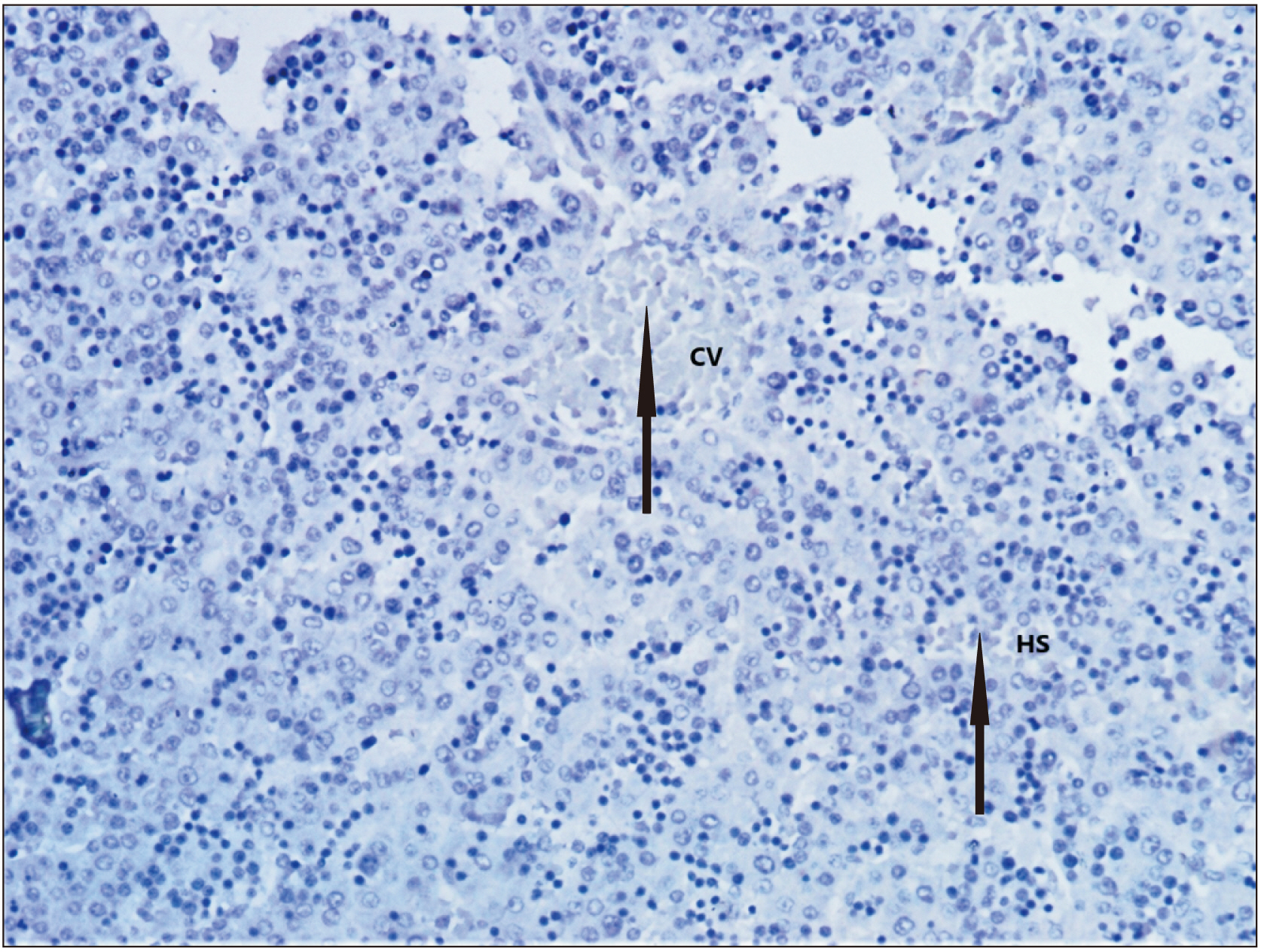
Fig. 5 Photomicrograph (×10) of immunohistochemistry with c-myc: brown discolouration of developing central vein (CV), hepatic sinusoids (HS) labelled with c-myc indicates immunopositivity which is more prominent than the Ber-H2.
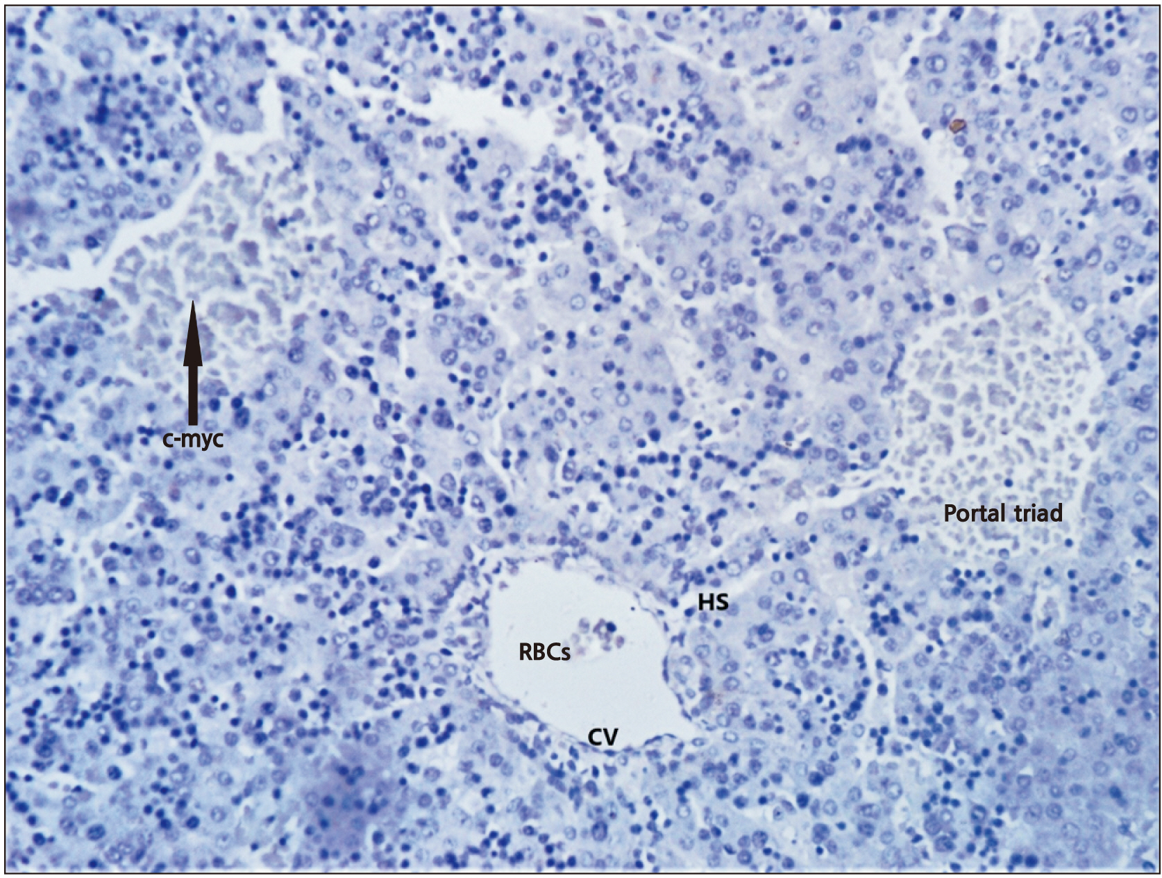
Fig. 6 Photomicrograph (×10) of immunohistochemistry with Ki-67: brown discolouration of developing hepatic parenchyma indicates immunopositivity which is of similar intensity as c-myc. RBCs, red blood cells; HS, hepatic sinusoids; CV, central vein.
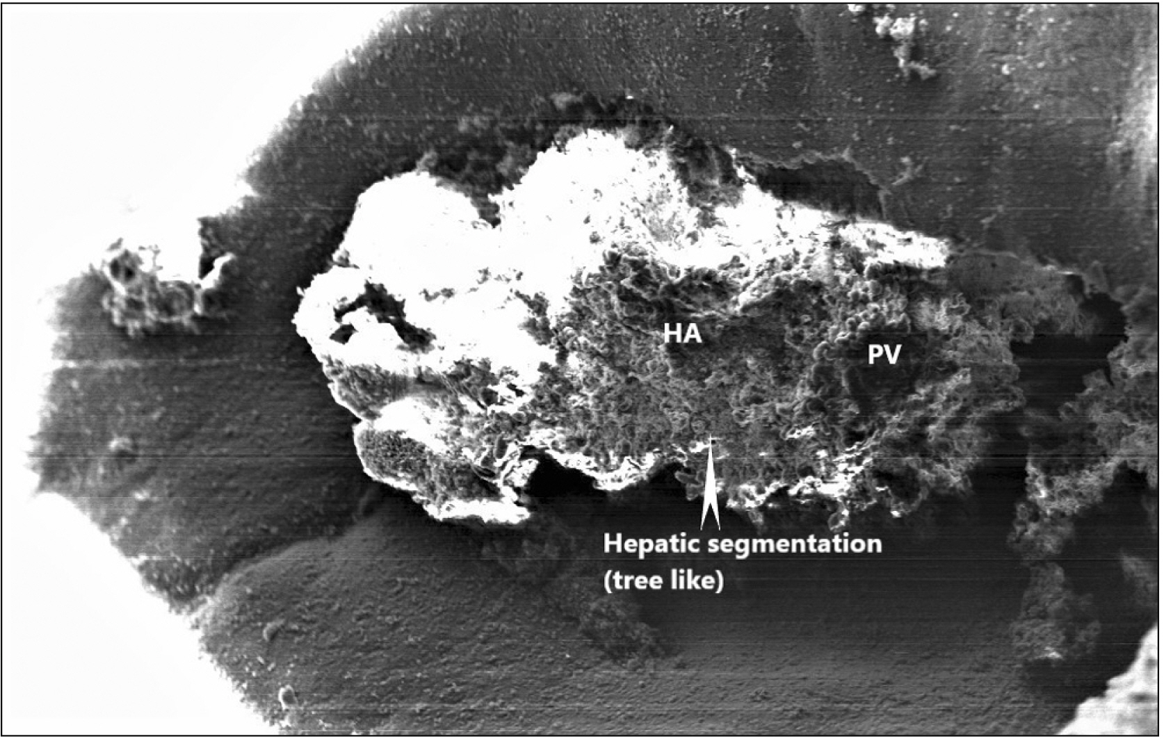
Fig. 7 Photomicrograph of the ultrastructure of the foetal liver obtained through scanning electron microscopy: showing tree-like branching of a portal canal indicating hepatic segmentation of liver. HA, hepatic artery; PV, portal vein.
Reference
-
References
1. Michalopoulos GK. 2007; Liver regeneration. J Cell Physiol. 213:286–300. DOI: 10.1002/jcp.21172. PMID: 17559071. PMCID: PMC2701258.
Article2. Sadler TW. 2005. Langman's medical embryology. 7th ed. Lippincott Williams & Wilkins;Philadelphia: p. 198–201.3. Gil MN, Choi DR, Yu KS, Jeong JH, Bak DH, Kim DK, Lee NS, Lee JH, Jeong YG, Na CS, Na DS, Ryu KH, Han SY. 2016; Rhus verniciflua Stokes attenuates cholestatic liver cirrhosis-induced interstitial fibrosis via Smad3 down-regulation and Smad7 up-regulation. Anat Cell Biol. 49:189–98. DOI: 10.5115/acb.2016.49.3.189. PMID: 27722012. PMCID: PMC5052228.
Article4. Ajmani ML. Ajmani ML, editor. 1993. Practical embalming. Text Book Principles of Embalming Techniques in Autopsied Body. Jaypee Brothers Publishers;New Delhi: p. 131–4.5. Naik KS, Lokanadham S, Gurushanthaiah M. 2020; Different gestational age related histogenesis of human foetal liver. Int J Anat Res. 8:7408–11. DOI: 10.16965/ijar.2020.115.
Article6. Terada T, Nakanuma Y, Ohta G. 1987; Glandular elements around the intrahepatic bile ducts in man; their morphology and distribution in normal livers. Liver. 7:1–8. DOI: 10.1111/j.1600-0676.1987.tb00308.x. PMID: 3553822.
Article7. Kieusseian A, Brunet de la Grange P, Burlen-Defranoux O, Godin I, Cumano A. 2012; Immature hematopoietic stem cells undergo maturation in the fetal liver. Development. 139:3521–30. DOI: 10.1242/dev.079210. PMID: 22899849.
Article8. Jaiswal A, Sinha DN, Singh AK. 2015; A study on histology of fetal liver. Natl J Clin Anat. 4:26–9. DOI: 10.4103/2277-4025.297268.
Article9. Zorn AM. Harvard Stem Cell Institute. 2008. Liver development. StemBook. Harvard Stem Cell Institute;Cambridge: DOI: 10.3824/stembook.1.25.1. PMID: 20614624.
Article10. Moore KL, Persaud TVN, Torchia MG. 2008. The developing human: clinically oriented embryology. 8th ed. Elsevier Saunders;Philadelphia: p. 211–42.11. Naito M, Hasegawa G, Takahashi K. 1997; Development, differentiation, and maturation of Kupffer cells. Microsc Res Tech. 39:350–64. DOI: 10.1002/(SICI)1097-0029(19971115)39:4<350::AID-JEMT5>3.0.CO;2-L.
Article12. Lipp W. 1952; The early structural development of the liver parenchyma in man. Z Mikrosk Anat Forsch. 59:161–86. German.13. Severn CB. 1972; A morphological study of the development of the human liver. II. Establishment of liver parenchyma, extrahepatic ducts and associated venous channels. Am J Anat. 133:85–107. DOI: 10.1002/aja.1001330106. PMID: 5008885.
Article14. Villeneuve J, Pelluard-Nehme F, Combe C, Carles D, Chaponnier C, Ripoche J, Balabaud C, Bioulac-Sage P, Lepreux S. 2009; Immunohistochemical study of the phenotypic change of the mesenchymal cells during portal tract maturation in normal and fibrous (ductal plate malformation) fetal liver. Comp Hepatol. 8:5. DOI: 10.1186/1476-5926-8-5. PMID: 19602240. PMCID: PMC2721154.
Article15. Zamboni L. 1965; Electron microscopic studies of blood embryogenesis in humans. II. The hemopoietic activity in the fetal liver. J Ultrastruct Res. 12:525–41. DOI: 10.1016/S0022-5320(65)80045-4.16. Hamilton WJ, Boyd JD, Mossman HW. 1978. Human embryology: prenatal development of form and function. 4th ed. Macmillan;London: p. 339–48.17. Slayton WB, Juul SE, Calhoun DA, Li Y, Braylan RC, Christensen RD. 1998; Hematopoiesis in the liver and marrow of human fetuses at 5 to 16 weeks postconception: quantitative assessment of macrophage and neutrophil populations. Pediatr Res. 43:774–82. DOI: 10.1203/00006450-199806000-00010. PMID: 9621987.
Article18. Couvelard A, Scoazec JY, Dauge MC, Bringuier AF, Potet F, Feldmann G. 1996; Structural and functional differentiation of sinusoidal endothelial cells during liver organogenesis in humans. Blood. 87:4568–80. DOI: 10.1182/blood.V87.11.4568.bloodjournal87114568. PMID: 8639825.
Article19. Emura I, Sekiya M, Ohnishi Y. 1983; Two types of immature erythrocytic series in the human fetal liver. Arch Histol Jpn. 46:631–43. DOI: 10.1679/aohc.46.631. PMID: 6673688.
Article20. Miranda RN, Omurtag K, Castellani WJ, De las Casas LE, Quintanilla NM, Kaabipour E. 2006; Myelopoiesis in the liver of stillborns with evidence of intrauterine infection. Arch Pathol Lab Med. 130:1786–91. DOI: 10.5858/2006-130-1786-MITLOS. PMID: 17149951.
Article21. Balis JU, Chan A, Conen PE. 1964; Electron microscopy study of the developing human liver. Tijdschr Gastroenterol. 7:133–47. DOI: 10.1159/000387692. PMID: 14264616.22. Potter EL, Craig JM. Potter EL, Craig JM, editors. 1975. Weights standards for organs for early human fetuses. Pathology of the Fetus and the Infant. 3rd ed. Year Book Medical Publishers;Chicago: p. 15–24.23. Notenboom RG, de Boer PA, Moorman AF, Lamers WH. 1996; The establishment of the hepatic architecture is a prerequisite for the development of a lobular pattern of gene expression. Development. 122:321–32. DOI: 10.1242/dev.122.1.321. PMID: 8565845.
Article24. Payushina OV. 2012; Hematopoietic microenvironment in the fetal liver: roles of different cell populations. Int Sch Res Not. 2012:979480. DOI: 10.5402/2012/979480.
Article25. Valdes-Dapena MA. 1957. An atlas of foetal and neonatal histology. J.P. Lippincott Co;Philadelphia, PA: p. 75–87. DOI: 10.1016/0002-9378(58)90623-9.26. Abbaszadeh H, Ghorbani F, Derakhshani M, Movassaghpour A, Yousefi M. 2020; Human umbilical cord mesenchymal stem cell-derived extracellular vesicles: a novel therapeutic paradigm. J Cell Physiol. 235:706–17. DOI: 10.1002/jcp.29004. PMID: 31254289.
Article27. Giancotti A, Monti M, Nevi L, Safarikia S, D'Ambrosio V, Brunelli R, Pajno C, Corno S, Di Donato V, Musella A, Chiappetta MF, Bosco D, Panici PB, Alvaro D, Cardinale V. 2019; Functions and the emerging role of the foetal liver into regenerative medicine. Cells. 8:914. DOI: 10.3390/cells8080914. PMID: 31426422. PMCID: PMC6721721.
Article28. El Baz H, Demerdash Z, Kamel M, Hammam O, Abdelhady DS, Mahmoud S, Hassan S, Mahmoud F, Atta S, Riad NM, Gaafar T. 2020; Induction of hepatic regeneration in an experimental model using hepatocyte-differentiated mesenchymal stem cells. Cell Reprogram. 22:134–46. DOI: 10.1089/cell.2019.0076. PMID: 32243193.
Article
- Full Text Links
-
- Actions
-
Cited
- CITED
-
- Close
- Share
-
- Similar articles
-
- Comparison of survival outcomes after anatomical resection and non-anatomical resection in patients with hepatocellular carcinoma
- Resection of Colorectal Liver Metastases
- Long Term Effects of IUD on the Human Endometrium: Histologic, Histochemical and Ultrastructural Studies
- Experience of laparoscopic liver resection for various liver diseases
- How to minimize conversion to open surgery during laparoscopic liver resection: the point of view of hemostasis
