Multiple ancient schwannomas of the ulnar nerve at distant sites: a case report
- Affiliations
-
- 1Department of Plastic and Reconstructive Surgery, Seoul National University Hospital, Seoul National University College of Medicine, Seoul, Korea
- KMID: 2526655
- DOI: http://doi.org/10.12790/ahm.21.0136
Abstract
- In rare cases, schwannoma—a benign peripheral nerve tumor—undergoes chronic degenerative changes and progresses to ancient schwannoma. Herein, we report the first case of ulnar nerve-derived multiple ancient schwannomas, which uncommonly developed in the extremities and major nerves. A 76-year-old female patient presented with tingling sensations in her left ring and small fingers. She had a gradually enlarging mass that had developed 40 years ago in the proximal upper arm and a new mass that had grown on the wrist for the past few years. Based on physical examination, ultrasonography, and magnetic resonance imaging, ancient schwannomas of the ulnar nerve were suspected. The older and larger mass in the upper arm was more entangled with nerve fascicles, having necrotic changes. Through meticulous dissection of the nerve fascicles, both masses were successfully enucleated, and pathological examination confirmed ancient schwannoma. As ancient schwannomas grow, they become more entangled with the nerves; thus, early surgical removal is recommended.
Keyword
Figure
-
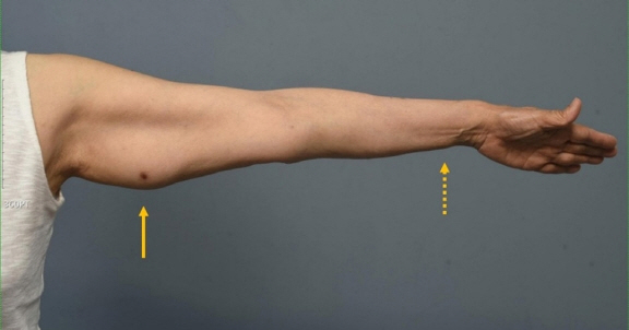
Fig. 1. Multiple masses presented in the posteromedial aspect of the patient’s left upper arm and wrist. A 4-cm firm, painless mass was palpated on the patient’s left upper arm (arrow). A 0.5-cm firm, painless mass was palpated on the patient’s wrist (dotted arrow).
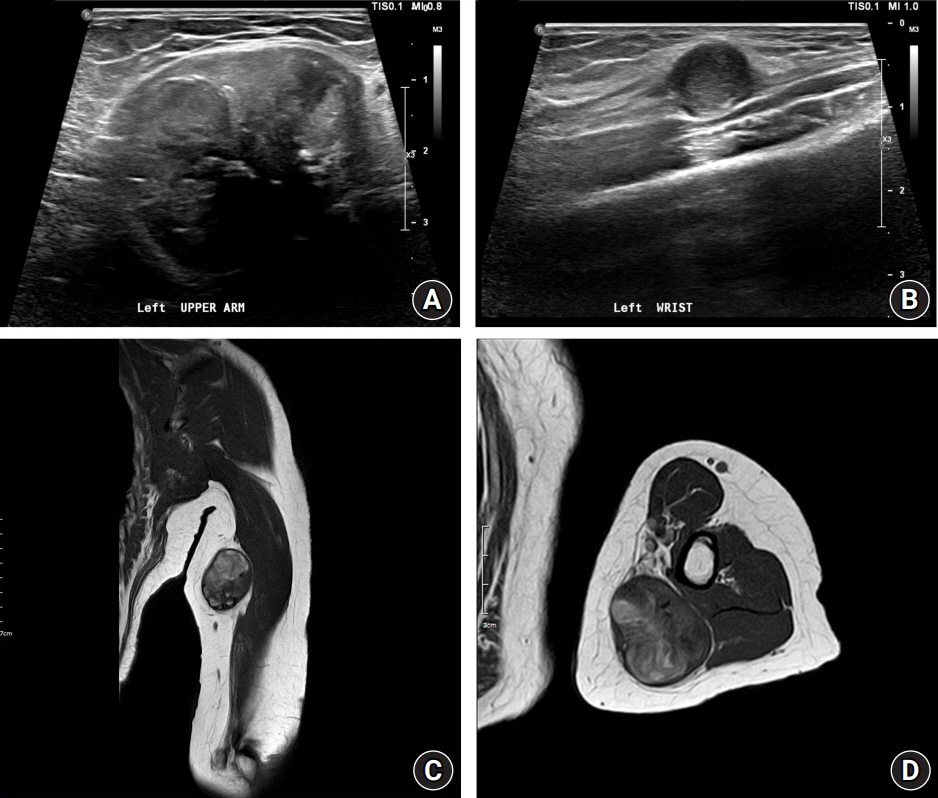
Fig. 2. Ultrasound and magnetic resonance T1 imaging. On ultrasound imaging, well-circumscribed heterogeneous echoic masses with internal calcification and internal vascularity originating from the ulnar nerve are observed in the subcutaneous layer of the upper arm (A) and wrist (B). The hyaline degeneration, cystic change, calcification, and hemorrhage observed on a magnetic resonance image of the upper arm mass are typical characteristics of ancient schwannoma (C, D).

Fig. 3. Multiple ancient schwannomas of the upper arm (A and B) and wrist (C and D) from the ulnar nerve. (A, C) Ancient schwannomas originating from the ulnar nerve are surrounded by nerve fascicles. (B, D) The masses are resected from the upper arm and forearm with minimal nerve damage through meticulous dissection. (E, F) Multiple ancient schwannomas are enucleated from the ulnar nerve of the left arm.
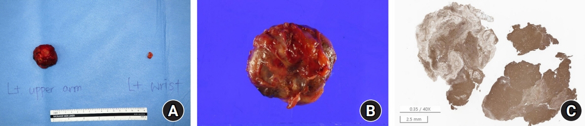
Fig. 4. Enucleated ancient schwannomas, magnified image, and immunohistochemical staining. The upper arm mass had developed for a longer period than the wrist mass. Hence, it is larger (A) and more entangled with the nerve fascicles (B). (C) Immunohistochemical staining is positive for S-100, a typical feature of schwannoma (×40).
Reference
-
References
1. Das Gupta TK, Brasfield RD, Strong EW, Hajdu SI. Benign solitary schwannomas (neurilemomas). Cancer. 1969; 24:355–66.
Article2. Bhattacharyya AK, Perrin R, Guha A. Peripheral nerve tumors: management strategies and molecular insights. J Neurooncol. 2004; 69:335–49.
Article3. Kehoe NJ, Reid RP, Semple JC. Solitary benign peripheral-nerve tumours. Review of 32 years’ experience. J Bone Joint Surg Br. 1995; 77:497–500.
Article4. Isobe K, Shimizu T, Akahane T, Kato H. Imaging of ancient schwannoma. AJR Am J Roentgenol. 2004; 183:331–6.
Article5. Mikami Y, Hidaka T, Akisada T, Takemoto T, Irei I, Manabe T. Malignant peripheral nerve sheath tumor arising in benign ancient schwannoma: a case report with an immunohistochemical study. Pathol Int. 2000; 50:156–61.
Article6. Lee YS, Kim JO, Park SE. Ancient schwannoma of the thigh mimicking a malignant tumour: a report of two cases, with emphasis on MRI findings. Br J Radiol. 2010; 83:e154–7.
Article7. Vlychou M, Dailiana ZH. Ancient schwannoma of the hand. J Hand Surg Am. 2011; 36:2030–3.
Article8. Shilpa B. Ancient schwannoma-a rare case. Ethiop J Health Sci. 2012; 22:215–8.9. Malizos K, Ioannou M, Kontogeorgakos V. Ancient schwannoma involving the median nerve: a case report and review of the literature. Strategies Trauma Limb Reconstr. 2013; 8:63–6.
Article10. Joseph CM, Cherian M, Mishra MN, Chase A. Ancient schwanomma of the distal ulnar nerve: a rare presentation. J Clin Mol Pathol. 2020; 4:24.
- Full Text Links
-
- Actions
-
Cited
- CITED
-
- Close
- Share
-
- Similar articles
-
- Surgical treatment of multiple plexiform schwannomas arising from the superficial radial nerve: a case report
- Ulnar Nerve Palsy due to Multiple Ganglion Cysts at the Elbow: A Case Report
- A Case of Ancient Schwannoma of the Lingual Nerve
- Tardy Ulnar Nerve Palsy with Recurrent Ulnar-Nerve Dislocation: Case Report
- Laparoscopic Resection of Ancient Shwannoma of Vagus Nerve in the Lesser Sac
