Prosthetic rehabilitation by obturator considering the biomechanics in partially edentulous patient after maxillectomy
- Affiliations
-
- 1Department of Prosthodontics, School of Dentistry and Institute of Oral Bio-Science, Jeonbuk National University, Jeonju, Republic of Korea
- KMID: 2526004
- DOI: http://doi.org/10.14368/jdras.2021.37.4.281
Abstract
- Patients who went through maxillectomy can have severely impaired swallowing, mastication, and pronunciation functions because of palatal defects. Leakage occurs through the nasal cavity while eating, chewing becomes difficult due to the loss of teeth and alveolar ridges, and oral and nasal passages are not separated, leading to hyper-nasal sound, and significantly reducing the quality of life. To prosthetically reconstruct the defect, the weight of the obturator should be reduced as much as possible to minimize dropout because of gravity, and the bulb of the obturator should be properly extended into the defect to get additional retention and stability. In this case of a partially edentulous patient who underwent additional maxillary resection because of tumor recurrence, a metal framework was designed by applying the basic design principles of removable partial dentures. An obturator with improved retention, stability, and support was fabricated through functional impressions. The patient was satisfied with the improved facial expression, mastication, swallowing, and pronunciation, and showed stable occlusion and oral hygiene management during the follow-up period.
Figure
-

Fig. 1 Intra-oral examination. (A) Maxillary occlusal view, (B) Right lateral view, (C) Frontal view, (D) Left lateral view, (E) Mandibular occlusal view.

Fig. 2 Pre-treatment panoramic radiograph.
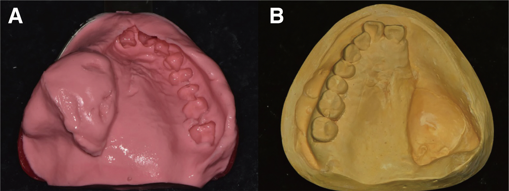
Fig. 3 (A) Preliminary impression with irreversible hydrocolloid material, (B) Diagnostic stone cast.
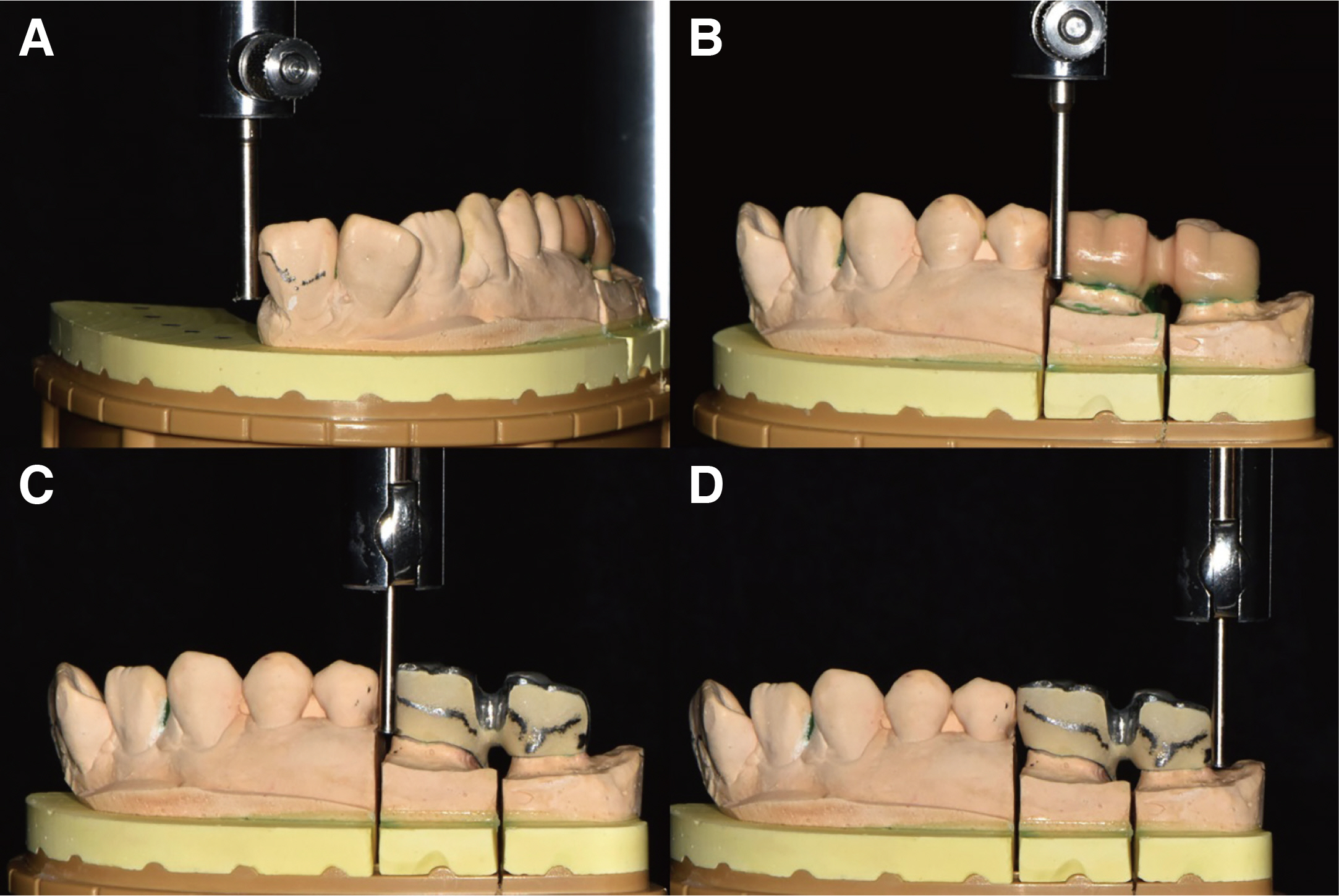
Fig. 4 (A), (B) Surveying of full contour wax-up cast, (C), (D) Surveying of porcelain build-up die cast.

Fig. 5 (A) Boder molding with modeling compound wax, (B) Final impression with Polyvinylsiloxane impression material.

Fig. 6 (A) Definitive cast, (B), (C), (D) Surveying for fabrication of metal framework.

Fig. 7 (A) Metal framework, (B) Occlusion rim.

Fig. 8 Arrangement of artificial teeth and formation of occlusion. (A) Maxillary occlusal view, (B) Right lateral view of centric occlusion, (C) Frontal view of centric occlusion, (D) Left lateral view of centric occlusion, (E) Right lateral view of left lateral movement, (F) Mandibular occlusal view of left lateral movement, (G) Left lateral view of right lateral movement.

Fig. 9 Delivery of definitive prosthesis. (A) Maxillary occlusal view, (B) Right lateral view, (C) Frontal view, (D) Left lateral view, (E) Definitive prosthesis.

Fig. 10 Extraoral Photograph. (A) Pretreatment, (B) Post-treatment.
Reference
-
References
1. 2017; The Glossary of Prosthodontic Terms: Ninth Edition. J Prosthet Dent. 117(5S):e62. DOI: 10.1016/j.prosdent.2016.12.001. PMID: 28418832.2. Marunick M. 2004; Hybrid gate design framework for the rehabilitation of the maxillectomy patient. J Prosthet Dent. 91:315–8. DOI: 10.1016/j.prosdent.2004.01.015. PMID: 15116031.3. Beumer J, Marunick MT, Esposito SJ. 2011. Maxillofacial rehabilitation: Prosthodontic and surgical management of cancer related, acquired, and congenital defects of the head and neck. 3rd ed. Quintessence Pub.;Illinois: p. 187–210.4. Desjardins RP. 1978; Obturator prosthesis design for acquired maxillary defect. J Prosthet Dent. 39:424–35. DOI: 10.1016/S0022-3913(78)80161-9. PMID: 273694.5. Schwartzman B, Caputo AA, Beumer J. 1990; Gravityinduced stresses by an obturator prosthesis. J Prosthet Dent. 64:466–8. DOI: 10.1016/0022-3913(90)90045-E. PMID: 2231457.6. Firtell DN, Grisius RJ. 1980; Retention of obturatorremovable partial denture: A comparison of buccal and lingual retention. J Prosthet Dent. 43:212. DOI: 10.1016/0022-3913(80)90189-4.7. Aramany MA. 1978; Basic principles of obturator design for partially edentulous patients. Part I: Classification. J Prosthet Dent. 40:554–7. DOI: 10.1016/0022-3913(78)90092-6. PMID: 364015.8. Rosenstiel SF, Land MF, Fujimoto J. 2017. Contemporary fixed prosthodontics. 5th ed. Elsevier Inc.;Amsterdam: p. 582–606.9. The council of professor of dental universities. 2019. Removable partial prosthodontics. Revision. Dental Wisdom Pub.;Seoul: p. 100–39. p. 256–65.10. King GE, Martin JW. 1983; Cast circumferential and wire clasps for obturator retention. J Prosthet Dent. 49:799–802. DOI: 10.1016/0022-3913(83)90352-9. PMID: 6348262.11. Lyons KM, Beumer J 3rd, Caputo AA. 2005; Abutment load transfer by removable partial denture obturator frameworks in different acquired maxillary defects. J Prosthet Dent. 94:281–8. DOI: 10.1016/j.prosdent.2005.06.005. PMID: 16126081.12. Berg T Jr, Caputo AA. 1978; Anterior rests for maxillary removable partial denture. J Prosthet Dent. 39:139–46. DOI: 10.1016/S0022-3913(78)80010-9. PMID: 340666.13. The council of professor of dental universities. 2014. Prosthodontic treatment for edentulous patients. 2nd ed. Dental Wisdom Pub.;Seoul: p. 566–83.14. Gay WD, King GE. 1980; Applying basic prosthodontic principles in the edentulous maxillectomy patient. J Prosthet Dent. 43:433–5. DOI: 10.1016/0022-3913(80)90216-4.15. Aramany MA. 1978; Basic principles of obturator design for partially edentulous patients. Part II: Design principles. J Prosthet Dent. 40:656–62. DOI: 10.1016/0022-3913(78)90065-3. PMID: 364026.16. Parr GR, Tharp GE, Rahn AO. 1989; Prosthodontic principles in the framework design of maxillary obturator prosthesis. J Prosthet Dent. 62:205–12. DOI: 10.1016/0022-3913(89)90315-6.17. Schwartzman B, Caputo AA, Beumer J. 1985; Occlussal force transfer by removable partial denture designs for a radical maxillectomy. J Prosthet Dent. 54:397–403. DOI: 10.1016/0022-3913(85)90560-8.18. Martin JW, King GE. 1984; Framework retention for maxillary obturator prostheses. J Prosthet Dent. 51:669–72. DOI: 10.1016/0022-3913(84)90415-3. PMID: 6374123.19. King GE, Martin JW. 1983; Cast circumferential and wire clasps for obturator retention. J Prosthet Dent. 49:799–802. DOI: 10.1016/0022-3913(83)90352-9. PMID: 6348262.20. Ahmad I, Sherriff M, Waters NE. 1992; The effect of reducing the number of clasps on removable partial denture retention. J Prosthet Dent. 68:928–33. DOI: 10.1016/0022-3913(92)90553-M. PMID: 1494122.21. Stewart KL, Rudd KD, Kuebker WA. 1983. Clinical removable partial prosthodontics. 1st ed. CV Mosby;St. Louis: p. 101–2.22. Oral K, Aramany MA, McWilliams BJ. 1979; Speech intelligibility with the buccal flange obturator. J Prosthet Dent. 41:323–8. DOI: 10.1016/0022-3913(79)90017-9. PMID: 283232.23. McAndrew KS, Rothenberger S, Minsley GE. 1998; An innovative investment method for the fabrication of a closed hollow obturator prosthesis. J Prosthet Dent. 80:129–32. DOI: 10.1016/S0022-3913(98)70098-8. PMID: 9656185.24. Yoon BK, Ko SO, Shin HK. 2001; A clinical study of palatal lift for treatment of velopharyngeal incompetency. J Korean Assoc Oral Maxillofac Surg. 27:92–6.25. Kummer AW. 2001. Cleft palate and craniofacial anomalies: the effects of speech and resonance. Singular Thomson Learning Inc.;San Diego: p. 145–76.26. Suh KS, Kim JY, Kim YK. 1994; Relationship between formants and constriction area of vocal tract in 9 Korean standard vowels. J Korean Soc Laryngol Phoniatr Logop. 5:44–58.27. Cho JH, Pyo WY, Choi HS, Choi BJ, Son HK, Sim HS. 2004; Physioantomy of nasopharyngeal space and hypernasality in cleft palate. J Korean Acad Pediatr Dent. 31:721–8.
- Full Text Links
-
- Actions
-
Cited
- CITED
-
- Close
- Share
-
- Similar articles
-
- Prosthetic rehabilitation using an obturator in a fully edentulous patient who had partial maxillectomy
- Prosthetic rehabilitation of a fully edentulous patient after maxillectomy: A case report
- Prosthetic rehabilitation of partially edentulous patient after maxillectomy: A case report
- A three-dimensional finite element analysis of obturator prosthesis for edentulous maxilla
- Prosthetic rehabilitation by double-processing technique for edentulous patient with soft palate defect after maxillectomy: A case report
