Full mounth rehabilitation using OP finder ® system for patient with inadequate occlusal plane and multiple occlusal wear tooth state: a case report
- Affiliations
-
- 1Department of Prosthodontics, Daejeon Dental Hospital, Wonkwang University, Daejeon, Republic of Korea
- KMID: 2525988
- DOI: http://doi.org/10.14368/jdras.2021.37.3.138
Abstract
- The diagnosis and treatment plan for forming ideal occlusal plane in full mouth rehabilitation are difficult because each process is complicated and information exchange between dentist and technician is subjective. The OP finder® system simplifies this process and helps to deliver more objective and accurate information. In this case, full mouth rehabilitation was performed using OP finder® system for patients with old bad fixed prosthesis and severely worn mandibular teeth, and reported that the result of proper occlusal plane setting and masticatory function recovery was obtained
Keyword
Figure
-

Fig. 1 Intraoral photograph before treatment. (A) Upper view, (B) Right view, (C) Frontal view, (D) Left view, (E) Lower view.
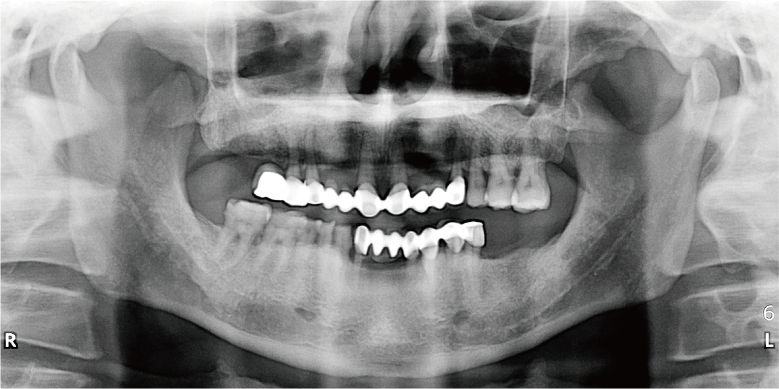
Fig. 2 Panoramic radiograph before treatment.

Fig. 3 Face bow transfer. (A) OP finder 1 fontal view, (B) OP finder 1 Later view, (C) OPUS articulator using OP finder 2.
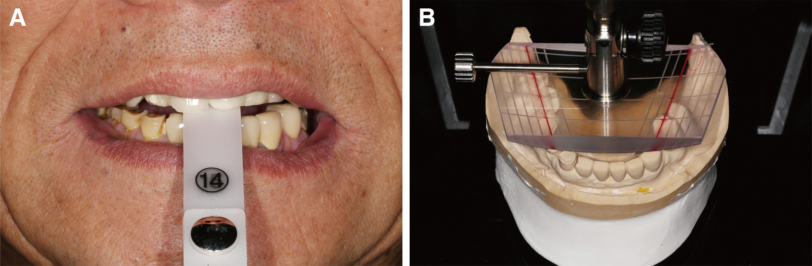
Fig. 4 Diagnosis. (A) Leaf guage, (B) OP finder 3.
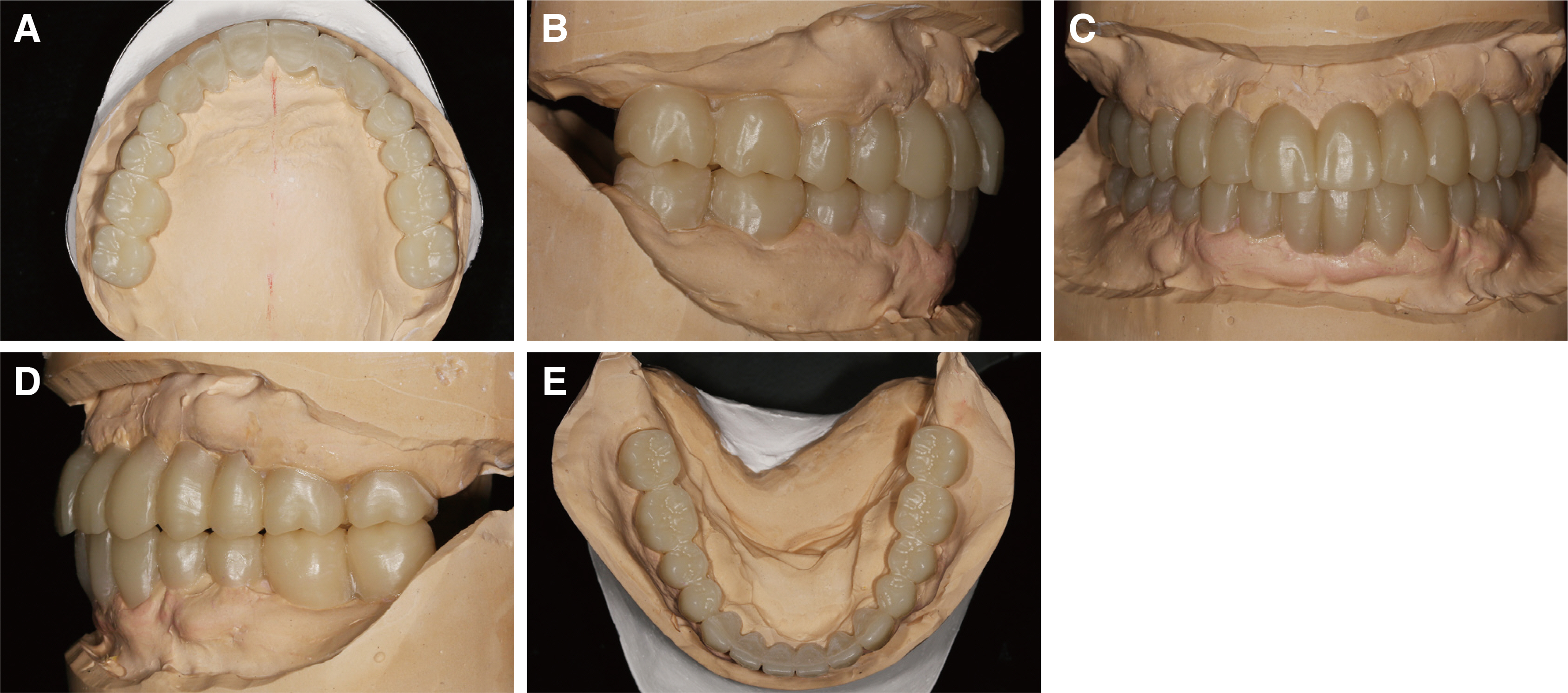
Fig. 5 Diagnostic wax-up cast model. (A) Upper view, (B) Right view, (C) Frontal view, (D) Left view, (E) Lower view.
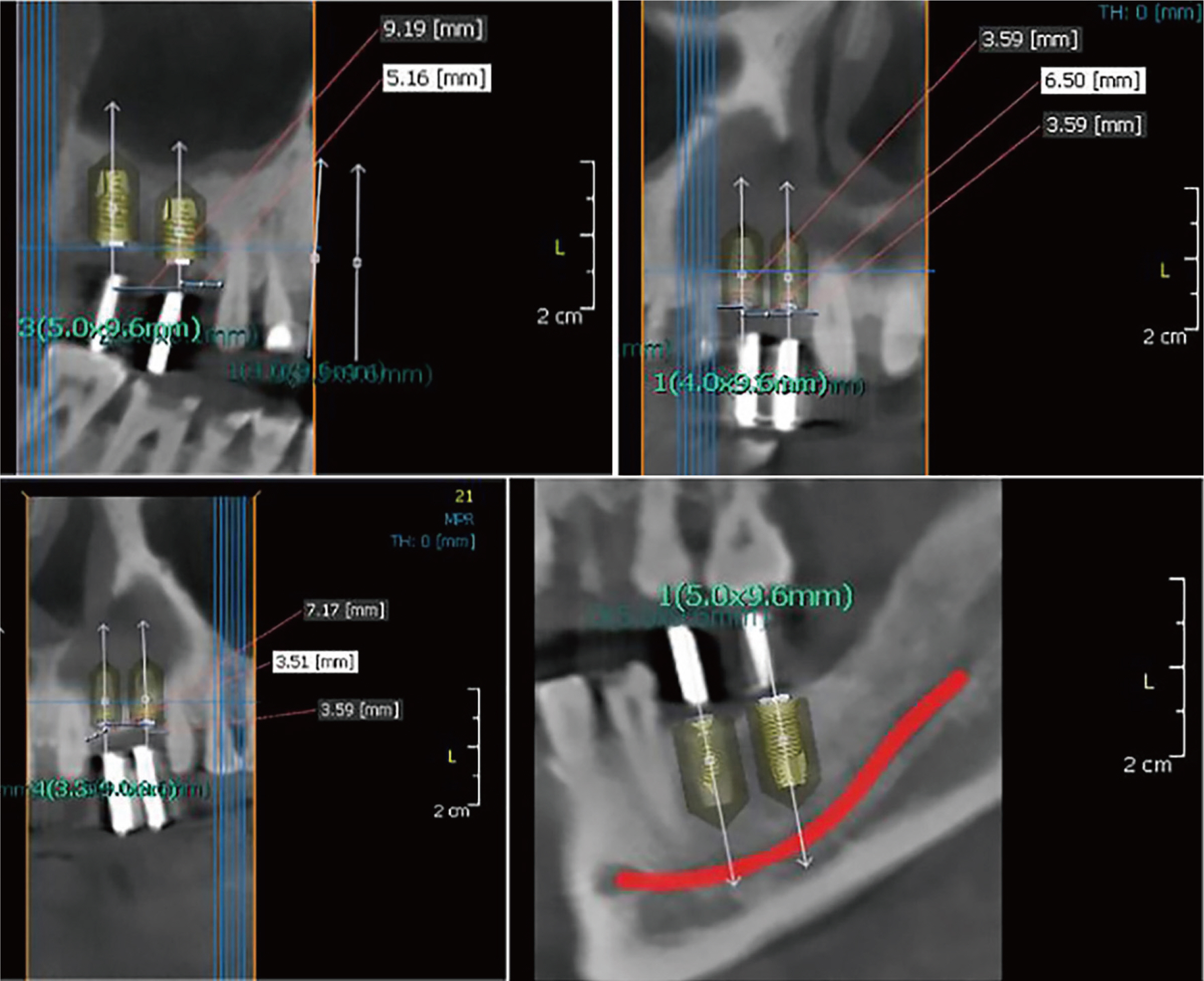
Fig. 6 Bone quality analysis and plan.

Fig. 7 Implant surgery. (A) Rt upper posterior, (B) Rt upper anterior, (C) Lt upper anterior.

Fig. 8 Provisional restoration. (A) Rt balancing, (B) Upper view, (C) Lt working, (D) Right view, (E) Frontal view, (F) left view, (G) Rt working, (H) Lower view, (I) Lt balancing.

Fig. 9 (A) Articulator adjustment value, (B) Customized anterior guidance table.

Fig. 10 CAD-CAM double scanning.

Fig. 11 Definitive restoration. (A) Rt balancing, (B) Upper view, (C) Lt working, (D) Right view, (E) Frontal view, (F) left view, (G) Rt working, (H) Lower view, (I) Lt balancing.
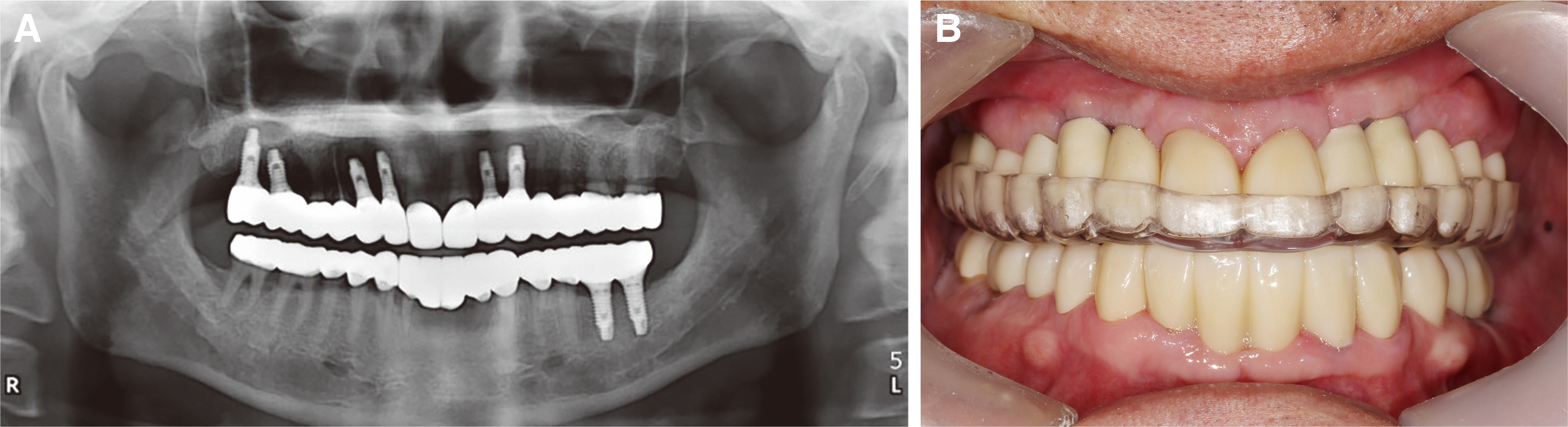
Fig. 12 (A) Panoramic radiograph after treatment, (B) Night guard.
Reference
-
References
1. Afsharzand Z, Rashedi B, Petropoulos VC. 2006; Communication between the dental laboratory technician and dentist: work authorization for fixed partial dentures. J Prosthodont. 15:123–8. DOI: 10.1111/j.1532-849X.2006.00086.x. PMID: 16650014.2. Turner KA, Missirlian DM. 1984; Restoration of the extremely worn dentition. J Prosthet Dent. 52:467–74. DOI: 10.1016/0022-3913(84)90326-3. PMID: 6389829.3. Rosenblum RH, Huffman RW. 1985; Leaf gauge with consecutively numbered leaves. J Prosthet Dent. 54:652–4. DOI: 10.1016/0022-3913(85)90242-2. PMID: 3863942.4. Ferrario VF, Sforza C, Miani A Jr. 1997; Statistical evaluation of Monson's sphere in healthy permanent dentitions in man. Arch Oral Biol. 42:365–9. DOI: 10.1016/S0003-9969(97)00021-6. PMID: 9233845.5. Farias-Neto A, Dias AH, de Miranda BF, de Oliveira AR. 2013; Face-bow transfer in prosthodontics: a systematic review of the literature. J Oral Rehabil. 40:686–92. DOI: 10.1111/joor.12081. PMID: 23829310.6. Paul SJ. 2001; Smile analysis and face-bow transfer: enhancing aesthetic restorative treatment. Pract Proced Aesthet Dent. 13:217–22. PMID: 11360768.7. Palik JF, Nelson DR, White JT. 1985; Accuracy of an earpiece face-bow. J Prosthet Dent. 53:800–4. DOI: 10.1016/0022-3913(85)90160-X. PMID: 3859649.8. Dawson PE. 2007. Functional Occlusion: From TMJ to Smile Design. Mosby;St. Louis: p. 199–206.9. Ogawa T, Koyano K, Suetsugu T. 1996; The relationship between inclination of the occlusal plane and jaw closing path. J Prosthet Dent. 76:576–80. DOI: 10.1016/S0022-3913(96)90432-1. PMID: 8957780.10. Joo HS, Park SW, Yun KD, Lim HP. 2016; Complete-mouth rehabilitation using a 3D printing technique and the CAD/CAM double scanning method: A clinical report. J Prosthet Dent. 116:3–7. DOI: 10.1016/j.prosdent.2016.01.007. PMID: 26946918.11. Papaspyridakos P, Chochlidakis K, Kang K, Chen YW, Alghfeli A, Kudara Y, Weber HP. 2020; Digital Workflow for Implant Rehabilitation with Double Full-Arch Monolithic Zirconia Prostheses. J Prosthodont. 29:460–65. DOI: 10.1111/jopr.13166. PMID: 32185825.12. Nazarian A. 2014; Utilizing an effective protocol for full-mouth reconstructions. Dent Today. 33:100. PMID: 24791321.13. Vafiadis D, Goldstein G, Garber D, Lambrakos A, Kowalski B. 2017; Immediate Implant Placement of a Single Central Incisor Using a CAD/CAM Crown-Root Form Technique: Provisional to Final Restoration. J Esthet Restor Dent. 29:13–21. DOI: 10.1111/jerd.12265. PMID: 27673748.
- Full Text Links
-
- Actions
-
Cited
- CITED
-
- Close
- Share
-
- Similar articles
-
- Full mouth rehabilitation of the patient with severely worn dentition using monolithic zirconia prosthesis: A clinical report
- Re-establishment of occlusal plane in a patient with a failed implant prosthesis
- Oral rehabilitation of excessive tooth wear patient usingzirconia fixed prosthesis with increased vertical dimension
- Full mouth rehabilitation of class III patient with disharmonious occlusal plane: A case report
- Full mouth rehabilitation of a patient with tooth wear and insufficient restorative space due to loss of posterior teeth support: a case report
