Cr-Co removable partial denture treatment fabricated by selective laser melting: a case report
- Affiliations
-
- 1Department of Prosthodontics, College of Dentistry, Dankook University, Cheonan, Republic of Korea
- KMID: 2525977
- DOI: http://doi.org/10.14368/jdras.2021.37.1.39
Abstract
- Compared to conventional method, if metal framework of removable partial denture is fabricated by selective laser melting, various laboratory works are omitted, saving time and simplifying the process. In addition, metal framework with homogeneous density can be obtained, expecting excellent mechanical properties, especially resistance to fatigue fracture. In these cases, impression were taken using conventional methods in partial edentulous patients, master casts were fabricated and scanned to obtain digital data. After designing the metal frameworks on the scanned data, removable partial dentures were fabricated using selective laser melting methods. Through these procedure, satisfactory outcomes were achieved both in functional and esthetic aspects.
Figure
-
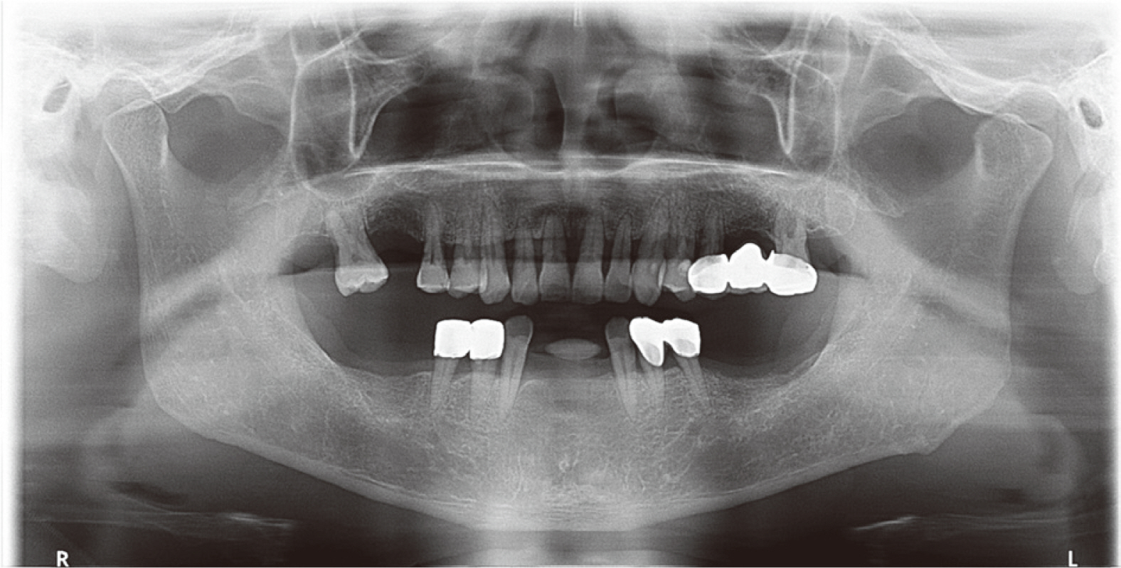
Fig. 1 Panoramic view of patient in case 1.
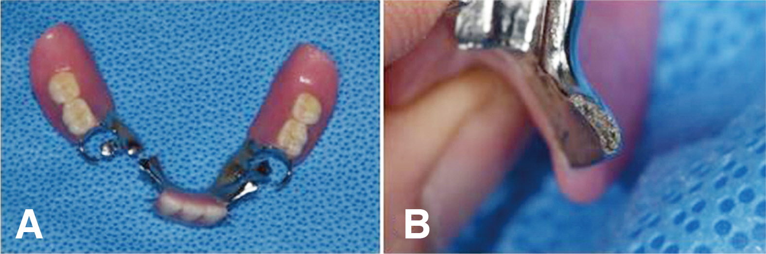
Fig. 2 Fracture of major connector in case 1. (A) Fractured position in major connector, (B) Fractured facet of major connector. Note the porous surface at the facet.
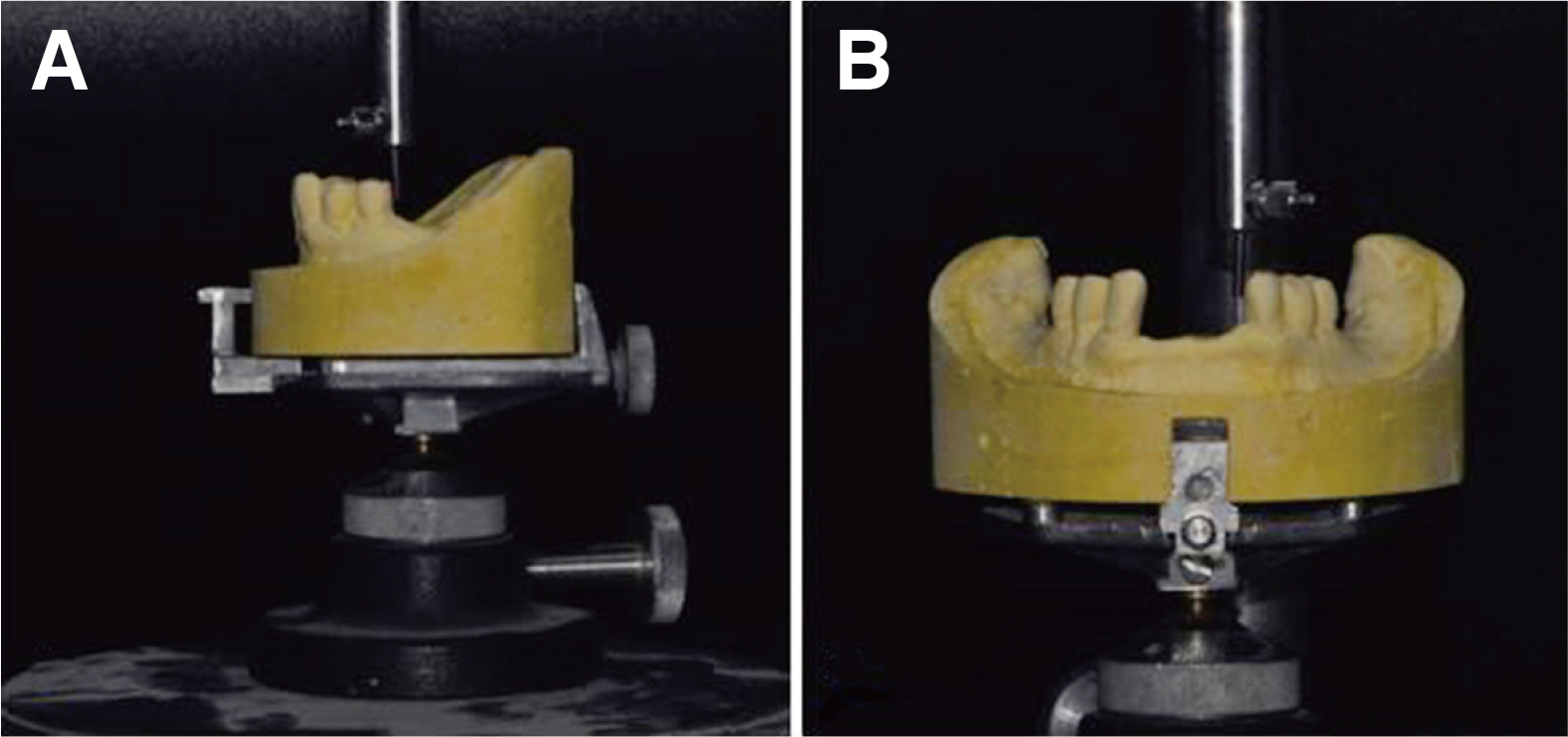
Fig. 3 Surveying procedure to identify proper path of insertion using dental surveyor in case 1. Prior to digital surveying, checking the path of the surveyed crowns. (A) Surveying of premolars, (B) Surveying of canines for checking path of insertion.
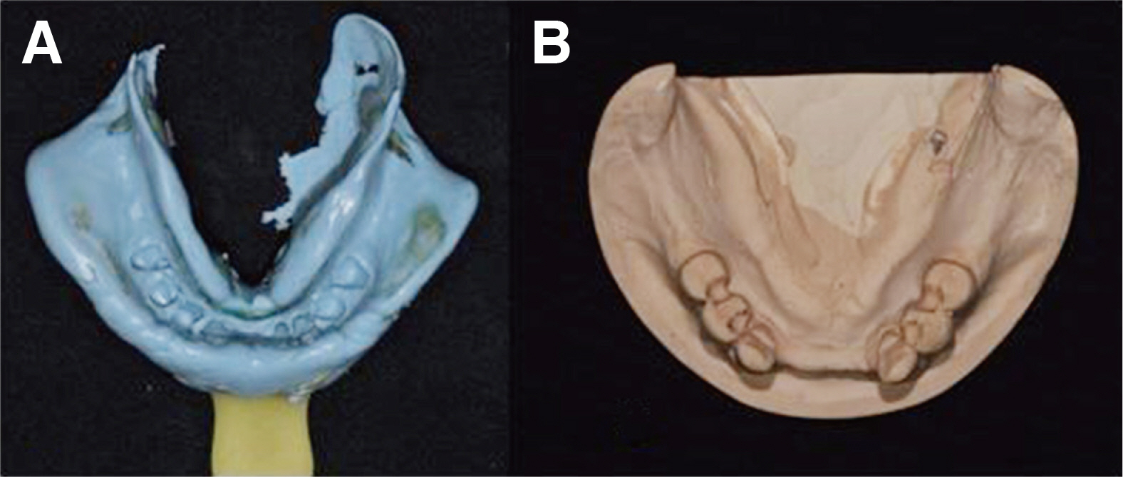
Fig. 4 Definitive impression in case 1. (A) Impression was taken by polyvinyl siloxane. (B) Impression was poured with improved dental stone.

Fig. 5 3D scan and design of metal framework in case 1. (A) Scanned master cast with determined path of insertion, (B) Designing retention grid and major connector, (C) Designing minor connector and clasp assemblies, (D) Pre-manufacturing of framework.
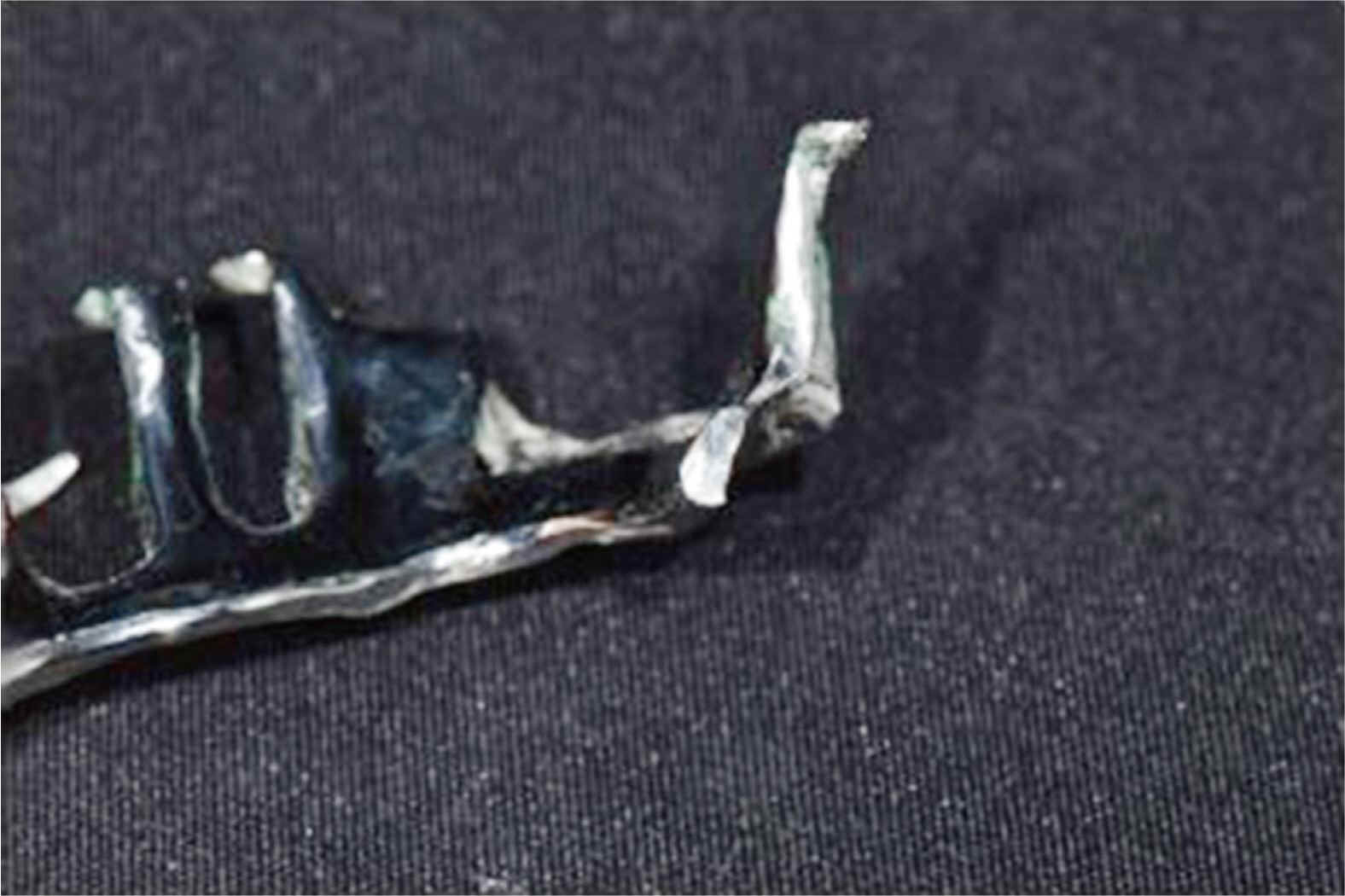
Fig. 6 Cut-off facet of metal framework fabricated by SLM in case 1. Note the dense surface at the facet showing more density than fractured porous surface.
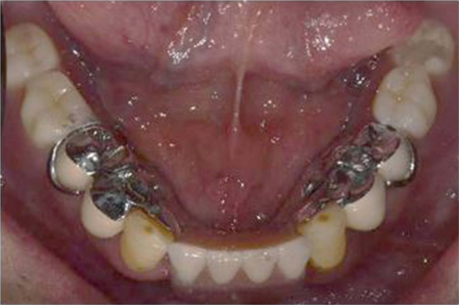
Fig. 7 Restoration of final denture in case 1. Showing proper adaptation of framework and comfort for patients.

Fig. 8 Panoramic view of patient in case 2.
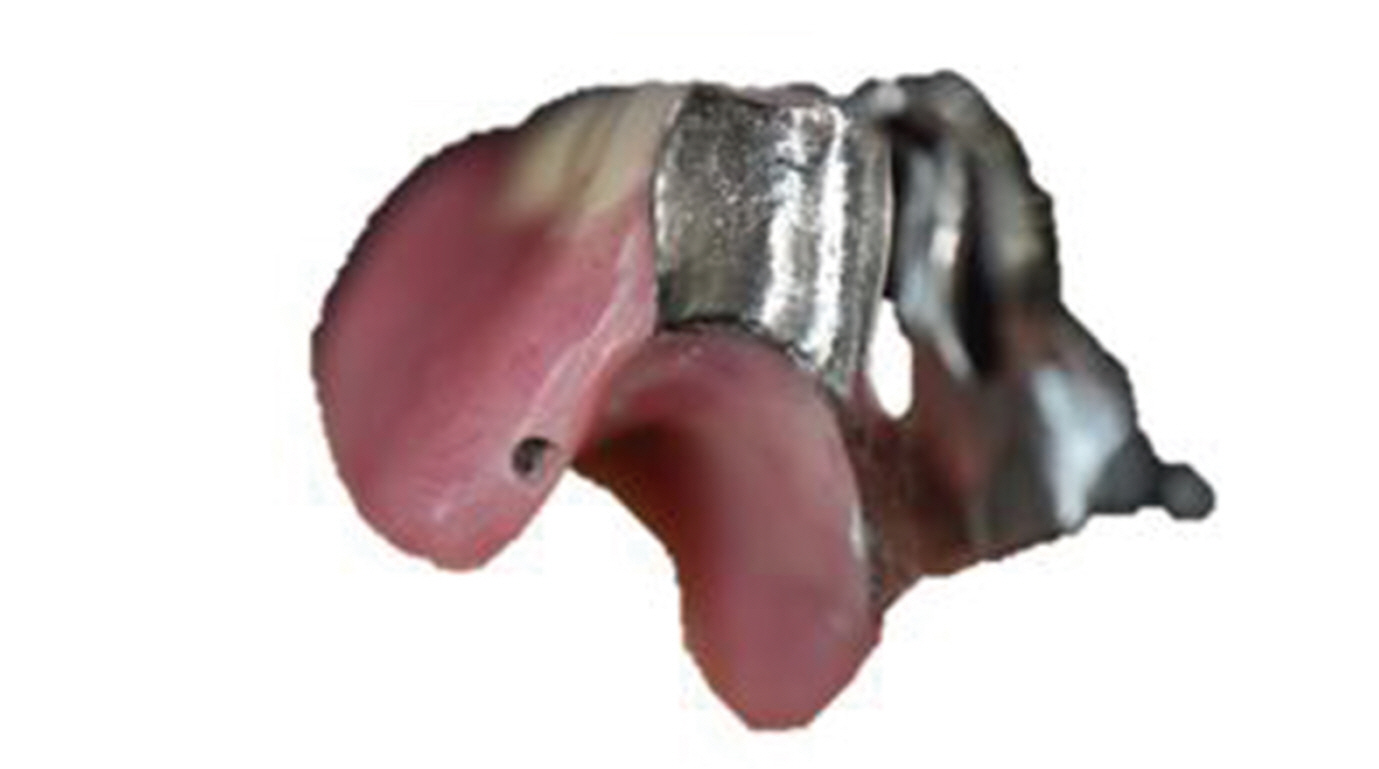
Fig. 9 Fractured facet of I-bar in case 2. The I-bar is entirely cut off from removable partial denture resulting from improper soft tissue undercut.
Fig. 10 Surveying procedure to identify proper path of insertion and undercut of soft tissue using dental surveyor in case 2. Prior to digital surveying, checking the path of the surveyed crowns.
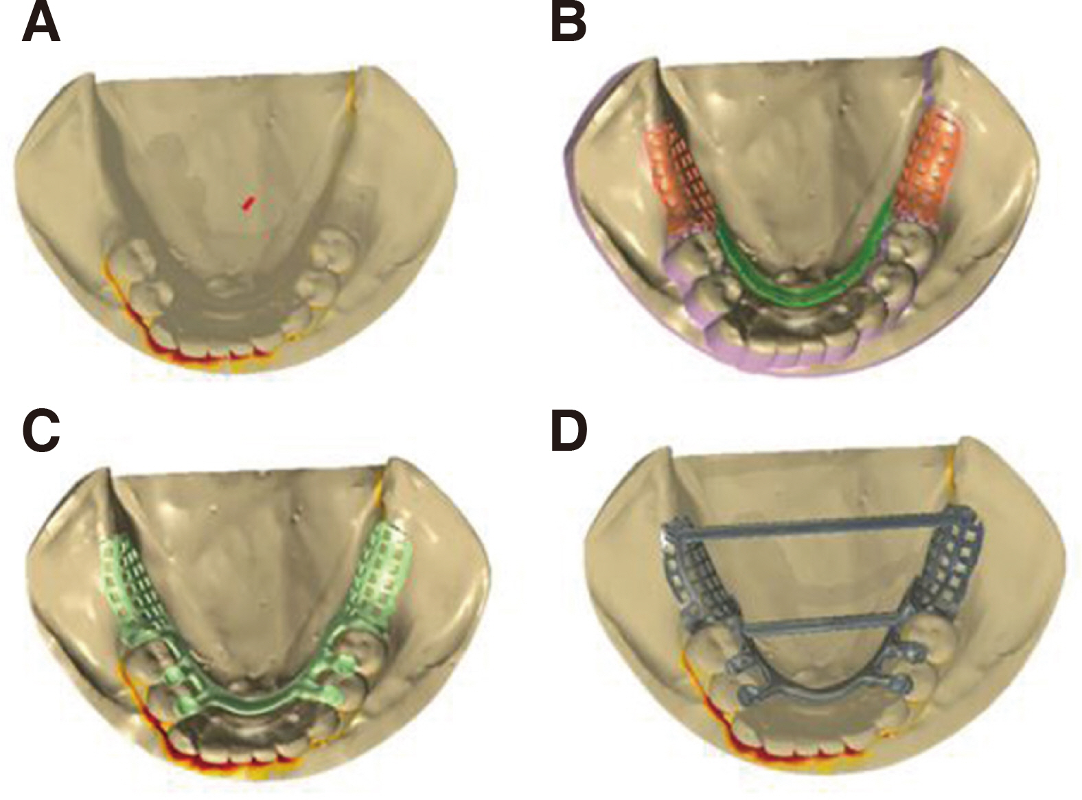
Fig. 11 3D scan and design of metal framework in case 2. (A) Scanned master cast with determined path of insertion, (B) Designing retention grid and major connector, (C) Designing minor connector and clasp assemblies, (D) Pre-manufacturing of framework.
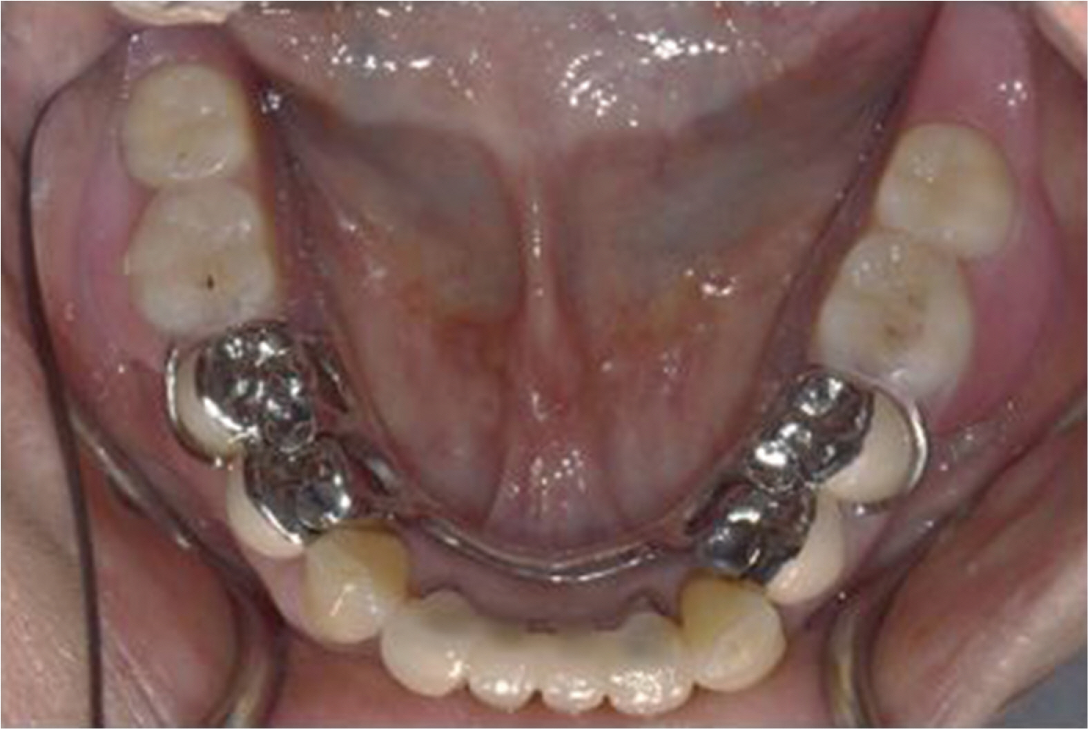
Fig. 12 Restoration of final denture in case 2. The patient showed satisfaction with new removable partial denture.
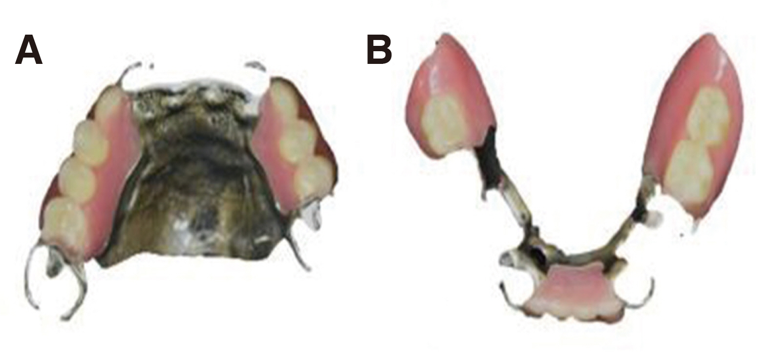
Fig. 13 Fractured metal framework in case 3. (A) Fracture at retentive arm in maxillary removable partial denture (#26), (B) Fracutre at minor connector and rest in mandibular removable partial denture (#46).
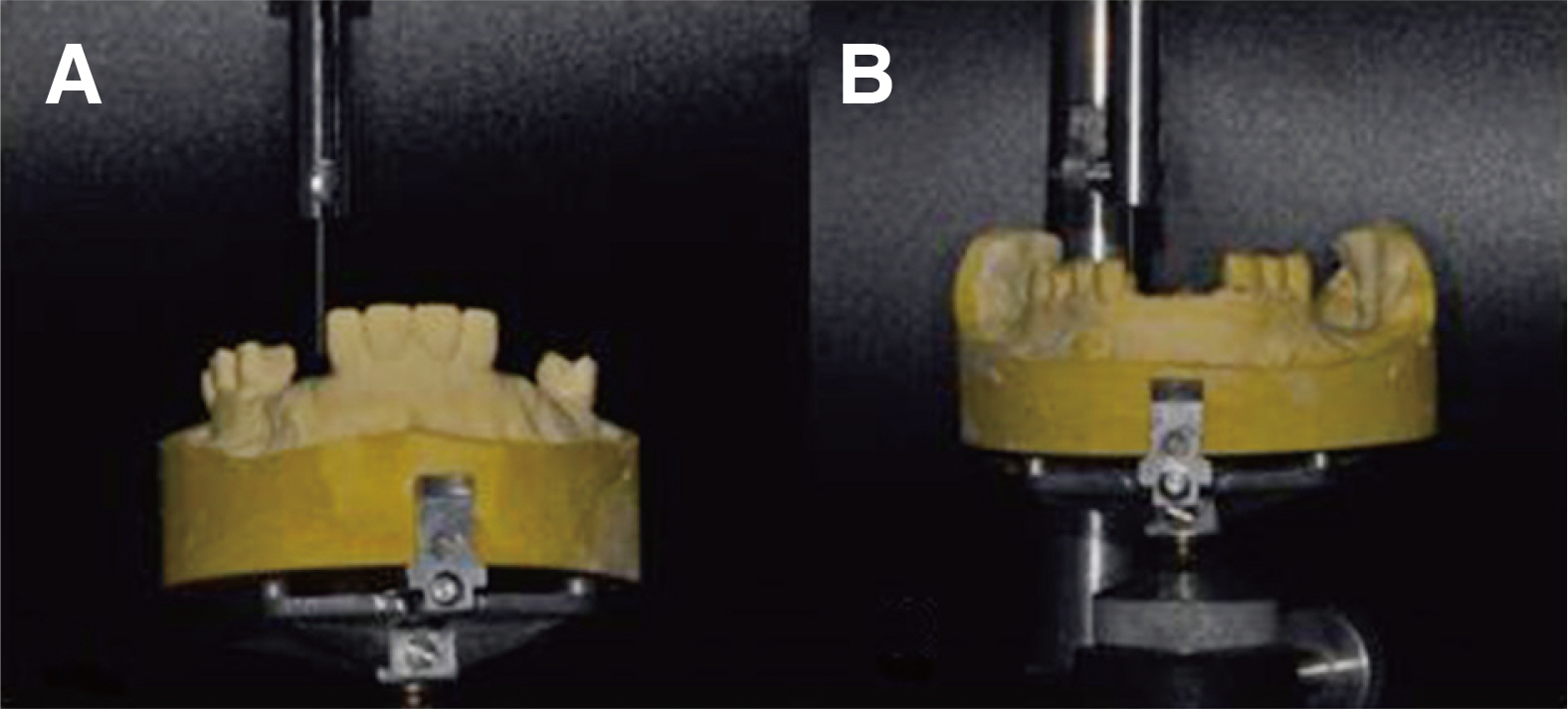
Fig. 14 Surveying procedure to identify proper path of insertion and undercut of pre-existed surveyed crowns and natural teeth in case 3. (A) Surveying of maxilla, (B) Sureveying of mandible.
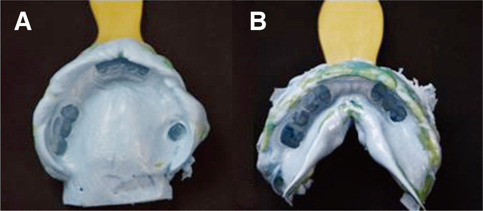
Fig. 15 Definitive impression in case 3. Impression was taken by polyvinyl siloxane. (A) Impression of maxilla, (B) Impression of mandible.
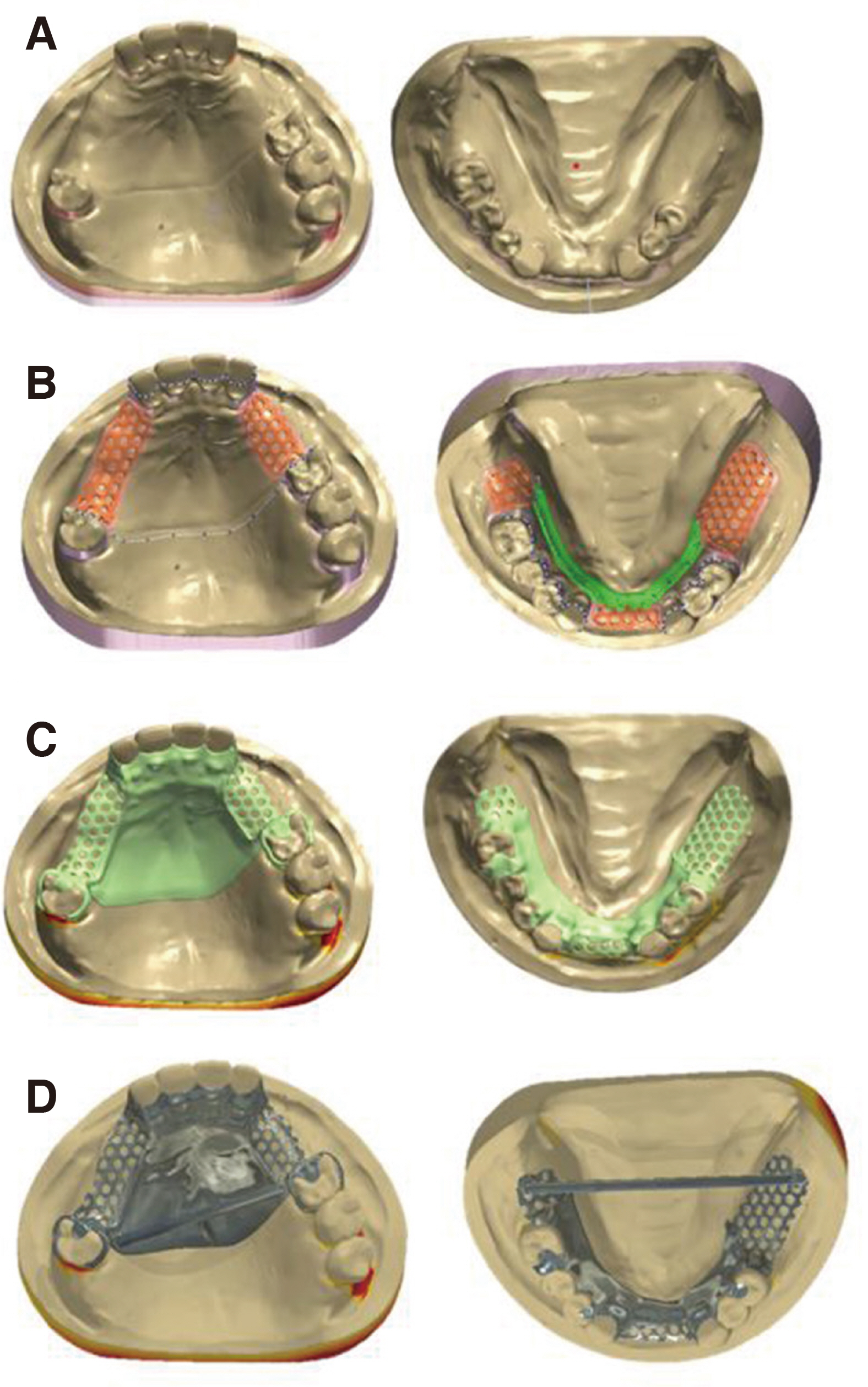
Fig. 16 3D scan and design of metal framework in case 3. (A) Scanned master cast with determined path of insertion, (B) Designing retention grid and major connector, (C) Designing minor connector and clasp assemblies, (D) Pre-manufacturing of framework.

Fig. 17 Restoration of final denture in case 3. (A) Maxilla, (B) Mandible. Kennedy class III case in maxillary restoration and short tissue supported area in mandibular restoration resulted in great stability and patient’s satisfaction.
Reference
-
References
1. Williams RJ, Bibb R, Rafik T. 2004; A technique for fabricating patterns for removable partial denture frameworks using digitized casts and electronic surveying. J Prosthet Dent. 91:85–8. DOI: 10.1016/j.prosdent.2003.10.002. PMID: 14739899.2. Williams RJ, Bibb R, Eggbeer D, Collis J. 2006; Use of CAD/CAM technology to fabricate a removable partial denture framework. J Prosthet Dent. 96:96–9. DOI: 10.1016/j.prosdent.2006.05.029. PMID: 16911885.3. Eggbeer D, Bibb R, Williams R. 2005; The computeraided design and rapid prototyping fabrication of removable partial denture frameworks. Proc Inst Mech Eng H. 219:195–202. DOI: 10.1243/095441105X9372. PMID: 15934395.4. Smith S, Dvorak D. 1998; Tool path strategies for high speed milling aluminum workpieces with thin webs. Mechatronics. 8:291–300. DOI: 10.1016/S0957-4158(97)00058-5.5. Dizon JRC, Espera AH, Chen Q, Advincula RC. 2018; Mechanical characterization of 3D-printed polymers. Addit Manufac. 20:44–67. DOI: 10.1016/j.addma.2017.12.002.6. Tasaka A, Shimizu T, Kato Y, Okano H, Ida Y, Higuchi S, Yamashita S. 2020; Accuracy of removable partial denture framework fabricated by casting with a 3D printed pattern and selective laser sintering. J Prosthodont Res. 64:224–30. DOI: 10.1016/j.jpor.2019.07.009. PMID: 31466919.7. Zhou Y, Li N, Yan J, Zeng Q. 2018; Comparative analysis of the microstructures and mechanical properties of Co-Cr dental alloys fabricated by different methods. J Prosthet Dent. 120:617–23. DOI: 10.1016/j.prosdent.2017.11.015. PMID: 29627206.8. Kim HR, Jang SH, Kim YK, Son JS, Min BK, Kim KH, Kwon TY. 2016; Microstructures and mechanical properties of Co-Cr dental alloys fabricated by three CAD/CAM-based processing techniques. Materials. 9:596. DOI: 10.3390/ma9070596. PMID: 28773718. PMCID: PMC5456947.9. Takaichi A, Nakamoto T, Joko N, Nomura N, Tsutsumi Y, Migita S, Doi H, Kurosu S, Chiba A, Wakabayashi N, Igarashi Y, Hanawa T. 2013; Microstructures and mechanical properties of Co-29Cr-6Mo alloy fabricated by selective laser melting process for dental applications. J Mech Behav Biomed Mater. 21:67–76. DOI: 10.1016/j.jmbbm.2013.01.021. PMID: 23500549.10. Schweiger J, Güth JF, Erdelt KJ, Edelhoff D, Schubert O. 2020; Internal porosities, retentive force, and survival of cobalt-chromium alloy clasps fabricated by selective laser-sintering. J Prosthodont Res. 64:210–16. DOI: 10.1016/j.jpor.2019.07.006. PMID: 31680054.11. Lapcevic AR, Jevremovic DP, Puskar TM, Williams RJ, Eggbeer D. 2016; Comparative analysis of structure and hardness of cast and direct metal laser sintering produced Co-Cr alloys used for dental devices. Rapid Prototyp J. 22:144–51. DOI: 10.1108/RPJ-04-2014-0051.12. Tuna SH, Pekmez NÖ, Kürkçüoğlu I. 2015; Corrosion resistance assessment of Co-Cr alloy frameworks fabricated by CAD/CAM milling, laser sintering, and casting methods. J Prosthet Dent. 114:725–34. DOI: 10.1016/j.prosdent.2015.02.031. PMID: 26187104.13. Hedberg YS, Qian B, Shen Z, Virtanen S, Wallinder IO. 2014; In vitro biocompatibility of CoCrMo dental alloys fabricated by selective laser melting. Dent Mater. 30:525–34. DOI: 10.1016/j.dental.2014.02.008. PMID: 24598762.14. Mansour M, Sanchez E, Machado C. 2016; The use of digital impressions to fabricate tooth-supported partial removable dental prostheses: A clinical Report. J Prosthodont. 25:495–7. DOI: 10.1111/jopr.12346. PMID: 26371612.15. Al Jabbari YS, Koutsoukis T, Barmpagadaki X, Zinelis S. 2014; Metallurgical and interfacial characterization of PFM Co-Cr dental alloys fabricated via casting, milling or selective laser melting. Dent Mater. 30:79–88. DOI: 10.1016/j.dental.2014.01.008. PMID: 24534375.16. Kajima Y, Takaichi A, Nakamoto T, Kimura T, Yogo Y, Ashida M, Doi H, Nomura N, Takahashi H, Hanawa T, Wakabayashi N. 2016; Fatigue strength of Co-Cr-Mo alloy clasps prepared by selective laser melting. J Mech Behav Biomed Mater. 59:446–58. DOI: 10.1016/j.jmbbm.2016.02.032. PMID: 26974490.17. Tregerman I, Renne W, Kelly A, Wilson D. 2019; Evaluation of removable partial denture frameworks fabricated using 3 different techniques. J Prosthet Dent. 122:390–5. DOI: 10.1016/j.prosdent.2018.10.013. PMID: 30948301.18. Ye H, Ning J, Li M, Niu L, Yang J, Sun Y, Zhou Y. 2017; Preliminary clinical application of removable partial denture frameworks fabricated using computer aided design and rapid prototyping techniques. Int J Prosthodont. 30:348–53. DOI: 10.11607/ijp.5270. PMID: 28697204.19. Tasaka A, Kato Y, Odaka K, Matsunaga S, Goto TK, Abe S, Yamashita S. 2019; Accuracy of Clasps Fabricated with Three Different CAD/CAM Technologies: Casting, Milling, and Selective Laser Sintering. Int J Prosthodont. 32:526–9. DOI: 10.11607/ijp.6363. PMID: 31664269.20. Kanazawa M, Iwaki M, Minakuchi S, Nomura N. 2014; Fabrication of titanium alloy frameworks for complete dentures by selective laser melting. J Prosthet Dent. 112:1441–7. DOI: 10.1016/j.prosdent.2014.06.017. PMID: 25258261.
- Full Text Links
-
- Actions
-
Cited
- CITED
-
- Close
- Share
-
- Similar articles
-
- Dental Co-Cr alloys fabricated by selective laser melting: A review article
- Restoration of bilateral distal extension removable partial denture using a fixed implant prosthesis in unilateral partial edentulous patient: A case report
- Esthetic removable partial denture with implants and resin clasp: Case report
- Rehabilitation of maxillary partial edentulous patients using implant assisted removable partial denture
- Implant-assisted removable partial denture using digital guide surgery in partially edentulous mandible: A case report
