A Rare Case of Pituicytoma Presenting Cystic Formation
- Affiliations
-
- 1Departments of Neurosurgery, Myongji Hospital, Hanyang University College of Medicine, Goyang, Korea
- 2Departments of Pathology, Myongji Hospital, Hanyang University College of Medicine, Goyang, Korea
- KMID: 2522222
- DOI: http://doi.org/10.14791/btrt.2021.9.e11
Abstract
- Pituicytoma is a rare solid benign tumor of the sellar and/or suprasellar region originating from the pituicytes of the neurohypophysis or infundibulum, which is not differentiated from a pituitary adenoma that is diagnosed mostly in the sellar and/or suprasellar region. In addition, cystic tumors are very rare and have not been reported due to their solid and hypervascular natures. A 33-year-old man presented with a chronic headache which exacerbated recently. MRI was performed and revealed a cystic tumor in the sellar and suprasellar regions with a small parenchymal island in the cyst compressing the optic chiasm. The endoscopic endonasal transsphenoidal approach was used to remove the tumor. Immunohistochemical staining was positive for thyroid transcription factor 1, S-100 protein, and glial fibrillary acidic protein. The pituicytoma was diagnosed based on histologic findings. The authors review herein the literature on clinical presentation, diagnosis, surgical management, and outcome.
Keyword
Figure
-
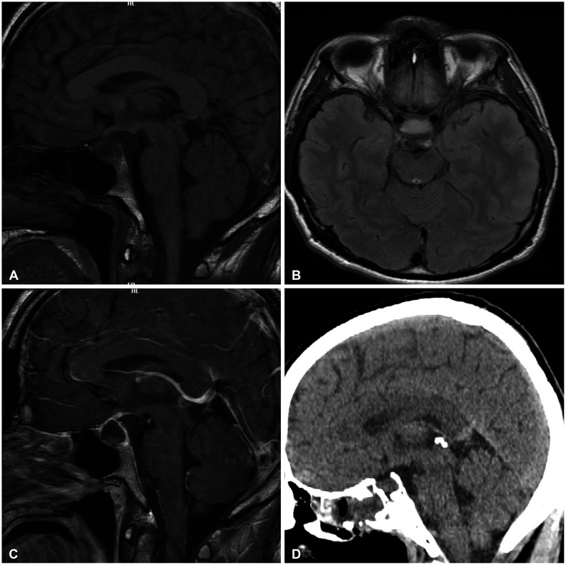
Fig. 1 Radiologic features of pituicytoma. A-C: Sellar MRI T1-weighted image shows isointense cyst wall with some parenchyma in depending position (A, sagittal view) and slightly hyperintense cyst content (B, axial view), with moderate hyperintensity of cyst wall at contrast image (C, sagittal). D: Postoperative precontrast CT scan in sagittal view.
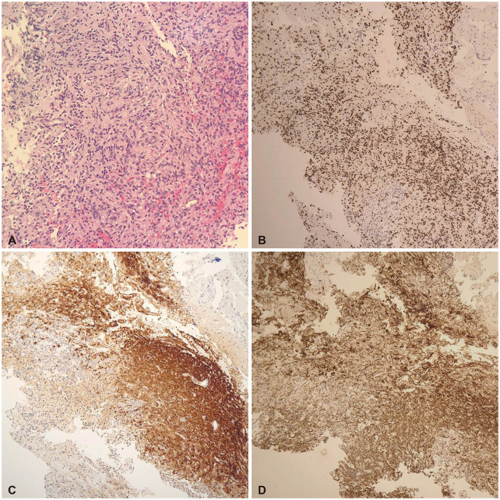
Fig. 2 Tissue morphology of pituicytoma. A: Tumor cells are arranged in sheets and fascicles. The tumor cells are spindled to epithelioid with abundant eosinophilic cytoplasm (hematoxylin and eosin, ×100). B-D: Immunohistochemistry of tumors. B: The cells shows diffuse, strong nuclear positivity for thyroid transcription factor 1 (×40). C: The tumor cells are S-100 positive (×40). D: Tumor cells shows positivity for glial fibrillary acidic protein (×40).
Reference
-
1. Fuller GN, Ribalta T, Wildrick DM. Brain tumors: an overview of current histopathologic and genentic classification. Winn HR, editor. Youmans and Winn neurological surgery. 7th ed. Vol 2. Philadelphia: Elsevier;2017. p. 774.2. Viaene AN, Lee EB, Rosenbaum JN, Nasrallah IM, Nasrallah MP. Histologic, immunohistochemical, and molecular features of pituicytomas and atypical pituicytomas. Acta Neuropathol Commun. 2019; 7:69. PMID: 31046843.3. Lee EB, Tihan T, Scheithauer BW, Zhang PJ, Gonatas NK. Thyroid transcription factor 1 expression in sellar tumors: a histogenetic marker? J Neuropathol Exp Neurol. 2009; 68:482–488. PMID: 19525896.4. Shim HK, Cha SH, Cho WH, Park SH. Pituicytoma with significant tumor vascularity mimicking pituitary macroadenoma. Brain Tumor Res Treat. 2017; 5:110–115. PMID: 29188213.5. Kowalski RJ, Prayson RA, Mayberg MR. Pituicytoma. Ann Diagn Pathol. 2004; 8:290–294. PMID: 15494936.6. Liss L, Kahn EA. Pituicytoma, tumor of the sella turcica; a clinicopathological study. J Neurosurg. 1958; 15:481–488. PMID: 13576191.7. Antoni N. Gliomas of the neurohypophysis and hypophysial stalk; a preliminary report. J Neurosurg. 1950; 7:521–531. PMID: 14795256.8. Scothorne CM. A glioma of the posterior lobe of the pituitary gland. J Pathol Bacteriol. 1955; 69:109–112. PMID: 13243176.9. Salge-Arrieta FJ, Carrasco-Moro R, Rodríguez-Berrocal V, et al. Clinical features, diagnosis and therapy of pituicytoma: an update. J Endocrinol Invest. 2019; 42:371–384. PMID: 30030746.10. Brat DJ, Scheithauer BW, Staugaitis SM, Holtzman RN, Morgello S, Burger PC. Pituicytoma: a distinctive low-grade glioma of the neurohypophysis. Am J Surg Pathol. 2000; 24:362–368. PMID: 10716149.11. Louis DN, Perry A, Reifenberger G, et al. The 2016 World Health Organization classification of tumors of the central nervous system: a summary. Acta Neuropathol. 2016; 131:803–820. PMID: 27157931.12. Shibuya M. Welcoming the new WHO classification of pituitary tumors 2017: revolution in TTF-1-positive posterior pituitary tumors. Brain Tumor Pathol. 2018; 35:62–70. PMID: 29500747.13. Zygourakis CC, Rolston JD, Lee HS, Partow C, Kunwar S, Aghi MK. Pituicytomas and spindle cell oncocytomas: modern case series from the University of California, San Francisco. Pituitary. 2015; 18:150–158. PMID: 24823438.14. Wang J, Liu Z, Du J, et al. The clinicopathological features of pituicytoma and the differential diagnosis of sellar glioma. Neuropathology. 2016; 36:432–440. PMID: 26919073.15. Feng M, Carmichael JD, Bonert V, Bannykh S, Mamelak AN. Surgical management of pituicytomas: case series and comprehensive literature review. Pituitary. 2014; 17:399–413. PMID: 24037647.16. Wolfe SQ, Bruce J, Morcos JJ. Pituicytoma: case report. Neurosurgery. 2008; 63:E173–E174. PMID: 18728556.17. Hammoud DA, Munter FM, Brat DJ, Pomper MG. Magnetic resonance imaging features of pituicytomas: analysis of 10 cases. J Comput Assist Tomogr. 2010; 34:757–761. PMID: 20861781.18. Gibbs WN, Monuki ES, Linskey ME, Hasso AN. Pituicytoma: diagnostic features on selective carotid angiography and MR imaging. AJNR Am J Neuroradiol. 2006; 27:1639–1642. PMID: 16971602.19. Benveniste RJ, Purohit D, Byun H. Pituicytoma presenting with spontaneous hemorrhage. Pituitary. 2006; 9:53–58. PMID: 16703409.20. Cossu G, Dimitriou J, Brouland JP, Daniel RT, Messerer M. An exceptional presentation of pituicytoma apoplexy: a case report. Oncol Lett. 2018; 16:643–647. PMID: 29928451.21. Kim YG, Park YS. Second-stage transsphenoidal approach (TSA) for highly vascular pituicytomas in children. Childs Nerv Syst. 2015; 31:985–989. PMID: 25771921.
