The Value of 18 F-FDG PET/CT in Evaluating Disease Severity and Prognosis in Idiopathic Pulmonary Fibrosis Patients
- Affiliations
-
- 1Department of Pulmonary and Critical Care Medicine, Asan Medical Center, University of Ulsan College of Medicine, Seoul, Korea
- 2Department of Nuclear Medicine, Asan Medical Center, University of Ulsan College of Medicine, Seoul, Korea
- 3Division of Nuclear Medicine, Department of Radiology, Kangnam Sacred Heart Hospital, Hallym University College of Medicine, Seoul, Korea
- KMID: 2521471
- DOI: http://doi.org/10.3346/jkms.2021.36.e257
Abstract
- Background
Several parameters are useful for assessing disease severity in idiopathic pulmonary fibrosis (IPF); however, the role of 18 F-fluorodeoxyglucose positron emission tomography/computed tomography ( 18 F-FDG PET/CT) is not well-defined. We aimed to evaluate the value of 18 F-FDG PET/CT for assessing disease severity and prognosis in IPF patients.
Methods
Clinical data of 89 IPF patients (mean age: 68.1 years, male: 94%) who underwent 18 F-FDG PET/CT for evaluation of lung nodules or cancer staging were retrospectively reviewed. Mean and maximal standardized uptake values (SUV mean , SUV max , respectively) were measured in the fibrotic area. Adjusted SUV, including SUV ratio (SUVR, defined as SUV max -to-liver SUV mean ratio), tissue fraction-corrected SUV mean (SUV meanTF ), and SUVR (SUVRTF ), and tissue-to-blood ratio (SUV max /SUV mean venous; TBR blood ) were obtained. Death was defined as the primary outcome, and associations between other clinical parameters (lung function, exercise capacity, C-reactive protein [CRP] level) were also investigated.
Results
All SUV parameters were inversely correlated with the forced vital capacity, diffusing capacity for carbon monoxide, and positively correlated with CRP level and the gender-agephysiology index. The SUV mean , SUV max , and SUV meanTF were associated with changes in lung function at six months. The SUVR (hazard ratio [HR], 1.738; 95% confidence interval [CI], 1.011–2.991), SUVR TF (HR, 1.441; 95% CI, 1.000–2.098), and TBR blood (HR, 1.377; 95% CI, 1.038–1.827) were significant predictors for mortality in patients with IPF in the univariate analysis, but not in the multivariate analysis.
Conclusion
18 F-FDG PET/CT may provide additional information on the disease severity and prognosis in IPF patients, and the SUVR may be superior to other SUV parameters.
Keyword
Figure
-
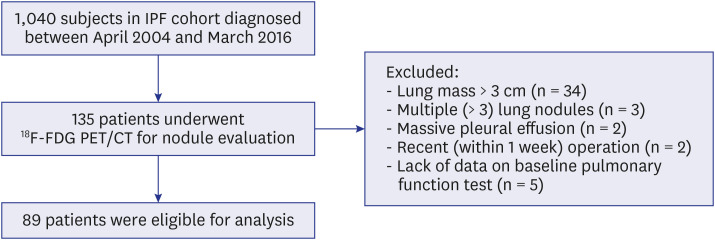
Fig. 1 Flowchart of the study.18F-FDG PET/CT = 18F-fluorodeoxyglucose positron emission tomography/computed tomography, IPF = idiopathic pulmonary fibrosis.
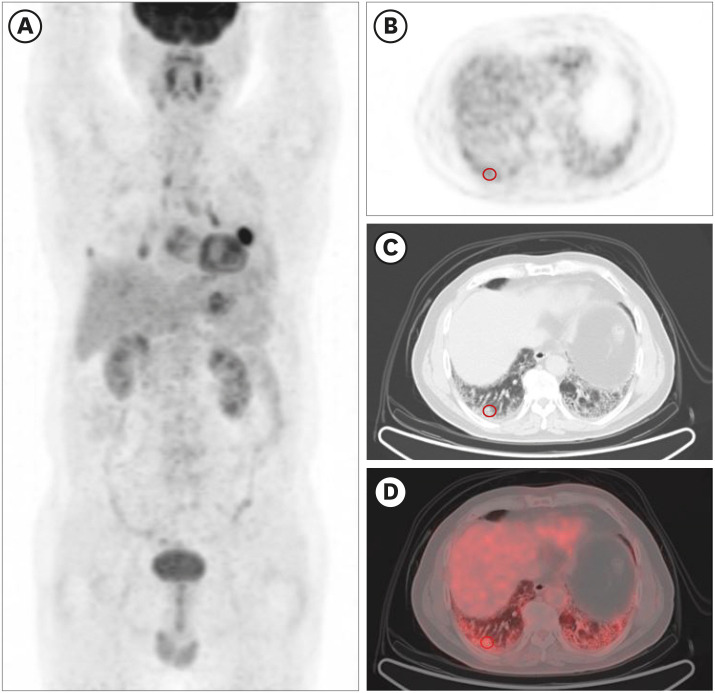
Fig. 2 Measurement of the standardized uptake value in the fibrotic area in 18F-FDG positron emission tomography/computed tomography images. (A) A coronal PET image. (B) A horizontal PET image. (C) A CT image. (D) A combined PET and CT fusion image. The red circles indicate the 1 cm diameter area centering on the highest 18F-FDG uptake in fibrotic areas. Both maximum standardized uptake value and mean standardized uptake value were measured in this circle.18F-FDG = 18F-fluorodeoxyglucose, PET = positron emission tomography, CT = computed tomography.
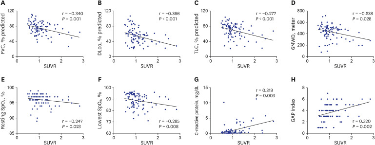
Fig. 3 Scatter plot depicting the correlation between standardized uptake value ratio and clinical parameters. (A) FVC. (B) DLco. (C) TLC. (D) 6MWD. (E) Resting SpO2. (F) Lowest SpO2. (G) C-reactive protein. H. GAP index.FVC = forced vital capacity, DLco = diffusing capacity for carbon monoxide, TLC = total lung capacity, 6MWD = six-minute walk distance, SpO2 = peripheral saturation of oxygen, GAP = gender-age-physiology, SUVR = standardized uptake value ratio.
Reference
-
1. Ley B, Collard HR, King TE Jr, King J. Clinical course and prediction of survival in idiopathic pulmonary fibrosis. Am J Respir Crit Care Med. 2011; 183(4):431–440. PMID: 20935110.
Article2. Raghu G, Rochwerg B, Zhang Y, Garcia CA, Azuma A, Behr J, et al. An official ATS/ERS/JRS/ALAT clinical practice guideline: treatment of idiopathic pulmonary fibrosis. An update of the 2011 clinical practice guideline. Am J Respir Crit Care Med. 2015; 192(2):e3–19. PMID: 26177183.
Article3. Shah NR, Noble P, Jackson RM, King TE Jr, Nathan SD, Padilla M, et al. A critical assessment of treatment options for idiopathic pulmonary fibrosis. Sarcoidosis Vasc Diffuse Lung Dis. 2005; 22(3):167–174. PMID: 16315778.
Article4. Collard HR, King TE Jr, Bartelson BB, Vourlekis JS, Schwarz MI, Brown KK. Changes in clinical and physiologic variables predict survival in idiopathic pulmonary fibrosis. Am J Respir Crit Care Med. 2003; 168(5):538–542. PMID: 12773325.
Article5. Flaherty KR, Mumford JA, Murray S, Kazerooni EA, Gross BH, Colby TV, et al. Prognostic implications of physiologic and radiographic changes in idiopathic interstitial pneumonia. Am J Respir Crit Care Med. 2003; 168(5):543–548. PMID: 12773329.
Article6. Caminati A, Bianchi A, Cassandro R, Mirenda MR, Harari S. Walking distance on 6-MWT is a prognostic factor in idiopathic pulmonary fibrosis. Respir Med. 2009; 103(1):117–123. PMID: 18786822.
Article7. Miller MR, Hankinson J, Brusasco V, Burgos F, Casaburi R, Coates A, et al. Standardisation of spirometry. Eur Respir J. 2005; 26(2):319–338. PMID: 16055882.8. McLean A, Warren PM, Gillooly M, MacNee W, Lamb D. Microscopic and macroscopic measurements of emphysema: relation to carbon monoxide gas transfer. Thorax. 1992; 47(3):144–149. PMID: 1519189.
Article9. Trip P, Nossent EJ, de Man FS, van den Berk IA, Boonstra A, Groepenhoff H, et al. Severely reduced diffusion capacity in idiopathic pulmonary arterial hypertension: patient characteristics and treatment responses. Eur Respir J. 2013; 42(6):1575–1585. PMID: 23949959.
Article10. Chugani HT, Phelps ME, Mazziotta JC. Positron emission tomography study of human brain functional development. Ann Neurol. 1987; 22(4):487–497. PMID: 3501693.
Article11. Erasmus JJ, Patz EF Jr. Positron emission tomography imaging in the thorax. Clin Chest Med. 1999; 20(4):715–724. PMID: 10587793.
Article12. Almuhaideb A, Papathanasiou N, Bomanji J. 18F-FDG PET/CT imaging in oncology. Ann Saudi Med. 2011; 31(1):3–13. PMID: 21245592.
Article13. Griffeth LK. Use of PET/CT scanning in cancer patients: technical and practical considerations. Proc (Bayl Univ Med Cent). 2005; 18(4):321–330. PMID: 16252023.
Article14. Groves AM, Win T, Screaton NJ, Berovic M, Endozo R, Booth H, et al. Idiopathic pulmonary fibrosis and diffuse parenchymal lung disease: implications from initial experience with 18F-FDG PET/CT. J Nucl Med. 2009; 50(4):538–545. PMID: 19289428.
Article15. El-Chemaly S, Malide D, Yao J, Nathan SD, Rosas IO, Gahl WA, et al. Glucose transporter-1 distribution in fibrotic lung disease: association with [18F]-2-fluoro-2-deoxyglucose-PET scan uptake, inflammation, and neovascularization. Chest. 2013; 143(6):1685–1691. PMID: 23699745.16. Lee EY, Wong CS, Fung SL, Yan PK, Ho JC. SUV as an adjunct in evaluating disease activity in idiopathic pulmonary fibrosis - a pilot study. Nucl Med Commun. 2014; 35(6):631–637. PMID: 24472818.
Article17. Nobashi T, Kubo T, Nakamoto Y, Handa T, Koyasu S, Ishimori T, et al. 18F-FDG uptake in less affected lung field provides prognostic stratification in patients with interstitial lung disease. J Nucl Med. 2016; 57(12):1899–1904. PMID: 27339874.
Article18. Justet A, Laurent-Bellue A, Thabut G, Dieudonné A, Debray MP, Borie R, et al. [18F]FDG PET/CT predicts progression-free survival in patients with idiopathic pulmonary fibrosis. Respir Res. 2017; 18(1):74. PMID: 28449678.
Article19. Castiaux A, Van Simaeys G, Goldman S, Bondue B. Assessment of 18F-FDG uptake in idiopathic pulmonary fibrosis: influence of lung density changes. Eur J Hybrid Imaging. 2018; 2(1):27.
Article20. Richeldi L, Costabel U, Selman M, Kim DS, Hansell DM, Nicholson AG, et al. Efficacy of a tyrosine kinase inhibitor in idiopathic pulmonary fibrosis. N Engl J Med. 2011; 365(12):1079–1087. PMID: 21992121.
Article21. Lee SH, Yeo Y, Kim TH, Lee HL, Lee JH, Park YB, et al. Korean guidelines for diagnosis and management of interstitial lung diseases: part 2. idiopathic pulmonary fibrosis. Tuberc Respir Dis (Seoul). 2019; 82(2):102–117. PMID: 30841014.
Article22. Wanger J, Clausen JL, Coates A, Pedersen OF, Brusasco V, Burgos F, et al. Standardisation of the measurement of lung volumes. Eur Respir J. 2005; 26(3):511–522. PMID: 16135736.
Article23. Macintyre N, Crapo RO, Viegi G, Johnson DC, van der Grinten CP, Brusasco V, et al. Standardisation of the single-breath determination of carbon monoxide uptake in the lung. Eur Respir J. 2005; 26(4):720–735. PMID: 16204605.
Article24. ATS Committee on Proficiency Standards for Clinical Pulmonary Function Laboratories. ATS statement: guidelines for the six-minute walk test. Am J Respir Crit Care Med. 2002; 166(1):111–117. PMID: 12091180.25. Ley B, Ryerson CJ, Vittinghoff E, Ryu JH, Tomassetti S, Lee JS, et al. A multidimensional index and staging system for idiopathic pulmonary fibrosis. Ann Intern Med. 2012; 156(10):684–691. PMID: 22586007.
Article26. Collard HR, Ryerson CJ, Corte TJ, Jenkins G, Kondoh Y, Lederer DJ, et al. Acute Exacerbation of Idiopathic Pulmonary Fibrosis. An International Working Group Report. Am J Respir Crit Care Med. 2016; 194(3):265–275. PMID: 27299520.
Article27. Thie JA. Understanding the standardized uptake value, its methods, and implications for usage. J Nucl Med. 2004; 45(9):1431–1434. PMID: 15347707.28. Watanabe H, Kanematsu M, Goshima S, Kondo H, Kawada H, Noda Y, et al. Adrenal-to-liver SUV ratio is the best parameter for differentiation of adrenal metastases from adenomas using 18F-FDG PET/CT. Ann Nucl Med. 2013; 27(7):648–653. PMID: 23625579.
Article29. Lambrou T, Groves AM, Erlandsson K, Screaton N, Endozo R, Win T, et al. The importance of correction for tissue fraction effects in lung PET: preliminary findings. Eur J Nucl Med Mol Imaging. 2011; 38(12):2238–2246. PMID: 21874321.
Article30. Win T, Screaton NJ, Porter JC, Ganeshan B, Maher TM, Fraioli F, et al. Pulmonary 18F-FDG uptake helps refine current risk stratification in idiopathic pulmonary fibrosis (IPF). Eur J Nucl Med Mol Imaging. 2018; 45(5):806–815. PMID: 29335764.31. Nathan SD, Albera C, Bradford WZ, Costabel U, du Bois RM, Fagan EA, et al. Effect of continued treatment with pirfenidone following clinically meaningful declines in forced vital capacity: analysis of data from three phase 3 trials in patients with idiopathic pulmonary fibrosis. Thorax. 2016; 71(5):429–435. PMID: 26968970.
Article32. Nakazawa S, Shimizu K, Mogi A, Kuwano H. Low diffusing capacity, emphysema, or pulmonary fibrosis: who is truly pulling the lung cancer strings? J Thorac Dis. 2018; 10(2):600–602. PMID: 29607119.
Article33. Boellaard R, Krak NC, Hoekstra OS, Lammertsma AA. Effects of noise, image resolution, and ROI definition on the accuracy of standard uptake values: a simulation study. J Nucl Med. 2004; 45(9):1519–1527. PMID: 15347719.34. Bural GG, Torigian DA, Chen W, Houseni M, Basu S, Alavi A. Increased 18F-FDG uptake within the reticuloendothelial system in patients with active lung cancer on PET imaging may indicate activation of the systemic immune response. Hell J Nucl Med. 2010; 13(1):23–25. PMID: 20411166.35. Liu G, Hu Y, Zhao Y, Yu H, Hu P, Shi H.Variations of the liver standardized uptake value in relation to background blood metabolism: an 2-[18F]Fluoro-2-deoxy-D-glucose positron emission tomography/computed tomography study in a large population from China. Medicine. 2018; 97(19):e0699. PMID: 29742723.
- Full Text Links
-
- Actions
-
Cited
- CITED
-
- Close
- Share
-
- Similar articles
-
- The Usefulness of F-18 FDG PET to Discriminate between Malignant and benign Nodule in Idiopathic Pulmonary Fibrosis
- F-18 FDG PET/CT Findings of Subcutaneous Panniculitis - Like T- Cell Lymphoma : A Case Report
- A Case of Incidentally Detected Nasopharyngeal Tuberculosis on F-18 FDG PET/CT
- 18F-FDG PET/CT Imaging of Peritoneal Fibrosis Mimicking Persistent Metastatic Ovarian Carcinoma
- Abdominal Mesh Implant Showing FDG Uptake on PET/CT
